INotes - Chapter 13
advertisement

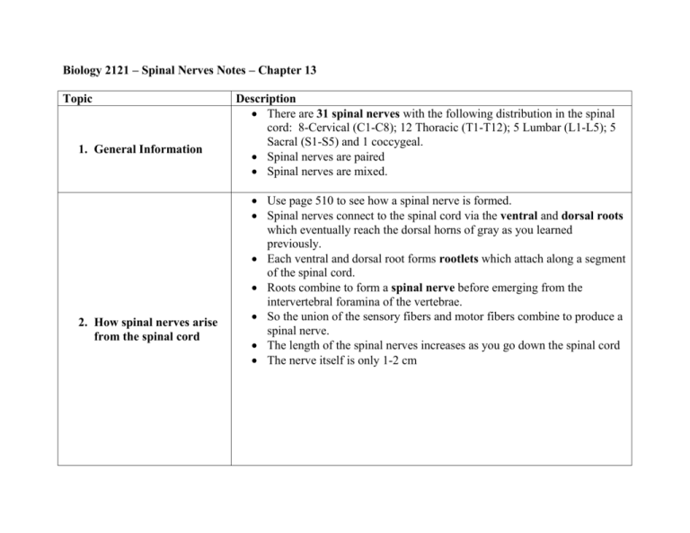
Biology 2121 – Spinal Nerves Notes – Chapter 13 Topic 1. General Information 2. How spinal nerves arise from the spinal cord Description There are 31 spinal nerves with the following distribution in the spinal cord: 8-Cervical (C1-C8); 12 Thoracic (T1-T12); 5 Lumbar (L1-L5); 5 Sacral (S1-S5) and 1 coccygeal. Spinal nerves are paired Spinal nerves are mixed. Use page 510 to see how a spinal nerve is formed. Spinal nerves connect to the spinal cord via the ventral and dorsal roots which eventually reach the dorsal horns of gray as you learned previously. Each ventral and dorsal root forms rootlets which attach along a segment of the spinal cord. Roots combine to form a spinal nerve before emerging from the intervertebral foramina of the vertebrae. So the union of the sensory fibers and motor fibers combine to produce a spinal nerve. The length of the spinal nerves increases as you go down the spinal cord The nerve itself is only 1-2 cm 3. Division of the spinal nerve 4. Innervation of the Back, Anterolateral Thorax and Abdominal Wall 5. The Nerve Plexuses After the spinal nerve is formed it branches into the dorsal ramus and a large ventral ramus and a small meningeal branch (goes back into the vertebral canal to innervate the meninges and blood vessels. Rami are also mixed nerves Rami communicantes are joined to the ventral rami of the thoracic spinal nerves (autonomic-visceral nerve fibers) The ventral rami will form the large plexuses of the body. All rami except for T1-T12 form plexuses. The dorsal rami innervates the posterior body trunk Each dorsal ramus innervates a thin strip of muscle and skin in line with its emergence point from the spinal cord. The ventral rami emerging from T1-T12 eventually reach each rib and are called the intercostals nerves These branch off into cutaneous branches that innervate the skin The intercostals nerves also serve the intercostals muscles and skin of the anterolateral thorax and abdominal wall. Remember that ventral rami form the plexuses except in T1-T12. In a plexus numerous ventral rami crisscross and then are distributed to their destined area. This is important due to the following reasons: (1). Each branch of the plexus contains fibers from several spinal nerves (2). Fibers from the ventral rami travel to the body by several routes (3). Each muscle receives nerve supply from several different fibers 6. Cervical Plexus 7. Brachial Plexus Formed from C1-C4 ventral rami Split into the cutaneous branch(superficial) and motor branch (deep) Branches from these nerves are mostly cutaneous nerves that supply the skin of the neck, ear, back of the head and shoulders Significant nerves-Cutaneous (1). Lesser occipital – cutaneous – serves skin on posterolateral part of the neck (2). Greater auricular – cutaneous – skin of ear and over parotid gland (3). Supraclavicular –cutaneous – skin of shoulder and clavicle region. Significant nerves of the motor branch (1). Phrenic – serves the diaphragm (only nerve supply to this area) C5-C8 ventral rami (intermixes); most of T1 ramus May receive from C4 and T2 or both From these cervical and thoracic vertebrae, branching will occur in the following order: (1). Begin with 5 roots which are the ventral rami (C5-C8 and T1) (2). Trunks are formed from the roots (3). Divisions then cords and finally branches make their way to the upper limb area. Along each part of the plexus small branching nerves appear. These make their way to the muscles and skin of the shoulder and upper thorax. Significant nerves: (1). Axillary – innervates the deltoid and teres minor; skin and joint capsule (2). Musculocutaneous – serves the biceps brachii and brachialis muscles; cutaneous service to the lateral forarm. (3). Median – anterior forearm skin and most flexor muscles (4). Ulnar – serves flexor carpi ulnaris and flexor digitorum; innervates most hand muscles; muscles that produce wrist and finger flexion (5). Radial- posterior limb skin; extensor muscles of upper limb 8. Lumbar Plexus 9. Sacral Plexus L1-L4 serves abdominal wall muscles and the psoas muscle; anterior and medial thigh Significant nerves: (1). Femoral – serves skin of anterior and medial thigh, medial leg and foot, hip and knee joint; motor service to the quadriceps and Sartorius of the thigh. (2). Obturator – serves adductor magnus, longus and brevis, gracilis; skin of medial thigh and hip and knee joints. Nerves L4-S4 12 named branches Serves the buttock and lower limb; other pelvic structures Sciatic nerve is the largest branch. Thickest and longest nerve in the body. Combination of two nerves the tibial and common fibular. Serves the lower limb. Other nerves: (1). Tibial – posterior muscles of leg and sin of posterior calf and sole of the foot. Divides into the plantar and sural nerve. (2). Plantar nerves- serves the foot (3). Sural nerve – serves part of the leg (4). Gluteal nerves- buttock and tensor fasciae latae muscles (5). Fibular nerve- knee joint; skin of lateral calf; muscles that dorsiflex the foot. 10.Dermatomes 11.Joints 12.Motor Endings Area of the skin innervated by the cutaneous branches of a single spinal nerve. All spinal nerves except for C1 innervates dermatomes. Body trunk dermatomes are uniform in width (horizontal) Joints are innervated by any nerve serving a muscle that produces movement at a joint and the skin over the joint. This is called Hilton’s law. Refers to the end (axon terminal) of a motor neuron (peripheral nerve). Neurotransmitters are released at the ends of these nerves and affect the action of skeletal muscles NOTE: This is the same process we discussed earlier in chapter 9. ACh is released into the synaptic cleft and binds to a receptor on the motor plate. Na and K channels are opened and a depolarization event is produced. This causes Ca to be released from the SR and then Ca binds to troponin and then the binding sites are uncovered. Myosin heads then attach to the actin binding sites and the actin is pulled across the myosin. Innervation of Visceral Muscle and Glands. The junctions between motor peripheral nerves and the muscle is simple compared to the junction involving skeletal muscle. Motor axons branch repeatedly and forms synapses with the effector cells. Described as knoblike swellings called varicosities. NT released will be either ACh or norepinephrine. Remember the NT binding in smooth muscle is a much slower process as compared to the process in skeletal muscle. 13.Motor Control 1. Reflex arc There are three levels of motor control concerning peripheral nerves: (1). Segmental level- lowest level; composed of spinal cord circuits. Circuit activates a network of ventral horn neurons which then stimulates a group of muscles. Central Pattern Generators are circuits that control locomotion and repeated motor activities. (2). Projection Level – consist of upper motor neurons which activate the direct pyramidal descending motor pathway (these axons produce discrete voluntary movements) and brain stem motor areas which oversee the indirect descending pathways (these axons control reflex). (3). Precommand Level- includes the basal nuclei and cerebellum. They regulate muscle motor activity. Coordinate movements with posture, stop unwanted movements and monitor muscle tone. Cerebellum is the most important part of the control center. Ascending sensory information eventually reach the cerebellum. Also receives information from descending pathways via the pyramidal tracts and brain stem nuclei. Body control system May be inborn or learned/acquired Components include the receptor, sensory neuron, integration center, motor neuron and effector (muscle) (13.14) Inborn or intrinsic reflexes are unlearned or inate responses. A response you do not have to think about (keeps us alive) Learned reflexes are acquired by repetition and practice. Driving a car for example. Reflexes are classified as either somatic (involving skeletal muscle) or autonomic (involving internal organs). 2. Spinal Reflexes 3. Anatomy of Muscle Spindles Somatic (skeletal muscle) reflexes mediated by the spinal cord. Do not depend on higher brain functioning. You see them in decerebrate animals which are animals who have had their brain destroyed. Reflexes still take place do to the spinal cord control. It is important to note that even though the higher brain centers are not directly involved, they are notified of most activity. Normal brain input is needed for proper spinal cord functioning. Before we can examine a specific reflex you must understand the anatomy of a muscle spindle. The nervous system must receive information about muscles for the appropriate reflex response to take place. The upper CNS needs to be notified of muscle tendon tension (provided by the Golgi Tendon Organs) and the length of a muscle via the muscle spindles. Both of these are ‘proprioceptors’ and are necessary for a complete and proper reflex . You can see what a spindle looks like in figure 13.15. It contains the following: (1). Intrafusal muscle fibers – enclosed in connective tissue; central portions are non-contractile; these serve as the receptive centers of the spindle; they are wrapped by primary sensory endings of type Ia fibers (stimulated by both rate and degree of stretch) and secondary sensory endings of type II fibers are at the spindle ends and are stimulated by degree of stretch only. The ends of the intrafusal muscle fibers are contractile. They contain gamma and alpha efferent fibers. The alpha fibers of the alpha motor neurons; stimulate the muscle to contract. (2). Extrafusal muscle fibers – effector fibers of the muscle. 4. Stretch Reflex The muscle spindle is stretched by an external force (carrying something heavy) that lengthens the muscle or by activation of the gamma efferent fibers which stimulate the distal ends of the intrafusal fibers to contract (internally stretches the spindle) When this occurs, the brain will set the muscle’s length. The stretch reflex makes sure the muscle stays at the appropriate length. Knee-Jerk is an example. (13.16b) Keeps your knees from buckling when standing upright. As your knees begin to buckle and the quads lengthens, the reflex causes the quads to contract to keep you standing. Important in controlling the large extensor muscles that help us to keep upright; muscle s of the trunk; spine muscles How does it work? (1). Muscle spindles are activated by stretch. (2). Sensory neurons transmit impulses to the spinal cord. (3). Sensory neurons synapse with alpha motor neurons (4). These neurons excite the extrafusal muscle fibers of the stretched muscle. (5). The reflexive muscle contraction resist further muscle stretching. (6). Antagonistic muscles are innervated with other sensory neurons. (7). Impulses to the antagonistic muscles cause inhibition of these muscle (8). Referred to as ‘reciprocal inhibition’ which causes the antagonistic muscles to relax and are unable to resist the shortening of the stretched muscle 5. Golgi Tendon Reflex 6. Flexor and CrossedExtensor Reflex 7. Superficial Reflex Stretch reflexes cause muscle contraction Golgi tendon reflex cause muscle relaxation It is exactly the opposite effect caused by the stretch reflex. See figure 13.18 This reflex is important because it keeps muscles from overstretching. Keeps muscles from tearing; tendon damage Initiated by painful stimulus Figure 13.19 Extensor reflex accompanies the flexor reflex. Brought on by cutaneous stimulation Plantar reflex- test to check the functioning of the corticospinal tracts. An sharp object is scraped across the sole of the foot. Normal response is downward movement of the toes. If there is damage to the corticospinal tracts a response know as Babinski’s sign (small toes fan out and the big toe dorsiflexes). All infants show this sign until one year of age. Abdominal reflex – if the skin of the abdomen is stroked, the umbilicus is moved by the abdominal muscles towards the stimulus site. This checks the integrity of the spinal cord and ventral rami from T8 to T12. If a corticospinal tumor is present this reflex does not occur.


