Incidence of second primary tumors in patients with carcinoma of the
advertisement

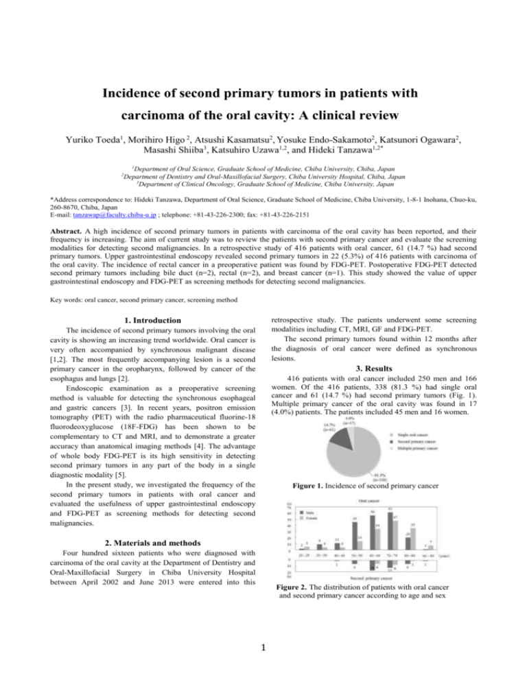
Incidence of second primary tumors in patients with carcinoma of the oral cavity: A clinical review Yuriko Toeda1, Morihiro Higo 2, Atsushi Kasamatsu2, Yosuke Endo-Sakamoto2, Katsunori Ogawara2, Masashi Shiiba3, Katsuhiro Uzawa1,2, and Hideki Tanzawa1,2* 1 Department of Oral Science, Graduate School of Medicine, Chiba University, Chiba, Japan Department of Dentistry and Oral-Maxillofacial Surgery, Chiba University Hospital, Chiba, Japan 3 Department of Clinical Oncology, Graduate School of Medicine, Chiba University, Japan 2 *Address correspondence to: Hideki Tanzawa, Department of Oral Science, Graduate School of Medicine, Chiba University, 1-8-1 Inohana, Chuo-ku, 260-8670, Chiba, Japan E-mail: tanzawap@faculty.chiba-u.jp ; telephone: +81-43-226-2300; fax: +81-43-226-2151 Abstract. A high incidence of second primary tumors in patients with carcinoma of the oral cavity has been reported, and their frequency is increasing. The aim of current study was to review the patients with second primary cancer and evaluate the screening modalities for detecting second malignancies. In a retrospective study of 416 patients with oral cancer, 61 (14.7 %) had second primary tumors. Upper gastrointestinal endoscopy revealed second primary tumors in 22 (5.3%) of 416 patients with carcinoma of the oral cavity. The incidence of rectal cancer in a preoperative patient was found by FDG-PET. Postoperative FDG-PET detected second primary tumors including bile duct (n=2), rectal (n=2), and breast cancer (n=1). This study showed the value of upper gastrointestinal endoscopy and FDG-PET as screening methods for detecting second malignancies. Key words: oral cancer, second primary cancer, screening method retrospective study. The patients underwent some screening modalities including CT, MRI, GF and FDG-PET. The second primary tumors found within 12 months after the diagnosis of oral cancer were defined as synchronous lesions. 1. Introduction The incidence of second primary tumors involving the oral cavity is showing an increasing trend worldwide. Oral cancer is very often accompanied by synchronous malignant disease [1,2]. The most frequently accompanying lesion is a second primary cancer in the oropharynx, followed by cancer of the esophagus and lungs [2]. Endoscopic examination as a preoperative screening method is valuable for detecting the synchronous esophageal and gastric cancers [3]. In recent years, positron emission tomography (PET) with the radio pharmaceutical fluorine-18 fluorodeoxyglucose (18F-FDG) has been shown to be complementary to CT and MRI, and to demonstrate a greater accuracy than anatomical imaging methods [4]. The advantage of whole body FDG-PET is its high sensitivity in detecting second primary tumors in any part of the body in a single diagnostic modality [5]. In the present study, we investigated the frequency of the second primary tumors in patients with oral cancer and evaluated the usefulness of upper gastrointestinal endoscopy and FDG-PET as screening methods for detecting second malignancies. 3. Results 416 patients with oral cancer included 250 men and 166 women. Of the 416 patients, 338 (81.3 %) had single oral cancer and 61 (14.7 %) had second primary tumors (Fig. 1). Multiple primary cancer of the oral cavity was found in 17 (4.0%) patients. The patients included 45 men and 16 women. Figure 1. Incidence of second primary cancer 2. Materials and methods Four hundred sixteen patients who were diagnosed with carcinoma of the oral cavity at the Department of Dentistry and Oral-Maxillofacial Surgery in Chiba University Hospital between April 2002 and June 2013 were entered into this Figure 2. The distribution of patients with oral cancer and second primary cancer according to age and sex 1 The percentage of male was higher in patients with second primary cancer. The distribution of age is shown in Figure 2. The mean age of patients with a primary oral cancer only and second primary cancer was 70.3 years and 62.5 years respectively. The number of combined second primary cancers in each site of first primary cancer of the oral cavity is shown in Table 1. Of the 61 cases, 31 had first primary cancer of the oral cavity. endoscopy still plays a decisive role in management of oral cancer. FDG-PET has been shown to be able to detect second primary tumors in patients with carcinoma of the oral cavity with a high sensitivity and specificity [9]. In the current study, the incidence of rectal cancer in a preoperative patient was found by FDG-PET. Postoperative FDG-PET detected second primary tumors including bile duct (n=2), rectal (n=2), and breast cancer (n=1). FDG-PET might be considered as a first diagnostic modality for detecting second primary tumors in patients with oral primary cancer. This study showed the value of upper gastrointestinal endoscopy and FDG-PET as screening methods for detecting second malignancies. We conclude that these modalities should be included in the routine examination in patients with oral cancer for the early detection of second primary tumors. Table 1. The number of second primary cancers in patients with first primary cancer of the oral cavity Competing interests The authors declare that they have no competing interests. Acknowledgement The tongue, gingiva, and buccal mucosa represented the most commonly involved site of first primary cancer of the oral cavity, making up 9 of the 31 patients respectively. The distribution of second primary cancer according to site of involvement was as follows: stomach in 12 cases (39%); esophagus in 8 (26%); large intestine in 4 (13%); larynx in 3 (10%); bile duct in 2 (6%); breast in 2 (6%). Synchronous lesions were found in 26 (42.6%) of 61 cases. Of these, 20 were detected as first primary cancer of the oral cavity accompanied by second primary cancer, using preoperative screening modalities. The screening modalities, identifying the second primary cancer in patients with first primary cancer of the oral cavity, are upper gastrointestinal endoscopy (n=22), FDG-PET (n=6) and CT (n=3). FDG-PET detected preoperative second primary cancer in a case and postoperative second primary cancer in 5 cases We thank Lynda C. Charters for editing the manuscript. References [1] Crosher R, McIlroy R. The incidence of other primary tumors in patient with oral cancer in Scotland. Br J Oral Maxillofac Surg 36: 58–62, 1998. [2] Winn DM, Blot WJ. Second cancer following cancers of the buccal cavity and pharynx in Connecticut, 1935–1982. Natl Cancer Inst Monogr 68: 25–48, 1985. [3] Fukuzawa K, Noguchi Y, Yoshikawa T, Saito A, Doi C, Makino T, Takanashi Y, Ito T, Tsuburaya A. High incidence of synchronous cancer of the oral cavity and the upper gastrointestinal tract. Cancer Letters 144: 145-151, 1999. [4] Ng SH, Yen TC, Liao CT, Chang JT, Chan SC, Ko SF, Wang HM, Wong HF. 18F-FDG PET and CT/MRI in oral cavity squamous cell carcinoma: a prospective study of 124 patients with histologic correlation. J Nucl Med 46: 1136–1143, 2005. [5] Krabbe CA, Balink H, Roodenburg JLN, Dol J, Visscher JGAM. Performance of 18F-FDG PET/contrast-enhanced CT in the staging of squamous cell carcinoma of the oral cavity and oropharynx. Int J Oral Maxillofac Surg 40: 1263-1270, 2011. [6] Li Z, Seah TE, Tang P, Ilankovan V. Incidence of second primary tumours in patients with squamous cell carcinoma of the tongue. Br J Oral Maxillofac Surg 49: 50-52, 2011. [7] Sessions DG, Spector GJ, Lenox J, Haughey B, Chao C, Marks J. Analysis of treatment results for oral tongue cancer. Laryngoscope 112: 616–625, 2002. [8] McGuirt WF. Panendoscopy as a screening examination for simultaneous primary tumors in head and neck cancer: A prospective sequential study and review of the literature. Laryngoscope 92: 569, 1982. [9] Krabbe CA, Pruim J, Laan BFAM, Rödiger LA, Roodenburg JLN. FDG-PET and detection of distant metastases and simultaneous tumors in head and neck squamous cell carcinoma: A comparison with chest radiography and chest CT. Oral Oncology 45: 234-240, 2009. 4. Discussion The high incidence of second primary tumors in patients with oral cancer has been well established worldwide, although the exact incidence varies from 9 [6] to 21 % [7] depending upon the report. Our results showed that the incidence of second primary tumors was 14.7 %, showing same trend in previous reports. The presence of second primary tumors in patients with carcinoma of the oral cavity is an important factor determining therapeutical approach. Preoperative screening methods are essential for detecting second primary tumors. Screening for second primary tumors has changed during the past years. In 1979, endoscopy was postulated as the method of choice [8]. Recently, FDG-PET has received more focus. In the present study, stomach was the most common site of second primary tumors. Upper gastrointestinal endoscopy revealed second primary tumors in 22 (5.3%) of 416 patients with carcinoma of the oral cavity. This screening method provided the opportunity to identify second malignancies, including gastric and esophageal carcinoma. Although new imaging techniques have been available, upper gastrointestinal 2 3


