Fluorescent Speckle Microscopy and Cell Division
advertisement
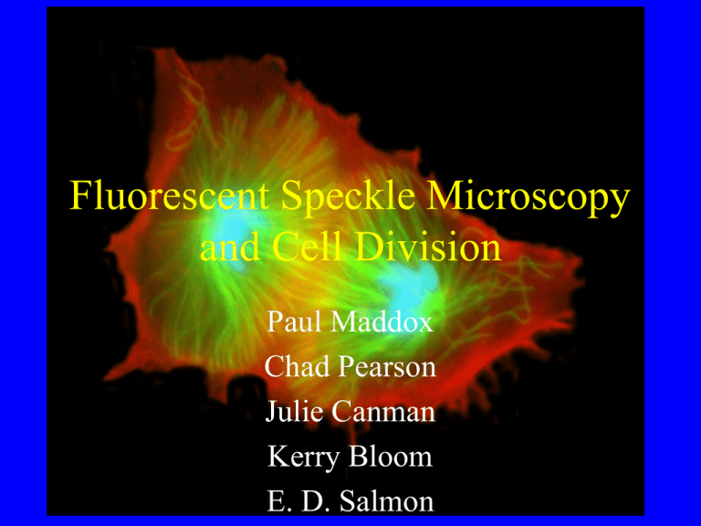
Fluorescent Speckle Microscopy and Cell Division Paul Maddox Chad Pearson Julie Canman Kerry Bloom E. D. Salmon What is Fluorescent Speckle Microscopy (FSM)? • Fluorescent discontinuities, “speckles” in biological polymers (e.g. microtubules, actin filaments) • Caused by stochastic incorporation of fluorescently tagged subunits into the polymer • Allows visualization of assembly dynamics and motility of the polymer C. M. Waterman-Storer and E. D. Salmon. (1998). How microtubules get fluorescent speckles. Biophys Journal 75, 2059-2069. ~5% ~0.5% Sites of Microtubule Assembly/Disassembly Microtubule Translocation How Do Microtubules get Fluorescent Speckles? How Do Microtubules get Fluorescent Speckles? • For resolution of 1.4 NA obj, r = 0.27 mm (N = 440 dimers) • Fraction of labeled tubulin, f = 1% • Mean # of fluorophores = M = fN = 4.4 • Standard deviation, SD = (fN(1-f))0.5=2.15 • Note: Microscope Magnification = (3*Pixel-width)/resolution = (3*6.7 mm) / 0.27 = 74.4 How Do Microtubules get Fluorescent Speckles? FSM Posters and Talks ASCB 99 • Gaudenz Danuser: Sat., 2:20 pm, rm 38, “Analysis of Cytoskeletal (actin) Dynamics” • Bill Bement and Clare Waterman-Storer: Sun., 5:25 pm, rm 31, “Actin and Microtubule Interactions” • Arshad Desai: Mon., 4:45 pm, rm 20, “Microtubule Motility in Xenopus and Drosophila Spindles” FSM Posters and Talks ASCB 99 • Tarun Kapoor et al. Mon. Session 2. “Investigating bipolar spindle formation and a kinesin inhibitor”. Poster #B-30. • Chloe Bulinski et al. Wed. Session 4. “Sparkling Speckles of GFP-Ensconsin on Microtubules” Poster #B-107 • Paul Maddox et al. Wed. Poster # B-108 Examples of FSM of Microtubule Poleward Flux in Spindles with Self-Organized Poles • Xenopus Extract Spindles • Ptk1 cells ~ 5 % labeled Tubulin ~ 0.05 % labeled Tubulin Waterman-Storer, C. M., A. Desai, J. C. Bulinski and E. D. Salmon. 1998. Fluorescent speckle microscopy: Visualizing the movement, assembly and turnover of macromolecular assemblies in living cells. Current Biology. 8:1227-1230. ~ 0.3 % labeled Tubulin Analysis of the polarity of assembly dynamics of astral and nuclear microtubules in the vegetative cell cycle P. Maddox, K. Bloom, and E.D. Salmon. 2000. The polarity and dynamics of microtubule assembly in the budding yeast S. cerevisiae. Nature Cell Biology, 2:36-41 Astral and Nuclear Microtubule Organization During the Yeast Cell Cycle Questions: • Where do assembly dynamics of astral microtubules occur? • What are the dynamic properties of microtubules in the Metaphase spindle? The Anaphase spindle? • How does the central spindle break down in telophase? Why worry about minus end assembly dynamics? Minus ends are at the SPB. Several microtubule motor proteins (Kar3p, Kip3p, Kip2p, Dhc1p) are localized to the SPB and have been proposed to function there to control microtubule length and produce force. e.g. Huyett et al. (1998). J. Cell Science 111, 295-301 Imaging System(s) Where do assembly dynamics of astral microtubules occur? Astral microtubules are dynamic at their plus ends and not their minus ends. What are the dynamic properties of microtubules in the Metaphase spindle? 66 % of metaphase spindle microtubules turnover with a half-life of 53 sec. 33% are much more stable. There are 24 microtubules per half spindle. 16 (66 % ) are kinetochore microtubules. While 8 (33 %) are overlapping interpolar microtubules. Winey et al. (1995) Journal of Cell Biology. 129(6):1601-1615. Therefore we conclude that the kinetochore microtubules are dynamic while the interpolar microtubules are stable. What are the dynamic properties of the microtubules in the Anaphase spindle? Anaphase microtubules are stable and grow from their plus ends. How does the mitotic spindle breakdown in telophase? Spindle disassembly occurs by plus end depolymerization. Summary • FSM is a powerful tool for studying polymer dynamics in living cells • FSM allows visualization of microtubule assembly dynamics and motility within the mitotic spindle • In vertebrates: Poleward flux and minus end disassembly occurs for non-centrosomal spindle microtubules; Centrosomal microtubule minus ends are stable (WatermanStorer et al., 1998 Cur. Biol. 8:1227-1230.) • In yeast: Astral and Anaphase spindle microtubules extend from the Spindle Pole Body and have stable minus ends Acknowledgements: • Ted Salmon – Clare Waterman-Storer – Julie Canman – Bonnie Howell • Kerry Bloom – – – – – Sid Shaw Elaine Yeh Dale Beach Chad Pearson Doug Thrower • Tim Mitchison – Aneil Mallavarapu – Aaron Straight • • • • • • • Arshad Desai Shinya Inoue MBL Nikon Hamamatsu Photonics Universal Imaging Yokagawa
