1
2
3
4
4
5
6
7
8
Pigmented Skin Lesions
MELANOMA
MELANOMA
Malignant melanoma is a skin cancer due
to uncontrolled growth of pigment cells melanocytes.
Melanocytes
• Normal melanocytes occur in the basal layer of the
epidermis
• They produce melanin
• Melanin (a protein) protects the skin by absorbing
ultraviolet (UV) radiation
• Melanocytes are found in equal numbers in black and in
white skin
• Melanocytes in black skin produce much more melanin
• Non-cancerous growth of melanocytes results in moles
(benign melanocytic naevi) and freckles
• Cancerous growth of melanocytes results in melanoma
Risk Factors for Melanoma
•
•
•
•
•
Sun exposure, particularly during childhood
Fair skin that burns easily
Blistering sunburn, especially when young
Previous melanoma
Previous non-melanoma skin cancer (BCC,
SCC)
• Family history of melanoma
• Large numbers of moles (esp if > 100)
• Abnormal moles (atypical or dysplastic naevi)
Epidemiology of Melanoma
•
•
•
•
3% of all cancers and 10% of skin cancers.
Incidence 1:10,000 per annum
Incidence is increasing in developed countries
Incidence rises with age, rare in children,
commonest in over 75s
• 3rd commonest cancer in young people.
• In UK 2002 - 1,640 deaths from malignant melanoma
Over 65% of deaths from malignant melanoma were
in the over 65s.
• It is commoner in women than in men but men have
a worse prognosis.
Melanoma in situ
• Superficial forms of melanoma spread out
within the epidermis (horizontal growth).
• If all the melanoma cells are confined to
the epidermis, it is melanoma in situ.
• Lentigo maligna is a special case of
melanoma in situ that occurs around hair
follicles on the sun damaged skin of the
face or neck.
• Melanoma in situ is cured by excision
Invasive Melanoma
• When the cancerous cells have grown through
the basement membrane into the deeper layer
of the skin (the dermis), it is known as invasive
melanoma (vertical growth)
• Nodular melanoma appears to be invasive from
the beginning, and has little or no relationship to
sun exposure.
• Metastatic disease increases in likelihood with
increasing depth of the melanoma.
• 15% of people with invasive melanoma will die
from it.
Where do melanomas occur?
• Melanoma can arise from otherwise normal appearing skin (50%)
• Or from within a mole or freckle, which starts to grow larger and
change in appearance. Precursor lesions include:
– Congenital melanocytic naevus (brown birthmark)
– Atypical or dysplastic naevus (funny-looking mole)
– Benign melanocytic naevus (normal mole)
• Melanomas occur anywhere on the skin, not only in sun-exposed
areas. Commonest sites: men - back (40%), women - leg (40%).
• Melanomas can also occur on mucous membranes (lips, genitals).
• May also occurs in other parts of the body such as the eye, brain,
mouth or vagina.
Moles (Melanocytic Naevi)
•
•
•
•
Very common
May be flat or protruding
Vary in colour from pink to black
Brown or black coloured moles are also
called ‘pigmented naevi’.
• Mostly round or oval in shape
• Range in size from 2mm to several cm
Moles
• Most frequently moles arise during
childhood or early adult life (acquired
melanocytic naevi).
• Exposure to sunlight increases the number
of moles.
• Teenagers and young adults tend to have
the greatest number of moles.
Classification
• Junctional naevi
Groups or nests of naevus cells at the junction of the
epidermis and dermis. Tend to be flat colourful moles.
• Dermal/Intradermal naevi
Nests of naevus cells in the dermis. These moles are
thickened and often protrude from the skin surface
(papillomatous naevi).
• Compound naevi
Nests of naevus cells at the epidermal-dermal junction
as well as within the dermis. These moles have a central
raised area surrounded by flat pigmentation.
Junctional Naevus
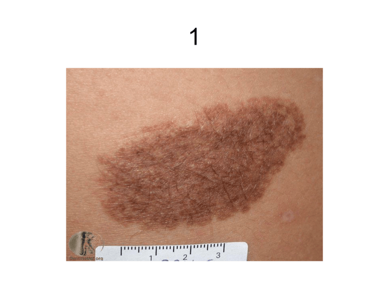
Congenital Melanocytic Naevus
• Brown or black naevi
• Present at birth or develop in the first year or so
of life
• Moles that look like birthmarks but were not
present at birth may be called ‘congenital
naevus-like’ naevi or ‘congenital-type’ naevi.
• About one baby in 100 has a small or medium
sized congenital naevus, so they are quite
common.
• Very large, giant or bathing trunk naevi are very
rare.
Types of congenital melanocytic naevus
• Typically multi-shaded, oval, fairly uniform pigmented patches
• Most grow with the child but become proportionally smaller and less
obvious with time.
• May darken, become bumpy or hairy especially at puberty.
• Rarely fade away or disappear.
• Congenital melanocytic naevi in adults are classed as ‘small’ (<
1.5cm di), ‘medium’ (>1.5 <10cm) or ‘large’ (>10cm)
• ‘Giant’ congenital naevi are greater than 20cm in diameter. Often
found on the buttocks (‘bathing trunk’ naevi)
• Café-au-lait macule - a flat tan mark, usually oval (inherited).
Multiple café-au-lait macules may be a sign of neurofibromatosis.
• Speckled lentiginous naevus (naevus spilus) has dark spots
scattered on a flat tan background.
Risk of Melanoma
• The risk of melanoma in a small or
medium-sized congenital melanocytic
naevus is very small (< 1%)
• Melanoma never arises from café-au-lait
macules
• Melanoma is more likely in the giant naevi
(~ 5% over a lifetime) especially in those
that lie across the spine
Congenital Melanocytic Naevus
Café au lait Macule
Giant Melanocytic Naevus
Speckled Melanocytic Naevus
Atypical Naevi
• Melanocytic naevi with unusual features eg indistinct
edge, larger size.
• May resemble Malignant Melanomoa but are benign
• Sometimes called dysplastic naevi, active junctional
naevi, B-K moles and Clark's naevi.
• May be familial or sporadic.
• The inherited form is usually part of a syndrome Familial Atypical Mole and Melanoma (FAMM) syndrome
(formerly dysplastic naevus syndrome).
– One or more first-degree or second-degree relative with
malignant melanoma
– A large number of naevi (often more than 50) some of which are
atypical naevi
– Naevi that show certain histological features.
Atypical Naevi
Fair-skinned individuals with light coloured
hair and freckles are most at risk of getting
atypical naevi, especially if they have been
frequently exposed to the sun or have a
family history of atypical naevi.
Atypical naevi may develop at any time but
most develop during the first 15 years of
life.
Atypical Naevi
• People with one to four atypical naevi have a
slightly higher risk than the general population of
developing malignant melanoma
• People with FAMM syndrome are significantly
more at risk of developing melanoma.
• Atypical naevi are harmless (benign) and do not
need to be removed. However, it is not always
easy to tell whether a lesion is an atypical
naevus or a melanoma, so if in doubt, it should
be removed by excision biopsy.
Atypical Naevus
Atypical Naevus
Glasgow 7-point Checklist
• Major features
– Change in size
– Irregular shape
– Irregular colour
• Minor features
– Diameter >7mm
– Inflammation
– Oozing
– Change in sensation
ABCDE of Melanoma
A.
B.
C.
D.
E.
Asymmetry
Border - irregularity
Colour - variation
Diameter - over 6 mm
Evolving - (enlarging, changing)
Types of Melanoma
• Flat patches (horizontal slow growth)
– Superficial spreading melanoma (SSM)
– Lentigo maligna melanoma (sun damaged skin of face, scalp
and neck)
– Acral lentiginous melanoma (on soles of feet, palms of hands or
under the nails – the subungual melanoma)
• Nodules (vertical rapid growth)
– Nodular melanoma
– Mucosal melanoma (arising on lips, eyelids, vulva, penis, anus)
– Desmoplastic melanoma (fibrous tumour with a tendency to grow
down nerves)
• Combinations occur e.g. nodular melanoma arising
within a superficial spreading melanoma.
Typical Superficial Spreading Melanoma
Superficial Spreading Melanoma with regression
Amelanotic Melanoma
Lentigo Maligna
Lentigo Maligna Melanoma
Lentigo Maligna
•
•
•
•
•
•
Sun-exposed areas of the face and neck
Elderly
Slow growing
Often quite large (>20mm).
Pre-cancerous
Conversion to a lentigo maligna melanoma
occurs in ~ 5% of patients
• Identifying lesions that require referral is not
easy – but see ABCDE
Nodular Melanoma in Lentigo Maligna
Acral Lentiginous Melanoma
Subungual Melanoma
Amelanotic Subungual Melanoma
Nodular Melanoma
Nodular Melanoma
Nodular Melanoma
Diagnosis
• Excision biopsy with a 2 to 3-mm margin
• Breslow depth - thickness of the
melanoma in mm
• Clark's level - describe which layer of the
skin has been breached. Clark’s level 1
refers to melanoma in situ. Invasive
melanoma may reach Clark's level 2 (thin)
to 5 (reaching the subcutaneous fat layer).
• Systematic search for metastasis
Prognosis
Death is unlikely if a melanoma has a
Breslow thickness of less than 1mm
50% dead within 5 years if >4mm


