Velscope Powerpoint
advertisement
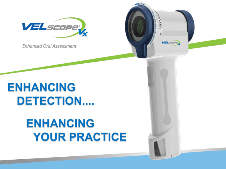
ENHANCING DETECTION.... ENHANCING YOUR PRACTICE Staggering Statistics on Oral Cancer • 1 person dies from oral cancer each hour of every day • The Mortality Rate for oral cancer has not decreased in over 30 years • 72% of the cases of oral cancer are not diagnosed until stage two or later • Late detection results in a five year survival rate of only 52% • Cases of oral cancer are increasing at an alarming rate Public Awareness of Oral Cancer Must Improve • 41,350 new cases of oral cancer were discovered in 2013 • Oral cancer impacted 3X as many people in 2013 when compared to cervical cancer (41,350 cases versus 12,340 cases respectively) • HPV is increasing the number of oral cancer cases • There has been a 60% increase in oral cancer in adults under the age of 40 (25% of these cases have no traditional risk factors (smoking, drinking, etc.) New England Journal of Medicine, 2007 “Oral HPV infection is strongly associated with oropharyngeal cancer among subjects with or without the established risk factors of tobacco and alcohol use.” EARLY DETECTION IS THE KEY 67% of all oral cancer is currently discovered beyond this stage. (Stage II) Appropriate Stage for Discovery & Intervention Early Dysplasia Moderate Dysplasia Severe Dysplasia Potentially Malignant Disease Stages Carcinoma-In-Situ (CIS) 5 Invasive Squamous Cell Carcinoma (SCC) VELscope VX – The Technology • A safe blue light (no radiation) shines through the epithelial tissue and basement membrane to the stromal collagen • Patented technology filters out everything except the fluorescence of the tissues • Healthy tissue reflects back through the scope as a brilliant green color • Suspicious tissue will absorb the light and not fluoresce, appearing extremely dark, almost black Normal Floor of the Mouth Obvious Dysplasia? Images courtesy of the British Columbia Oral Cancer Prevention Program Fluorescence Visualization Carcinoma-In-Situ (CIS) Clinical Appearance (Visible White Light) Loss of Fluorescence Copyright ® 2002-2007 by Oral Health Study, Oral Oncology/Dentistry, BCCA Severe Dysplasia on Alveolar Ridge Clinical Impression: Denture Trauma? Inflammation resolved in 2 weeks after removal of denture Excisional Biopsy: Severe Dysplasia Images courtesy of the University of Washington Oral Medicine Program When Do Observe or Refer? Images courtesy of the British Columbia Oral Cancer Prevention Program Observe or Refer? Excisional Biopsy: Severe Dysplasia Oral Lesions May Show as Irregular, Dark Areas Images courtesy of the British Columbia Oral Cancer Prevention Program Observe or Refer? Hyperplasia An Oral Lesion that Shows No Change in Autofluorescence Appearing Pale Green Images courtesy of the British Columbia Oral Cancer Prevention Program Moderate Dysplasia Dept of Oral Medicine, University of Washington Pre-clinical discovery Left palate : low-grade mucoepidermoid carcinoma Blanching under Diascopic Pressure Using an instrument to blanch is a technique1 for indicating whether or not an abnormality has an inflammatory component. This particular area is inflammation and blanches completely Treatment and follow-up in two weeks showed complete resolution 1. Rudd M, Eversole R, Carpenter W. “Diascopy: a clinical technique for the diagnosis of vascular lesions”, Gen Dent. 2001 Mar-Apr; 49(2):206-9. EXTENSIVE CLINICAL SUPPORT 10+ Years of Research in the Oral Cavity • • • • • 2011 General Dentistry – E. Truelove, et al. 2009 General Dentistry – K. Huff, et al 2007 Head & Neck – P.M. Williams, et al. 2006 Clinical Cancer Research – C.F. Poh, et al 2005 Oral Oncology – deVeld, et al. Proven Efficacy www.VELscope.com/research 19 “Since we have incorporated the VELscope in our practice we have had three positive oral carcinomas treated by oral and plastic surgeons in 2 1/2 years. Numerous biopsies have resulted in the removal of worrisome and non malignant growths which has increased our patient acceptance and referrals. Thanks for the technology.” Thomas R. Leischner DDS Elk Grove Village, IL Portable Cordless Affordable: $3,299.00 THANK YOU!
