ED Ultrasound
advertisement
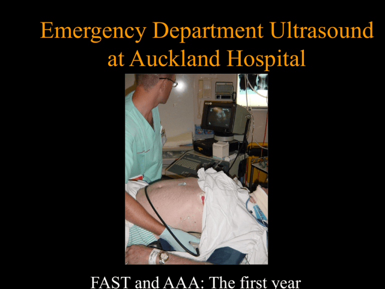
Emergency Department Ultrasound at Auckland Hospital FAST and AAA: The first year Objectives • The role of FAST • History of ED ultrasound at Auckland Hospital • The ultrasound credentialling process • How we performed in the first year • How we compare to the rest of the world • Where we go from here FAST • • • • Focused Assessment Sonography Trauma FAST • Integral part of initial trauma workup • Proven – – – – – Quick Safe Reliable Reproducible Repeatable FAST • Pitfalls – Poor sonographer – Poor scan • Air • Obesity – Negative FAST doesn’t exclude injury! – Failure to serially examine the patient History • 1998 Purchased portable ultrasound machine • 1998 First Australasian FAST course • 1999-2001 Sporadic use of ultrasound • Dec 2000 Formal Emergency Ultrasound credentialling program • Feb 2001 1st credentialled ED sonographers The Credentialling Process Background Radiologist Clinician Radiologist Clinician The Credentialling Process Background • Much debate in literature last 10 years • Consensus meeting • Each department decide own credentialling process • 200 scans and ongoing audit • Subsequent literature – Shackford 1999 4 yr experience • 50 scans • Suggests acceptable error rates The Credentialling Process Background • Workshop beneficial – Rozycki 1996 • Exit exam – Sisley 1999 The Credentialling Process Background • American College of Emergency Physicians 2001 – 8 workshop hours – 25 scans in each of 6 areas – Can be partially credentialled • Only 1/76 departments met criteria – Boulanger 2000 The Credentialling Process Background • Australasian College for Emergency Medicine – – – – – 16 workshop hours 25 Accurate scans for FAST 15 Accurate scans for AAA >50% clinically indicated Proctored by credentialled/ultrasound qualified person – Exit exam Auckland ED • Adopted ACEM guideline December 2000 • 4 sonographers – – – – Satisfied workshop requirement Scans should not alter management All measured against ‘gold standard’ Proctored by radiologist – Standardised form – Monthly/bimonthly – Modified criteria for scans – 100% clinically indicated – Exit examination February 2001 Results FAST • 1 ED registrar ‘credentialled’ by June 2001 – 79% Indicated scans • 2/3 ED Specialists credentialled by Feb 2002 – All scans clinically indicated Results FAST • For Detection Any Free Fluid • 113 scans in 102 patients over 13 months – 9 scanned by 2 sonographers – 1 scanned by 3 sonographers Results FAST (Any Free Fluid) n=113 • • • • TP TN FP FN 20 83 3 7 • • • • • Sn Sp PPV NPV 74.1% 96.5% 87% 92% Accuracy 91.2% Results FAST (Laparotomy or Extra Investigation) • • • • TP TN FP FN 11 89 5 2 • • • • • Sn Sp PPV NPV n=107 84.6% 94.7% 68.8% 97.8% Accuracy 93.5% Results FAST Existing literature • vs gold standard, novice sonographers • 3 studies • Sn 69-79% • Sp 96-98% • vs clinical observation and experienced sonographers • Sn • Sp 80-98% >90% Errors FAST • 7 FN – 5/7 Trivial fluid, conservative management – 1 penetrating trauma with minor injury – 1 blunt trauma bladder injury, stable • All views adequate and correct interpretation according to radiologist Errors FAST • 3 FP – 1 “ascites” – 1 “?pericardial effusion” – 1 Retroperitoneal and abdominal wall haematomas • Adequate views but incorrect interpretation Result of errors FAST • 1 CT scan thorax for “?Pericardial effusion” Emergency Department Ultrasound for AAA • 2 Case series in literature Results AAA • 66 Scans in 58 Patients in 12 months – 5 Scanned by 2 sonographers – 1 Scanned by all 4 • 3/4 sufficient scans to meet requirement Results AAA n=66 • • • • TP TN FN FP 26 39 1 0 • • • • • Sn SP PPV NPV 96.3% 100% 96.3% 97.5% Accuracy 98.3% “Error” AAA • Free air obscured 6cm AAA • Free fluid detected in Morison’s and Splenorenal recesses • Found to have perforated DU AAA Existing Literature • Shuman 1998 – n=60 • Sn 97% • Sp 100% • Kuhn 2000 – n=68 • Sn 100% • Sp 95% Time Taken to Scan • FAST median 5min (1-20) • AAA median 3.5 (1-16) • Similar to literature published FAST Learning Curve • Debate about this • Shackford only author to look at initial experience – – – – Suggests 10 scans before proficient Showed ‘Institutional learning curve’ 12 Individuals = wide variation in error rates Only 4/12 had >25 scans in 4 years FAST Learning Curve Pooled Error Rates for EU 0.12 Error Rate 0.10 0.08 Any Free Fluid 0.06 Clinically Significant 0.04 0.02 0.00 5 10 15 20 25 Number of Scans 30 33 FAST Learning Curve • Error rate <10% • Most ‘errors’ clinically insignificant • Individual variation Potential Bias • Patients not consecutive – Opportunity for pre-selection of patients • Individual sonographers could discard unsatisfactory scans prior to proctoring Summary • Emergency Department Ultrasound is established in Auckland Hospital • Accuracy mirrors existing literature • Pitfalls exist and should be considered The future • • • • • Credentialling continues Credentialled sonographers record in notes Clinical management may alter Ongoing audit Expanded indications – Unstable patient with abdominal pain • Is there free fluid? Case 1 • 37f – 4hr Abdominal pain – Collapse and seizure – Shock – Arrives ED 1755 – SLOH 1806 Case 1 • OT 1815 Case 2 • 28f – 1/2 hr Abdominal pain – HR 84, SBP 90, RR 16 – Arrives ED 0910 – S/B registrar 1000 – SLOH 1018 Case 2 • Urine pregnancy test 1025, positive Case 2 • OT 1055 Case 3 • 19m – – – – – Fall from tree Collapse at home Fighting en route Arrives ED 1635 FAST 1645 Case 3 • OT for thoracotomy


![Jiye Jin-2014[1].3.17](http://s2.studylib.net/store/data/005485437_1-38483f116d2f44a767f9ba4fa894c894-300x300.png)
