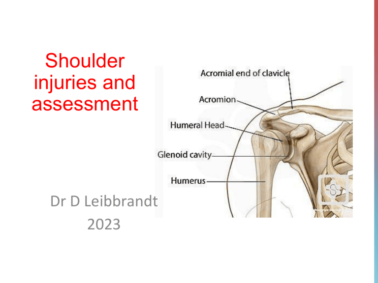
Shoulder injuries and assessment Dr D Leibbrandt 2023 Anatomy Biomechanics • GHJ is a ball and socket joint • The glenoid cavity is shallow (not like in the hip which has a deeper socket) • The shoulder stability therefore comes from other structures not necessarily the shoulder itself – Glenohumeral ligaments, glenoid labrum, capsule (static components) – Rotator cuff and stabilizing muscles (dynamic components) Stability • Anterior and posterior bands of inferior glenohumeral ligament attach to labrum to provide static stabilization • The anterior band of the inferior glenohumeral ligament prevents anterior translation and the posterior translation of the humeral head • Shoulder stability is also enhanced by the glenoid labrum Stability Labrum • Ring of fibrous tissue attached to rim of glenoid Dynamic stabilizers • Rotator cuff muscles Muscle Origin on scapula Attachment on humerus Function Supraspinatus muscle supraspinous fossa superior facet of the greater tubercle abducts the humerus Infraspinatus muscle infraspinous fossa posterior facet of the greater tubercle externally rotates the humerus Teres minor muscle middle half of lateral border inferior facet of the greater tubercle externally rotates the humerus lesser tubercle internally rotates the humerus Subscapularis muscle subscapular fossa Rotator cuff muscles • Control position of the humeral head • Adequate scapulohumeral rhythm is necessary for full upper limb elevation, it enhances joint stability at greater than 90˚ • Abnormal scapulohumeral rhythm may predispose one to injury – normally due to weakness of the scapular stabilizers Scapula stabilizers • Trapezius (upward rotation of the scapula) • Serratus anterior (protraction of the scapula) • Rhomboids maj and min (elevate, retract and downwardly rotate the scapula) Scapulohumeral rhythm https://www.youtube.com/watch?v=uOTO6YtfFUA Physical examination • • • • • • Observation Posture Functional demonstration Active physiological movements Passive physiological movements Muscle tests – – – Length Strength Control Physical examination • Neurological tests – – • Palpation – – – • Neurodynamic tests Conduction test Muscles General Passive accessory movements Joint integrity tests Active physiological movements • • • Flexion/ extension Abd/ Add IR/ER Functional tests: • HBB • HBH Muscle testing • • Isometric testing - test rotator cuff | stabilizers https://youtu.be/zW8SE6WOxD0 • Test supraspinatus by resisting the initiation of shoulder abduction (place a hand on the lateral aspect of the lower humerus). Alternatively, test supraspinatus by placing the straight arm into 90° of elevation in the scapular plane and 45° of lateral rotation then isometrically resist further elevation in the scapular plane. Muscle testing • Isometric testing - test rotator cuff | stabilizers • Test subscapularis by resisting shoulder medial rotation (elbow at 90° and into the side; place a hand against the inner aspect of the forearm). Alternatively, test subscapularis by placing the arm into the hand behind-back position with the wrist over L5 then isometrically resist medial rotation. Muscle testing • Isometric testing - test rotator cuff | stabilizers • Test infraspinatus and teres minor by resisting shoulder lateral rotation (start as above and place a hand against the outside of the forearm). Alternatively, place the arm into 45° of medial rotation by taking the arm across the body then isometrically resist lateral rotation. Isometric testing Impingement • More a clinical sign rather than a diagnosis • Suggestion that rotator cuff tendons are impinged as they pass through the subacromial space (formed between the acromion, coracoacromial arch, AC joint) Impingement • External vs Internal • External/subacromial – Is the mechanical encroachment of the soft tissue (bursa, rotator cuff tendons) in the subacromial space between the humeral head and the acromial arch. This encroachment particularly takes place in the midrange of motion, often causing a ‘‘painful arc’’ during active abduction. • Internal – Internal impingement comprises encroachment of the rotator cuff tendons between the humeral head and the glenoid rim. Internal impingement • Occurs mainly in overhead athletes • Impingement of rotator cuff against posteriorsuperior surface of the glenoid External impingement • In primary impingement, a structural narrowing of the subacromial space causes pain and dysfunction, such as acromioclavicular arthropathy, type I acromion, or swelling of the soft tissue in the subacromial space. • In secondary impingement, there are no structural obstructions causing the encroachment, but rather functional problems, occurring only in specific positions. Secondary impingement may occur in the subacromial space as well as internally in the glenohumeral joint. Testing for impingement • Hawkins-Kennedy test • Neer’s Test • Jobe’s empty can (RC impingement) Hawkins-Kennedy • • • Description of test – Patients shoulder (& elbow) flexed to 90˚ – Shoulder then passively moved into internal rotation by physio Interpretation – Positive for pain in subacromial area – -ve for internal impingement Sensitivity 88.7% Neer’s test • • Description – Patient seated – Physio stabilize scapula (to prevent scapula rotation) – Physio then moves the shoulder into maximal forward flexion Interpretation – Subacromial impingement if test positive for pain in anterior shoulder – Internal impingement if test positive for pain in posterior shoulder Jobe’s/Empty can test • Description – Patient in standing both shoulders elevated to 90˚ in scapular plane – Physio maximally internally rotates shoulders then places downward pressure over lower arm and resists further elevation • Interpretation – Positive for pain in subacromial impingement – Negative in patients with internal impingement Rotator cuff injuries • Tendinopathy • Strains/tears • Assessment – Palpation – Active movement – Isometric testing – Gerbers lift off Full can test • Description – Patient in standing. Both shoulders elevated to 90˚ in the scapula plane. Shoulders are maximally externally rotated. Then places downward pressure over lower arm and resists further elevation. • Interpretation – Very active rotator cuff in this test. Pain, indicates rotator cuff pathology. But if –ve, with a positive empty can – possibly impingement Empty can and full can tests Instability • Identify any joint instability • Laxity may imply the ability to sublux or dislocate Anterior drawers test • Description – Patient supine – Shoulder abducted to 80-120˚, 20˚ horizontal flexion, 30˚ lateral rotation. Physio must stabilize the scapula with one hand and glide the humeral head anterior • Interpretation – Excessive ROM, patient anxious or click +ve for anterior instability – https://youtu.be/G8s_7Q5zfTM Posterior instability • Jerk test • Description – Patient supine or sitting. Shoulder is abducted to 90˚ and medially rotated. Physio applies a longitudinal cephalad force to elbow while humerus moved in horizontal flexion • Interpretation – Positive test indicated by click or jerk when the arm moves into and out of horizontal flexion – https://youtu.be/mn7f6KVvScE Inferior instability • • • • Sulcus sign Description – The patient sits or stands. The physio applies a longitudinal force to the patients humerus. Interpretation Positive for inferior instability if there is sulcus or indentation (of about 5mm) below the acromion Biceps tendinopathy • Occurs in athletes who perform weight training • Overuse injury • Examination – Tender on palpation – Speeds test Speeds test • • Description – The patient’s arm elevated to 90˚ in forward flexion, the elbow extended, forearm supinated. Physio applies resistance distal to the elbow in the direction of arm extension. Interpretation – Positive when there is localized pain over biciptal groove Biceps load test II • Description – The patient is supine with the shoulder at 120˚ of the abduction and the elbow at 90˚ flexion. Physio applies resistance to elbow flexion. • Interpretation – Positive if the patients complains of pain when resistance is applied. – https://www.youtube.com/watch?v=h2IyvaCEYpk Glenoid labrum injuries • Injuries to the glenoid labrum are divided into superior labrum anterior to posterior (SLAP) or non-SLAP SLAP Non-SLAP Injuries to the labrum that extend from anterior to the biceps tendon to posterior to the tendon Degenerative flap, vertical labral tears and Bankart lesions |(injury to anterior labrum following anterior dislocation) Glenoid labrum injuries • Common in repetitive overhead injuries • Pain often poorly localized but exacerbated by overhead activities • Popping or catching may be present O’Brien’s test • Description – Arm flexed to 90˚ forward flexion and 10˚ adduction. Arm is internally rotated and pronated with thumb pointing down. A downward force is applied by the physio. Test is then repeated with thumbs up. • Interpretation – Positive if pain located at joint line and was elicited in the internally rotated position and reduced or eliminated in the externally rotated position – https://youtu.be/v_EL9XqTJQQ Active physiological movements GHJ | • Shoulder flexion |extension • Shoulder abduction • Hand behind back | Hand behind head • Horizontal adduction/ flexion • The acromioclavicular joint can be 'squeezed' or the acromiohumeral joint compressed during the painful movement as with flexion and abduction. If pain is localized to the posterior area of the gleno-humeral joint and the head of humerus seems to sub-lux posteriorly during the movement, a posterior capsular instability may be suspected and investigated further. Active physiological movements | GHJ • Lateral rotation • If this movement is accompanied by apprehension and the appearance of the head of the humerus subluxing anteriorly, an anterior capsular instability should be suspected and investigated further. • Medial rotation Active physiological movements (with O/P) | GHJ GHJ Accessory movements • https://youtu.be/hKjQw-blrZ4 Maitland PAMs of the shoulder (practical) Glenohumeral joint: PA: arm by side, abduction, prone AP: arm by side, abduction Longitudinal movement caudad Lateral transverse: arm by side, 90°flexion. GHJ | Posteroanterior movement Uses: Mobilizing or stretching of the anterior and posterior parts of the glenohumeral capsule. GHJ PA- Abduction, Arm by side, prone GHJ | Anteroposterior movement Uses: • Not as useful as the longitudinal movement or posteroanterior movement in treating very painful shoulders. • Grades III and IV are more useful when the joint is stiff. • Stretching the posterior glenohumeral capsule, or relocating an anteriorly subluxed humeral head. • A means of encouraging proprioceptive recovery in unstable shoulders. • A means of mobilizing the interface surrounding local nerve tissue. GHJ AP-arm by side, Abduction GHJ | Longitudinal and Lateral movement Uses • Pain and stiffness in conjunction with other techniques. • Capsular tightness. • Joint surface pain (as a through-range grade I, II or III-). • To increase the range of flexion and abduction at the limit of range • To help regain shoulder abduction after fracture of the neck of the humerus. GHJ Longitudinal- arm by side GHJ Longitudinal- Abd, Full flex GHJ lateral movement- arm by side, flexion Maitland PAMs of the shoulder (practical) AC joint: AP PA Longitudinal movement caudad ACJ | Anteroposterior movement Uses - Acromioclavicular pain after injury, subluxation or fracture of the clavicle. - Stiffness and pain especially with horizontal adduction and hand-behind-back movements. ACJ | Posteroanterior movement Uses - - - Acromioclavicular pain. To improve the range of movement of horizontal adduction (flexion) or extension. Stiffness at the end of range of shoulder movements. ACJ | Longitudinal movement caudad Uses -Painful acromioclavicular joint -Painful arc of flexion. -Stiffness at the limit of flexion, abduction and horizontal adduction. ACJ- AP, PA, longitudinal
