Neurochem. Int. Vol. 26, No. 5, pp. 455~464, 1995
~
Pergamon
0197-0186(94)00157-X
Copyright © 1995 ElsevierScienceLtd
Printed in Great Britain. All rights reserved
01974) 186/95 $9.50+ 0.00
D I F F E R E N T I A L EXPRESSION OF P R O T E I N K I N A S E C
ISOFORMS IN GLIAL A N D N E U R O N A L CELLS.
T R A N S L O C A T I O N A N D D O W N - R E G U L A T I O N OF PKC
ISOFORMS IN C6 G L I O M A A N D N G 108-15 H Y B R I D CELLS :
EFFECTS OF E X T R A C E L L U L A R Ca2+-DEPLETION
LIN
C H I N G - C H O W C H E N , * J A C Q U E C H A N G and W A N - W A N
Institute of Pharmacology, College of Medicine, National Taiwan University, Taipei 10018, Taiwan
(Received 13 September 1994" accepted 16 November 1994)
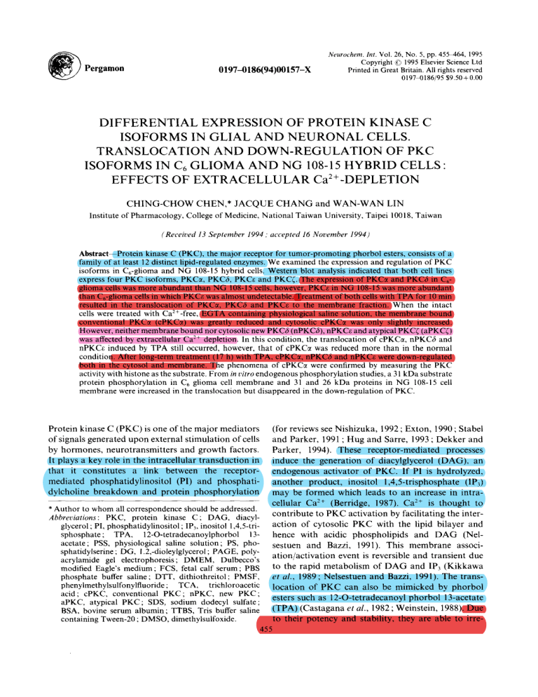
Abstrae~Protein kinase C (PKC), the major receptor for tumor-promoting phorbol esters, consists of a
family of at least 12 distinct lipid-regulated enzymes. We examined the expression and regulation of PKC
isoforms in C6-glioma and NG 108-15 hybrid cells. Western blot analysis indicated that both cell lines
express four PKC isoforms, PKCct, PKCb, PKCe and PKC~. The expression of PKC~ and PKC6 in C6glioma cells was more abundant than NG 108-15 cells, however, PKCe in NG 108-15 was more abundant
than C6-glioma cells in which PKC~, was almost undetectable. Treatment of both cells with TPA for 10 min
resulted in the translocation of PKCct, PKC6 and PKCc to the membrane fraction. When the intact
cells were treated with Ca2+-free, EGTA containing physiological saline solution, the membrane bound
conventional PKCct (cPKCc0 was greatly reduced and cytosolic cPKC~ was only slightly increased.
However, neither membrane bound nor cytosolic new PKC3 (nPKC6), nPKCe and atypical PKC~ (aPKC~)
was affected by extracellular Ca :+ depletion. In this condition, the translocation of cPKCc¢, nPKC6 and
nPKCe induced by TPA still occurred, however, that of cPKC~ was reduced more than in the normal
condition. After long-term treatment (17 h) with TPA, cPKC~, nPKC3 and nPKCe were down-regulated
both in the cytosol and membrane. The phenomena of cPKC~ were confirmed by measuring the PKC
activity with histone as the substrate. From in vitro endogenous phosphorylation studies, a 31 kDa substrate
protein phosphorylation in C6 glioma cell membrane and 31 and 26 kDa proteins in NG 108-15 cell
membrane were increased in the translocation but disappeared in the down-regulation of PKC.
Protein kinase C (PKC) is one of the major mediators
of signals generated upon external stimulation of cells
by hormones, neurotransmitters and growth factors.
It plays a key role in the intracellular transduction in
that it constitutes a link between the receptormediated phosphatidylinositol (PI) and phosphatidylcholine breakdown and protein phosphorylation
(for reviews see Nishizuka, 1992 ; Exton, 1990 ; Stabel
and Parker, 1991 ; Hug and Sarre, 1993 ; Dekker and
Parker, 1994). These receptor-mediated processes
induce the generation of diacylglycerol (DAG), an
endogenous activator of PKC. If P1 is hydrolyzed,
another product, inositol 1,4,5-trisphosphate (IP3)
may be formed which leads to an increase in intracellular Ca -,+ (Berridge, 1987). Ca 2+ is thought to
* Author to whom all correspondence should be addressed.
contribute to P K C activation by facilitating the interAbbreviations: PKC, protein kinase C; DAG, diacylaction of cytosolic P K C with the lipid bilayer and
glycerol ; PI, phosphatidylinositol ; IP3, inositol 1,4,5-trisphosphate; TPA, 12-O-tetradecanoylphorbol 13- hence with acidic phospholipids and D A G (Nelacetate; PSS, physiological saline solution; PS, phosestuen and Bazzi, 1991). This membrane associsphatidylserine; DG, 1.2,-dioleylglycerol; PAGE, polyation/activation event is reversible and transient due
acrylamide gel electrophoresis; DMEM, Dulbecco's
modified Eagle's medium; FCS, fetal calf serum; PBS to the rapid metabolism of D A G and IP3 (Kikkawa
phosphate buffer saline; DTT, dithiothreitol; PMSF,
et al., 1989; Nelsestuen and Bazzi, 1991). The transphenylmethylsulfonylfluoride; TCA, trichloroacetic location of P K C can also be mimicked by phorbol
acid; cPKC, conventional PKC; nPKC, new PKC;
esters such as 12-O-tetradecanoyl phorbol 13-acetate
aPKC, atypical PKC; SDS, sodium dodecyl sulfate;
(TPA) (Castagana et al., 1982 ; Weinstein, 1988). Due
BSA, bovine serum albumin; TTBS, Tris buffer saline
to their potency and stability, they are able to irrecontaining Tween-20 ; DMSO, dimethylsulfoxide.
455
456
Chmg-('hov: Chcn el a/.
versibly insert P K C into the lipid bilaycr thereby causing a cumulative and long-term stimulation of the
enzyme (Nelsestuen and Bazzi, 1991). This activation is
eventually terminated by subsequent proteolytic degradation (down-regulation) of PKC (Young et al., 1987).
Molecular cloning analysis has shown that P K C is
a family of at least 12 isozymes, all having closely
related structures but differing in their individual
properties. They are divided into three groups ; group
A contains the putative Ca -~~-binding region C-2 in
the regulatory d o m a i n and is Ca~ C-responsive (conventional PKCx, ill, fill and 7)- group B lacks this
region and is Ca :~ -unresponsive (new PKC6, ¢:, q, 0),
and group C also lacks this region and has only one
cysteine-rich zinic finger-like m o t i f in the region (7-I
(atypical PKC~, 2) (Nishizuka, 1991 ; C h a n g et al.,
1993). Two atypical members of the P K C family.
P K C / a n d PKCtz, are also reported (Selbie et al., 1993 :
J o h a n n e s el al., 1994). | s o f o r m s comprising this family
differ in activation requirements, cellular distribution,
susceptibility to proteolytic d e g r a d a t i o n and substrate
specificity. These differences have led to the hypothesis
that each isozyme responds differently to different
input signals (Nishizuka, 1992). The agonist-induced
PI b r e a k d o w n was negatively regulated by p h o r b o l
esters through activation of PKC, however, nucleotide
receptor mediated PI t u r n o v e r in C,, glioma and N G
108-15 cells showed differential susceptibility to these
P K C activators (Lin, 1994). In order to investigate
if P K C isozyme in these two cells was differentially
regulated by TPA. short-term a n d long-term exposure
to T P A was studied. F u r t h e r m o r e , p h o s p h o r y l a t i o n of
endogenous m e m b r a n e protiens after TPA treatment
was also investigated. In the cell free system, the effec{
of Ca e' on T P A induced-intracellular translocation of
conventional P K C was reported (Wolfe{ al., 1985a,b :
G o p a l a k r i s h n a et al.. 1986). However. in intact cells,
it was not addressed whether T P A - i n d u c e d translocation of wtrious P K C isozymes is affected by Ca-".
Therefore, to elucidate the role o f Ca: + in m o d u l a t i n g
T P A - i n d u c e d redistribution of conventional (c), new.
(n) and atypical (a) P K C isoforms in intact cells, these
two types of cells in physiological saline solution (PSS)
with and without Ca e~ were treated with TPA, and
Western blot analysis with isoform-specific antibodies
was performed.
EXPERIMENTAL PROCEDURES
were from L. C. Services Corp. (Woburn, Mass). HistoneIIIS, EGTA, phenylmehtylsulfonyl fluoride (PMSF) and
Triton X-100 were from Sigma (St Louis, Mo). Ultrapure
ATP and leupeptin were 1¥om Boehringer Mannheim
(Mannheim, Germany). Phosphatidylserine (PS) and 1,2dioleylglycerol (DG) were from Avanti Polar Lipids (Birmingham, AI). DEAE ~ellulose (DE-52) was from Whatman
(Clifton, N.J.). Reagents for SDS-PAGE were from BioRad. [7-3~P]ATP and ~251-proteinA were from Du Pont New
England Nuclear.
Stock solutions of TPA were made in dimethylsulfoxidc
(DMSO) and diluted just prior to use. DMSO up to a concentration of 0.1% had no effect on cells.
( "ell culture am/cell Irealmenl with various agenl.s
C, glioma cells from ATCC. which were kindly supplied
by D. M. Chuang (Molecular Neurobiology, NIMH, NIH)
were grown in DMEM supplemented with 10% FCS, 100
U/ml penicillin and 100 itg/ml streptomycin. NG 108-15 cells
Ineuroblastoma x glioma hybrid cells) obtained from Dr S.
H. Chueh (Department of Biochemistry, National Defense
Medical Center, Taipei) were grown in the same medium as
C~, glioma cells except for additional 0.1 mM hypoxanthine.
{1.4 I~M aminopterin and 16 I~M thymidine in growth
medium. All the cells were grown in 145 mm Petri dishes in
an atmosphere of 5% COx/95% humidified air at 37 C.
When the cells reached confluence, TPA, or DMSO was
added to the growth medium for 10 min or 17 h prior to the
harvest of cells. In the experiments for studying the effects of
Ca: + on PKC, confluent cells were washed three times with
PSS (118 mM NaCI, 4.7 mM KC1, 2.5 mM CaCI> 1.2 mM
MgCl> 1.2 mM KH:PO,, 11 mM glucose and 20 mM Hepes,
pH 7.4) or Ca2+-free PSS (CaCI2 was omitted and 0.5 mM
EGTA was added) and incubated for 20 rain at 3TC. Then
TPA. z-TPA or DMSO was added and incubated for another
l0 rain. After the incubation, the cells were rapidly washed
with ice-cold PBS and scraped, and were collected by centrifuging for l0 rain at 1000g.
/'reparation o/ ce// UXll'glCIS
For protein and enzyme assays, the collected cells were
lysed in ice-cold homogenizing buffer containing 20 mM
Tris CI, pH 7.5, 1 mM D T T , 5 mM EGTA, 2 mM EDTA.
10% glycerol., 0.5 mM PMSF and 5 gg/ml leupeptin by
a sonicator with lbur 10 s burst. The homogenates were
centrifuged at 45,000g for 1 h at 4 C to yield the supernatants
and pellets. The resulting pellets were resonicated in homogenizing buffer and centrifuged again at 45,000 g for 1 h.
These two supernatants were combined to get the crude
cytosolic extract. The pellets (membrane fractions) were divided into two parts. One part was suspended in Laemmli
sample buffer for Western blot analysis. The other part was
resonicated in the homogenizing buffer containing 1% Triton
X-100 and incubated for 1 h at 4 C and then recentrifuged
at 45,000 g for 40 min. The supernatants from this process
were designated as the crude membrane extract and prepared
lbr partial purification of PKC (see later).
MateriaA
hm~Tunoblot analysis
Rabbit polyclonal antibodies against pcptide sequence
unique to PKC:c 6, ~:and ~, DM EM and FCS were purchased
from GIBCO-BRL (Gaithersburg, Md). TPA and ~-TPA
The immunoblot analysis was pertbrmed as previously
described (Chen, 1993). The cytosolic extracts and membrane
pellets were denatured by heating in Laemmli stop solution
457
Protein kinase C isozymes in glial and neuronal cells
and subjected to SDS-PAGE using a 10% running gel. Proteins were transferred to nitrocellulose membrane and the
membrane was incubated successively with 1% BSA in TTBS
(50 mM Tris-4Sl, pH 7.5, containing 0.15 M NaCI and 0.05 %
Tween-20) at room temperature for 1 h, with rabbit antibodies to PKCct, PKC~, PKC~ and PKC~ diluted 1 : 250 in
TTBS containing 1% BSA for 3 h, and with [~25I]-protein A
(0.4 ~g. 4-6 gCi/20 ml) for 1 h. Following each incubation,
the membrane was washed extensively with TTBS. The
immunoreactive bands were visualized and quantitated by
Phosphor Imager-Image Quant (Molecular Dynamics,
Sunnyvale, Calif.).
Kodak XAR film for 3 days at -70°C or quantitated by
Phosphor Imager-Image Quant.
Partial purification of PKC
F r o m the same set of S D S - P A G E , electrotransfer
and immunoblot, we detected the expression of
cPKC~, nPKC6, nPKCe and aPKC~ in both Ca glioma and N G 108-15 cells with molecular mass of 80,
80, 90 and 80 k D a respectively by Western blot analysis (Fig. 1), cPKC/~ and cPKCy which expressed in
mouse brain (Chen, 1994) was not detected in these
two cells. Phosphor Imager analysis of the data
showed that the expression of cPKCct and nPKC6 in
C6 glioma cells was more abundant than N G 108-15
cells, however, nPKCe was almost undetectable and
only detected in some membrane preparations after
much longer time exposure than PKCct and PKC6.
On the other hand, the expression of aPKC~ in C0
glioma cells was only slightly higher than N G 108-15
cells (Fig. 1).
The crude cytosolic extracts of Triton X-100 membrane
extracts (2 mg protein) were applied to a DE-52 column (0.2
ml bed volume) pre-equilibrated in buffer A (20 mM TrisC1, pH 7.5, 1 mM DTT, 0.5 mM EGTA and 10% glycerol).
The column was washed with 2 ml buffer A and bound PKC
was stepwisely eluted with 0.6 ml buffer A containing 50 mM
KCI, then 0.6 ml buffer A containing 100 mM KC1 and
finally 0.6 ml buffer A containing 200 mM KCI as previously
described (Chen, 1994). Only the fractions eluted from 100
and 200 mM KC[ were used for PKC assay, because the
enzyme activity in 50 mM KCI eluates was very low (data
not shown).
PKC assay
PKC activity was measured as described previously (Chen,
1994). Reactions were carried out at 30'~Cfor 5 min in 25/d of
30 mM Tris-Cl buffer, pH 7.5 containing 6 mM magnesium
acetate, 0.12 mM [y-32p]ATP (1000 cpm/pmol), 0.4 mM
CaCI2, 1 mg/ml histone III S, 40/~g/ml PS, 8 ~g/ml DG and
enzyme preparations (0.5-1 /~g protein from 100 mM KCI
eluates and 1 2/~g protein from 200 mM KC1 eluates). The
Ca 2+- and phospholipid-independent activity was measured
under the same condition without Ca 2+ and phospholipid
but containing 2 mM EGTA. After termination of the reaction, 20 #1 of the reaction mixture was spotted to a ITLC
(Gelman instant thin-layer chromatography sheet) strip 1.5
cm from the bottom previously spotted with 20 #1 of 15%
TCA containing 50 mM ATP and followed by chromatography for 6 min in a beaker containing 5% TCA and
0.2 M KC1. After the strips were air dried, the origin which
contains the phosphorylated protein was excised for counting
in a scintillation counter. PKC activity was calculated by
subtracting the nonspecific kinase activity (cpm obtained in
the absence of Ca and P S + D G and in the presence of
EGTA) from the cpm obtained in the presence of Ca and
PS + DG.
In vitro phosphorylation of endogenous substrates
Equal protein concentrations of 200 mM KC1 eluates (1.52.5 pg protein) from DE-52 columns of crude membrane
extracts were carried out in 50 #1 of 30 mM Tris-Cl buffer,
pH 7.5 containing 6 mM magnesium acetate, 0.12 mM [),32p]ATP (1000 cpm/pmol), 0.4 mM CaCI: in the presence or
absence of 40 /~g/ml PS and 8 ttg/ml DG as previously
described (Chen, 1994). After a 5 rain incubation at 30°C,
the reaction was terminated by the addition of Laemmli
sample buffer. Proteins were separated on 13% acrylamide
gels. The gels were stained, destained, dried and exposed to
Statistics
Statistical analysis was by Student's t-test, and a value of
P < 0.05 was used as the criterion for statistical significance.
RESULTS
C6 glioma cells and NG 108-15 cells express four PKC
isoforms
Short-term and long-term effects of TPA
Responsiveness of various P K C isoforms to TPA
was evaluated in these two cell lines. A 10 rain
exposure of C6 glioma and N G 108-15 cells to 100 nM
TPA induced translocation o f cPKC~, nPKCO and
nPKCe, to the particulate fraction as reported previously in C6 glioma cells (Chen, 1993). However T P A
did not induce translocation or reduce the content of
aPKC~ [Fig. 2(A) and (B), lanes 2 and 5]. After a 17 h
exposure to 1 # M TPA, both cytosolic and membrane
cPKC~, n P K C 6 and nPKCe in these two cell lines
were down-regulated. On the other hand, the
expression of aPKC~ was unaltered [Fig. 2(A) and
(B), lanes 3 and 6]. P K C activity was assayed by using
histone as exogenous substrate, which is an effective
substrate for cPKC~ but not for a new and atypical
P K C isoforms (Schaap and Parker, 1990) (Fig. 3). In
Ca glioma cells, both 100 and 200 m M KCI eluates
from DE-52 columns contained P K C activity [Fig.
3(A) and (B)]. The P K C activity from 100 m M KC1
eluates was much higher than that from 200 m M KCI
eluates as previously reported (Chen, 1994). Ten min
exposure to TPA, this enzyme activity in the cytosol
was decreased and that in the membrane was dramatically increased, indicating the translocation of
Ching-Chow Chen
458
eta/.
( A ) C 6 glioma cell
Ct
8
97 - -
66-m
c
c
m
c
m
c
m
( B ) NG 108-15 cell
97--
66 - c
m
c
m
c
m
c
m
Fig. 1. Expression of the four P K C isot'orms in the c ) t o s o l and m e m b r a n e fractions of C~, g l i o m a (A) and
N G 108-15 (B) cells. Protein i m m u n o b l o t s show the relative levels of cPKC~, n P K C 6 , nPKC~: and a P K C (
in these two cells lines. Cytosolic (c) and m e m b r a n e (m) protein were p r e p a r e d and 60 #g proteins were
separated by 10'% SDS P A G E , transferred to nitrocellulose p a p e r and i m m u n o d e t e c t e d with PKC-specific
a n t i b o d i e s ( 1 : 250) as described in E x p e r i m e n t a l Procedures.
( B ) NG 108-15 cell
( A ) c 6 glioma cell
97-0~
66--
97 m
66--
97---
Oa
66--
97--
66-1
Ctrt
2
TIO'
[Cytosoll
3
T17h
4
Ctrl
5
TIO'
6
T17h
[Membrane]
1
Ctrl
2
TIO'
[Cytosoll
3
T17h
4
Ctrl
5
TIO'
6
T17h
[Membrane]
Fig. 2. T r a n s l o c a t i o n and d o w n - r e g u l a t i o n o f P K C i s o l o r m s in C,, g l i o m a (A) and N G 108-15 (B) cells in
responsc to TPA. Cells were i n c u b a t e d with 0. 1% D M S O (lanes I and 4) or 100 n M T P A for 10 rain (lanes
2 and 5) or 1 tiM T P A for 17 h (lanes 3 and 6), then l?actionated into cytosolic (lanes I 3) and m e m b r a n e
(lanes 4 6) fractions as described in E x p e r i m e n t a l Procedures. Each a u t o r a d i o g r a p h y from P h o s p h o r
l m a g e r Image q u a n t was separatcly magnified to get the clearest picture.
459
Protein kinase C isozymes in glial and neuronal cells
( A ) 100 mM KCI
35
-
30
-
eluates
( B ) 200 mM KCI eluates
Gm
..
~
25 -
C~
15
lO
°°
5
activity. After a 10 rain exposure to 100 n M T P A ,
the m e m b r a n e P K C activity was also increased. 17 h
exposure to 1 # M T P A , b o t h cytosolic a n d m e m b r a n e
P K C activity were almost depleted as well [Fig. 3(C)].
In order to explore which m e m b r a n e protein was
p h o s p h o r y l a t e d in the translocation a n d depleted in
the d o w n - r e g u l a t i o n of P K C , the 200 m M KC1 eluates
from D E - 5 2 columns which c o n t a i n e d more endogenous protein substrates (Chen, 1994) were chosen for
( A ) C 6 glioma cell
(c)
.~.
TPA 10 rain
Control
I~1 Control
TPA 10 rain
NGc
1TPA17h
NGm
9766-
io
Fig. 3. Protein kinase C activity of C6 glioma and NG 10815 cells in partially purified fractions eluted from DE-52
columns. Cytosolic and membrane fractions (2 mg of protein) were subjected to DE-52 chromatography as described
in Experimental Procedures. Cytosolic (Gc) and membrane
(Gm) fractions of C6 glioma cells eluted from 100 mM KC1
(A) and 200 mM KC1 (B) and those of NG 108-15 cells (NGc
and NGm, respectively) that eluted from 100 mM KCI (C)
were assayed. Data were presented as means+SE for
three experiments on separate culture preparations, each
performed in duplicate. P < 0.05 as compared with the
control.
c P K C ~ in this cell. After exposure to T P A for 17
h, b o t h cytosolic a n d m e m b r a n e P K C were a l m o s t
depleted [Fig. 3(A) a n d (B)]. In N G 108-15 cells, only
100 m M KCI eluates could be detected to have P K C
Fig. 4. Effect of TPA on the endogenous phosphorylation
of membrane proteins by PKC in C6 glioma (A) and NG
108-15 cells (B). Cells were treated with 0.1% DMSO or 100
nM TPA for 10 min or 1 #M TPA for 17 h at 37°C. After
washing in ice-cold PBS, the cells were homogenized and the
membrane fractions were prepared. Two mg protein from
membrane fractions were subjected to DE-52 chromatography and fractions eluted from 200 mM KCI were
used for endogenous phosphorylation studies. Equal
amounts of proteins (2.5 #g for C6 glioma cells and 1.5 #g for
NG 108-15 cells) were incubated in the presence or absence of
PS + DG (see legend under figure) as described in Experimental Procedures. Reactions were terminated by the SDS
sample buffer and the samples were analyzed on 13% acrylamide gels followed by autoradiography. Similar results
were obtained from at least three experiments.
45-
31-
21-
( B ) NG 108-15 cell
97
66
45
31
21
14
c~--
PS+DG
+
+
+
+
-
+
+
TPA 17h
460
Ching-Chow Chen ez a/.
hi Hlt'o phosphorylation study. In C,, glioma cells, the
phosphorylation of a 31 kDa membrane protein was
increased to 10-fold in PKC-translocated but disappeared in PKC down-regulated samples [Fig. 4(A)].
On the other hand, the phosphorylation of two membrane proteins (31 and 26 kDa) in N G 108-15 cells
was found, that of 31 kDa protein was increased to 4lbld and 26 kDa protein to 2-fold in translocation
(Fig. 4(b)].
EllS'el q / ('a-"
on TPA-mduced lraHs/ocatioH Of P K ( '
iSOQI'IllCS
When the intact cells were treated with C a : ' - f r e e
PSS containing 0.5 m M E G T A , cPKC7 in the inenlbrane fraction was decreased dramatically in both
cells (47% for Gm and 64% for N G m ) [~ isolkmn,
Fig. 5(A) and (B), lane 10 and Fig. 6 (A), G m and (B),
NGm). This isoform in cytosolic fraction of(?(, glioma
cells was only increased slightly [7 isoform, Fig. 5(A),
Gc], while that of N G 108-15 cells was unaltered [7
isofnrm, Fig. 5(B), lane 5 and Fig. 6(B), NGc]. In this
condition, 100 nM TPA still induced translocation of
cPKC7 (209% for Gm and 167% for NGm), however,
the extent was less than that induced by TPA in norreal PSS (445% for Gm and 292% for N G m ) [7
isol\~rm, Fig. 5(A) and (B), lane 9 and 7, and Fig. 6 (A),
Gm and (B) NGm]. When comparing to membrane
cPKC7 in Ca: ~ free, E G T A containing PSS as control
in which this isoform activity was already decreased,
the extent of translocation induced by TPA (469% for
Gm and 298% for N G m ) was still as prominent as
that in normal medium lee isoform, Fig. 5(A) and (B).
( B ) NG 108-15 cell
( A ) C 6 glioma cell
97-0t
66
97--
8
66 i
97-66--
97
66
1
2
3
4
[Cytosol]
5
6
7
8
9
[Membrane]
10
1
2
3
[Cytosol]
4
5
6
7
8
9
[Membrane]
Fig. 5. Translocation of PKC isoforms in C6 glioma (A) and NG 108-15 (B) cells induced by TPA in the
presence or absence of Ca z+ in PSS and effect of :~-TPA. Cells were equilibrated in normal (lanes 1-3 and
6 8) or Ca2+-free, EGTA containing PSS (lanes 4~5 and 9-10) for 20 min, then 0.1% DMSO (lanes 1,6
and 5,10), or 100 nM TPA (lanes 2,7 and 4,9) or inactive ~-TPA (lanes 3,8) was added and incubated for
another 10 rain. After washing with ice-cold PBS, the cells were homogenized and cytosolic (lanes I-5) and
membrane fractions (lanes 6-10) were prepared. Proteins were separated by 10% SDS PAGE, transferred
to nitrocellulose paper and immunodetected with PKC-specific antibodies as described in Experimental
Procedures. Each autoradiography from Phosphor lmage~ Image Quant was separately magnified to get
the clearest picture.
10
Protein kinase C isozymes in glial and neuronal cells
Gm
500
450
461
~_(A)
400
350
300
250
200
150
Gm
#
Gin,
I'-I Control,normalCa2+
1::::21TPA, normalCa2+
IIII ot-TPA,normalCa2+
F:~ TPA, Ca2+ free
.Contro,Ca2+fr~e
~
~
~
5O
~
0
a
8
NGm
~,
• T
250
I
r~Gm
* •
I
200
150
NGe
NGc
NGc
5O
0
~
Subtype of PKC
Fig. 6. Quantitative data of translocation of PKC isoforms in C6 glioma (A) and NG 108-15 cells (B)
induced by TPA in the presence or absence of Ca 2+ in PSS and effect of c~-TPA. Western blots were
analyzed by Phosphor Imager-Image Quant. Each PKC isoform in cytosolic (Gc, NGc) and membrane
(Gin, NGm) fractions of these two cells after various treatment was evaluated. Data are presented as
means + SE for at least four experiments. *P < 0.05 as compared with the control in normal Ca2+-PSS.
~P < 0.05 as compared with the control in Ca:+-free, EGTA containing PSS. Since nPKCe in the cytosol
is undetectable, data were only obtained from membrane.
lane 9 compared to lane 10 and lane 7 compared to
lane 6, and Fig. 6 (A), G m and (B), NGm]. Therefore,
extracellular Ca 2+ depletion changed the redistribution of cPKCc~ itself, especially decreased membrane bound cPKC~. TPA, in this condition, still
induced translocation of this conventional isoform,
indicating that the translocation of cPKCc~ induced
by T P A seemed to be independent on Ca 2÷. On the
other hand, for n P K C 6 and nPKCe, neither distribution in cytosol and membrane nor TPA-induced
translocation (about 2-fold) was affected by extracellular Ca 2+ depletion (Figs 5 and 6, 6 and e
isoforms). Again, a P K C ( was not translocated by
T P A in extracellular Ca 2÷ free condition. The inactive
phorbol ester, ~-TPA, did not induce translocation of
these isozymes in these two cells (Fig. 5, lanes 3 and
8, and Fig. 6). The P K C activity in 100 m M KC1
eluates partially purified from DE-52 columns was
presented and further supported the findings of
cPKC~ from Western blot analysis (Fig. 7).
DISCUSSION
In the present study we demonstrate that C6 glioma
and N G 108-15 neuroblastoma cells are heterogeneous with respect to P K C isoforms. These cells
express at least four different P K C isoforms : cPKC~,
nPKC6, n P K C e and a P K C ( . In an effort to assess if
the physiology of these multiple isoforms between two
cell lines is different, we have analyzed their relative
abundance, phorbol ester-induced translocation and
down-regulation and involvement of endogenous
membrane substrates, and Ca 2+ effect on the translocation of these various P K C isoforms induced by
TPA.
Due to differences in titer of the respective isoform
462
Ching-Chou Chen et a/.
(A)
Gm
45
"~
40
'°I
,
35
30
""
Gc
,
20
#
15
10
5
0
e~
(B)
10--
r=-I Control, normal Ca 2+
"~
"{
8 --
[ ~ l T P A , normal Ca 2+
I
c t - T P A , normal Ca 2+
~
TPA, Ca 2+ free
6 --
ll
~.)~
Control, Ca 2+ free
NGm
NGe
2
0
Fig. 7. Protein kinase C activity of (',, glioma (A) and NG
108-15 cells (B) in 100 mM KC1 eluates partially puritied
from DE-52 columns. Cytosolic (Go, NGc) and membrane
(Gin, NGm) fractions (2 mg protein) were subjected to
DE-52 chromatography as described in Experimental
Procedures. Data are presented as means_+SE for at least
four experiments. *P < 0.05 as compared with the control in
Ca-" ~-PSS. "P < 0.05 as compared with the control in Ca-' free, EGTA containing PSS.
antibodies, it was difficult to precisely quantitate the
relative levels of the expression of different isoforms
at the protein levels in one cell type. However, analyses
of the relative abundance of the same isoform in C~,
glioma and N G 108-15 cells could be achieved b3
autoradiographs from the same set of SDS PAGE.
electrotransfer and immunoblot. In this condition, the
expression of cPKC~ and nPKC6 in C, glioma cells
was in more abundance than N G 108-15 cells. On the
other hand, the expression of nPKC~¢ was higher in
N G 108-15 cells and almost undetectable in C~, glioma
cells. Therefore, different expression of PKC isoforms
might exist between glial and neuronal cells. Several
lines of evidence confirm this finding. The abundance
of PKC6 but undetectable PKCc was also found in the
primary cultures of rat astrocytes (Chen and Chang,
1994) and oligodendrocytes (Asotra and Macklin,
1993). On the other hand, abundance of PKC~: was
found in mice brain (Chen, 1994) and neuronal cell
lines, such as PC-12 (Messing et al., 1991 ). SH-SY5Y
{Jalava el al., 1993), N G 108-15 (present experiment),
SK-N-SH, Neura 2A and NCB-20 (unpubl. data). In
the last three neuronal cells, PKC6 was not expressed
(unpubl. data). Therefore, the PKC,4 in N G 108-15
cells might be due to glioma hybrid and the amount
is less than that in C~, glioma cells. Translocation or
down-regulation of PKC~, PKC6 and PKC~: but not
PKC~ induced by phorbol esters has been reported in
many different types of cells (Chen, 1993 ; C h e n and
Chang, 1994; Olivier and Parker, 1992; Gschwendt
et al., 1992; Ways et al., 1992), the exceptional cells
were rat fibroblasts, human platelets and rat cardiomyocytes in which PKC~ was translocated by phorbol
esters or agonists (Borner et al., 1992; Crabos c t a l . ,
1991: Baldassare et al., 1992 Church et al., 1993).
Although similar results were found in the present
experiment, further exploration of endogenous proreins was performed. The phosphorylation of a 31
kDa protein in C<, glioma cells and 31 and 26 kDa
proteins in N G 108-15 cells was increased in TPAinduced translocation but disappeared in the downregulation of PKC. The correlation of the presence o1"
the 26 kDa P K C substrate protein in membranes of
N G 108-15 cells with the presence of PKC~: in these
cells, and their respective absence in the C~, glioma
cells suggests that the 26 kDa protein may be a specific
substrate of PKCc. In addition, the extent of 31 kDa
protein phosphorylation stimulated by TPA-induced
translocation of P K C in C(~ glioma cells (10-fold) was
greater than that of N G 108-15 cells (4-fold). Receptor-mediated activation of PI hydrolysis is regulated
by a negative feedback mechanism triggered by PKC
{Nishizuka, 1986: Berridge, 1987). The PI turnover
mediated by nucleotide receptors in C,, glioma cells
was more susceptible to the PKC-dependent negative
regulation than N G 108-15 cells (Lin, 1994). Whether
the greater extent of 31 kDa membrane protein phosphorylation in the C~, glioma cells or specific 26 kDa
membrane protein phosphorylation in the N G 108-15
cells is related to this differential susceptibility to PKC
activators between these two cells lines remains to be
investigated.
In citro studies found that conventional PKC isoforms are Ca2+-responsive and dependent on Ca 2+
for activity, while new and atypical P K C isoforms are
Ca-'~-unresponsive and not dependent on Ca -~+ for
activity. In the present study, intact cells treated with
Ca e~ free, EGTA-containing PSS were performed.
The membrane-bound cPKC~ in both C6 glioma and
N G 108-15 cells was dramatically reduced as shown
from both Western blot analysis and P K C activity
measurement. However, membrane-bound nPKC6,
nPKC~: and a P K C ( were not affected. Although simi-
Protein kinase C isozymes in glial and neuronal cells
lar findings had been obtained from the cells lysed in
the presence or absence of Ca 2+ (Borner et al., 1992;
Kiley et al., 1990; Akita et al., 1990), results from
intact cells in the present experiment reflect more
physiological significance. The intracellular Ca 2+ level
was reduced from 150 to 50 nM in C6 glioma cells
(Lin et al., 1992). These results might imply that any
input signal that affects Ca 2+ levels may alter the
activation of conventioanl P K C isoform itself while it
leaves new and atypical isoforms unaffected. In this
condition, T P A still induced translocation of cPKC~
in C6 glioma and N G 108-15 cells, although the extent
of translocation was less than that in normal PSS.
However, comparing to membrane cPKC~ in extracellular Ca2+-depletion as control in which this isoform activity was already decreased, the extent of
translocation induced by T P A was still as prominent
as that in normal condition. Therefore, the translocation of Ca2+-dependent cPKCc~ in these two cell
lines induced by T P A seemed to be not dependent on
Ca 2+. Similarly, the translocation of Ca:+-inde pendent new P K C 6 and PKCe induced by T P A in
intact cells in this experiment was also independent of
Ca 2+"
In summary, these findings indicate that TPA
induced translocation and down-regulation of c P K C a
as well as n P K C 6 and nPKCe but not aPKC~ in C6
glioma and N G !08-15 cells. Therefore, all these P K C
isozymes between these two cell lines do not show
different dynamics in response to either short- or longterm treatment with T P A except that the greater
extent of phosphorylation of the 31 kDa membrane
protein was found in C6 glioma cells and 26 k D a
substrate protein only existed in N G 108-15 cells. The
decreased membrane bound cPKC~ but not n P K C 6
and nPKCe after treating intact cells with Ca 2+ -free,
E G T A containing PSS, indicating that in intact cells,
Ca2+-dependent conventional and Ca2+-independent
new P K C behave in the way predicted by their properties.
This work was supported by a research
grant from the National Science Council ofTaiwan (NSC842331-B002-100).
Acknowledqement
REFERENCES
Akita Y., Ohno S., Konno Y., Yano A. and Suzuki K. (1990)
Expression and properties of two distinct classes of the
phorbol ester receptor family, four conventional protein
kinase C types and a novel kinase C. J. biol. Chem. 265,
354-362.
Asotra K. and Macklin W. B. (1993) Use of affinity-purified
and protein G-purified antibodies to study protein kinase
463
C isozyme expression in oligodendrocytes. Focus 15, 9498.
Baldassare J. J., Henderson P. A., Burns D., Loomis C. and
Fisher G. J. (1992) Translocation of protein kinase C
isozymes in thrombin-stimulated human platelet. J. biol.
Chem. 267, 15,585-15,590.
Berridge M. J. (1987) lnositol trisphosphate and diacylglycerol: two interacting second messengers. Ann. Rev.
Biochem. 56, 159 163.
Borner C., Guadagno S. N., Fabbro D. and Weinstein I. B.
(1992) Expression of four protein kinase C isoforms in rat
fibroblasts. Distinct subcellular distribution and regulation by calcium and phorbol esters. J. biol. Chem. 267,
12,892-12,899.
Castagna M., Takai Y., Kaibuchi K., Sano K., Kikkawa U.
and Nishizuka Y. (1982) Direct activation of calciumactivated, phospholipid-dependent protein kinase by
tumor promoting phorbol esters. J. biol. Chem. 257, 78477851.
Chang J. D., Xu Y., Raychowdhury M. K. and Ware J.
A. (1993) Molecular cloning and expression of a cDNA
encoding a novel isoenzyme of protein kinase C (nPKC).
J. biol. Chem. 268, 14,208-14,214.
Chen C. C. (1993) Protein kinase C c~,6,e,and ~ in C6 glioma
cells. TPA induces translocation and down-regulation of
conventional and new PKC isoforrns but not atypical
PKC~. F E B S L e t t . 332, 169 173.
Chen C. C. (1994) Pentylenetretrazole-induced chemoshock
affects protein kinase C and substrate proteins in mice
brain. J. Neurochem. 62, 2308-2315.
Chen C~ C. and Chang J. (1994) Role of protein kinase C
subtypes ~ and/i in the regulation of bradykinin-mediated
phosphoinositide turnover in cerebellar astrocytes. Neurosci. Abstr. 20, 538.
Church D. J., Braconi S., Vallotton M. B. and Lang U. (1993)
Protein kinase C-mediated phospholipase A, activation,
platelet-activating factor generation and prostacyclin
release in spontaneously beating rat cardiomyoctes.
Biochem. J. 290, 477~482.
Crabos M., lmber R., Woodtli T., Fabbro D. and Erne
P. (1991) Different translocation of three distinct PKC
isoforms with tumor-promoting phorbol ester in human
platelets. Biochem. Biophys. Res. Commun. 178, 878-883.
Dekker L. V. and Parker P. J. (1994) Protein kinase C-a
question of specificity. Trends Biochem. Sci. 19, 73-77.
Exton J. H. (1990) Signaling through phosphatidylcholine
breakdown. J. biol. Chem. 265, 1~4.
Gopalakrishna R., Barsky S. H., Thomas T. P. and Anderson
W. B. (1986) Factors influencing chelator-stable, detergent-extractable, phorbol diester-induced membrane
association of protein kinase C. J. biol. Chem. 261, 16,43816,445.
Gschwendt M., Leibersperger H., Kittstein W. and Marks
F. (1992) Protein kinase C ~ and r/ in murine epidermis.
TPA induces down-regulation of PKCr/ but not PKC~.
FEBS Lett. 307, 151-155.
Hug H, and Sarre T. F. (1993) Protein kinase C isoenzymes :
divergence in signal transduction. Biochem. J. 291, 329343.
Jalava A., Lintunen M. and Heikkila J. (1993) Protein kinase
C-~ but not protein kinase C-c is differentially down-regulated by bryostatin 1 and tetradecanoyl phorbol 13-acetate
in SH-SY5Y human neuroblastoma cells. Biochem.
Biophys. Res. Commun. 191,472-478.
Johannes F. J., Prestle J., Eis S., Oberhagemann P. and
464
Ching-Chow ('hen c~"al.
Ptizenmaier K. 11994) PKCt~ is a novel, atypical lnember
of the protein kinase C family. ,1. biol. ('hem. 269, 6140
6148.
Kikkawa U., Kishimoto A. and Nishizuka Y. (1989) The
protein kinase C family : heterogeneity and its implication.
Ann. Rer. Bio<'hent. 58, 31 41.
Kiley S., Schaap D.. Parker P., Hsieh L. L. and Jaken S.
(1990) Protein kinase C heterogeneity in GH4C, rat pituitary cells. J. biol. Chem. 265, 15.704 15,712.
Lin W. W. (1994) Heterogeneity ofnucleotide receptor in NG
108-15 neuroblastoma and C,, glioma cells for mediating
phosphoinositide turnover. J. Neurochem. 62, 536 542.
Lin W. W.. Kiang J. G. and Chuang D M. (1992) Pharmacological characterization of endothelin-stimulated
phosphoinositide breakdown and cytosolic free Ca 2~ rise
in rat C~ glioma cells. J. Neurosci. 12, 1077 1085.
Messing R, O.. Peterson P. J. and Henrich C. J. (1991)
Chronic ethanol exposure increases levels of protein kinase
C¢5 and ~:and protein kinase C-mediated phosphorylation
in cultured neural cells. J. biol. Chem. 266, 23,428 23,432.
Nelsestuen G. L. and Bazzi M. D. (1991) Activation and
regulation of protein kinase C enzymes. ,I. Bioenerq.
Bionlemhr. 23, 43 61.
Nishizuka Y. (1986) Studies and perspectives of protein kinase C. Scietwe 233, 305 31 I.
Nishizuka Y. (I 992) lntracellular signaling by hydrolysis of
phospholipids and activation of protein kinasc C. Science
258, 607 614.
Olivicr A. R. and Parker P. J. (1992) Identification of multiple PKC isoforms in Swiss 3T3 cells: differential downregulation by phorbol ester. J. cell. PhvsioL 152, 240 244.
Schaap D. and Parker P. (1990) Expression, purification and
characterization of protein kinase C-~:. J. hiM. Chem. 265,
7301 7307.
Selbie k. A., Schmitz-Peiffer C., Sheng Y. and Biden T. J.
I 1993) Molecular cloning and characterization of PKCt, an
atypical isoform of protein kinase C derived from insulinsecreting cells. J. biol. Chem. 268, 24,29(>24,302.
Stabel S. and Parker P. J. ~1991) Protein kinase C. Pharmac.
fher. 51, 71 95.
Ways D. K.. Cook P. P., Webster C. and Parker P. J. 11992)
Effect ofphorbol esters on protein kinase C(. J. biol. Chem.
267, 4799~4805.
Weinstein 1. B. (1988) The origins of human cancer : Molecular mechanisms of carcinogenesis and their implication
for cancer prevention and treatment. Cancer Res. 48,
4135 4143.
Wolf M., Cuatrecasas P. and Sahyoun N. (1985a) Interaction
of protein kinase C with membranes is regulated by Ca-",
phorbol esters and ATP. J. hioL Chem. 260, 15,718 15,722.
Wolf M., LeVine Ill H., May Jr W. S., Cuatrecasas P. and
Sahyoun N. (1985b) A model for intracellular translocation of protein kinase C involving synergism between
( ' a : and phorbol esters. Nalure 317, 546 549.
Young S., Parker P. J_ Ullrich A. and Stabel S. (1987) Downregulation of protein kinase C is due to an increase rate of
degradation. Biochem..1. 244, 775-779.

