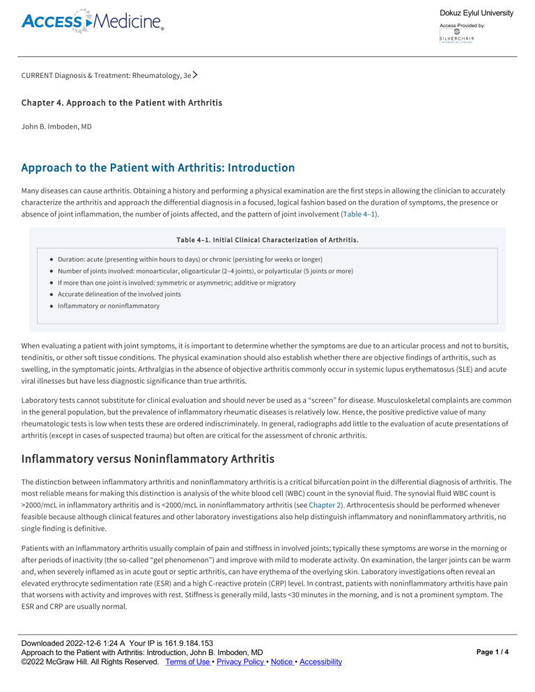
Dokuz Eylul University Access Provided by: CURRENT Diagnosis & Treatment: Rheumatology, 3e Chapter 4. Approach to the Patient with Arthritis John B. Imboden, MD Approach to the Patient with Arthritis: Introduction Many diseases can cause arthritis. Obtaining a history and performing a physical examination are the first steps in allowing the clinician to accurately characterize the arthritis and approach the differential diagnosis in a focused, logical fashion based on the duration of symptoms, the presence or absence of joint inflammation, the number of joints affected, and the pattern of joint involvement (Table 4–1). Table 4–1. Initial Clinical Characterization of Arthritis. Duration: acute (presenting within hours to days) or chronic (persisting for weeks or longer) Number of joints involved: monoarticular, oligoarticular (2–4 joints), or polyarticular (5 joints or more) If more than one joint is involved: symmetric or asymmetric; additive or migratory Accurate delineation of the involved joints Inflammatory or noninflammatory When evaluating a patient with joint symptoms, it is important to determine whether the symptoms are due to an articular process and not to bursitis, tendinitis, or other soft tissue conditions. The physical examination should also establish whether there are objective findings of arthritis, such as swelling, in the symptomatic joints. Arthralgias in the absence of objective arthritis commonly occur in systemic lupus erythematosus (SLE) and acute viral illnesses but have less diagnostic significance than true arthritis. Laboratory tests cannot substitute for clinical evaluation and should never be used as a “screen” for disease. Musculoskeletal complaints are common in the general population, but the prevalence of inflammatory rheumatic diseases is relatively low. Hence, the positive predictive value of many rheumatologic tests is low when tests these are ordered indiscriminately. In general, radiographs add little to the evaluation of acute presentations of arthritis (except in cases of suspected trauma) but often are critical for the assessment of chronic arthritis. Inflammatory versus Noninflammatory Arthritis The distinction between inflammatory arthritis and noninflammatory arthritis is a critical bifurcation point in the differential diagnosis of arthritis. The most reliable means for making this distinction is analysis of the white blood cell (WBC) count in the synovial fluid. The synovial fluid WBC count is >2000/mcL in inflammatory arthritis and is <2000/mcL in noninflammatory arthritis (see Chapter 2). Arthrocentesis should be performed whenever feasible because although clinical features and other laboratory investigations also help distinguish inflammatory and noninflammatory arthritis, no single finding is definitive. Patients with an inflammatory arthritis usually complain of pain and stiffness in involved joints; typically these symptoms are worse in the morning or after periods of inactivity (the so­called “gel phenomenon”) and improve with mild to moderate activity. On examination, the larger joints can be warm and, when severely inflamed as in acute gout or septic arthritis, can have erythema of the overlying skin. Laboratory investigations often reveal an elevated erythrocyte sedimentation rate (ESR) and a high C­reactive protein (CRP) level. In contrast, patients with noninflammatory arthritis have pain that worsens with activity and improves with rest. Stiffness is generally mild, lasts <30 minutes in the morning, and is not a prominent symptom. The ESR and CRP are usually normal. Constitutional Symptoms Downloaded 2022­12­6 1:24 A Your IP is 161.9.184.153 Page can 1/4 The presence of fever raises possibility of infection. Most patients with Approach to the Patient withthe Arthritis: Introduction, John B. Imboden, MD septic arthritis or disseminated gonococcal infection are febrile. Fever ©2022 McGraw arthritis Hill. All Rights Terms of Use (Table • Privacy Policy • Notice • Accessibility also accompany that is Reserved. not due to active infection 4–2). Indeed, intermittent high­grade fever ≥39°C is characteristic of Still disease. SLE can also cause fever ≥39°C. However, fever more often occurs when serositis, rather than polyarthritis, is the major manifestation of SLE. On the other hand, fever ≥38.3°C is unusual in rheumatoid arthritis, occurring in <1% of patients. and, when severely inflamed as in acute gout or septic arthritis, can have erythema of the overlying skin. Laboratory investigations often reveal an Dokuz Eylul University elevated erythrocyte sedimentation rate (ESR) and a high C­reactive protein (CRP) level. In contrast, patients with noninflammatory arthritis have pain Access Provided by: that worsens with activity and improves with rest. Stiffness is generally mild, lasts <30 minutes in the morning, and is not a prominent symptom. The ESR and CRP are usually normal. Constitutional Symptoms The presence of fever raises the possibility of infection. Most patients with septic arthritis or disseminated gonococcal infection are febrile. Fever can also accompany arthritis that is not due to active infection (Table 4–2). Indeed, intermittent high­grade fever ≥39°C is characteristic of Still disease. SLE can also cause fever ≥39°C. However, fever more often occurs when serositis, rather than polyarthritis, is the major manifestation of SLE. On the other hand, fever ≥38.3°C is unusual in rheumatoid arthritis, occurring in <1% of patients. Table 4–2. Fever and Arthritis. Active infection Septic arthritis Disseminated gonococcal infection Endocarditis Acute viral infections Mycobacterial Fungal Post infection Reactive arthritis (particularly in its early phases) Acute rheumatic fever and poststreptococcal arthritis Not due to infection Systemic lupus erythematosus Drug­induced lupus Still disease Gout Pseudogout Inflammatory bowel disease Paraneoplastic arthritis Acute sarcoidosis Systemic vasculitis Familial Mediterranean fever and other inherited periodic fever syndromes Significant weight loss is common at the initial presentation of reactive arthritis, systemic vasculitis, enteropathic arthritis, and paraneoplastic arthritis but is unusual in rheumatoid arthritis of recent onset. Constitutional symptoms rarely accompany noninflammatory forms of arthritis. Extra­Articular Manifestations Extra­articular manifestations, such as glomerulonephritis, pulmonary abnormalities, oral ulcerations, ocular inflammation, and peripheral neuropathy, may signal that arthritis is a manifestation of a systemic rheumatic disease or vasculitis. The presence of rash can be a very helpful clue to the diagnosis (Table 4–3). Table 4–3. Rash and Arthritis. Skin Manifestation Erythematous maculopapular rash Associated Conditions Superimposed drug reaction Still disease Viral syndromes Kawasaki disease Secondary syphilis Downloaded 2022­12­6 1:24 A Your IP is 161.9.184.153 Chronic meningococcemia Approach to the Patient with Arthritis: Introduction, John B. Imboden, MD ©2022 McGraw Hill. All Rights Reserved. Terms of Use • Privacy Policy • Notice • Accessibility Urticaria SLE Page 2 / 4 Erythematous maculopapular rash Superimposed drug reaction Dokuz Eylul University Still disease Access Provided by: Viral syndromes Kawasaki disease Secondary syphilis Chronic meningococcemia Urticaria SLE Hypocomplementemic urticarial vasculitis Serum sickness Acute hepatitis B Still disease Schnitzler syndrome Palpable purpura ANCA­associated vasculitis SLE Hypersensitivity vasculitis Cryoglobulinemia Henoch­Schonlein purpura Subacute bacterial endocarditis Papulosquamous lesions Psoriatic arthritis Reactive arthritis Discoid lupus Subacute cutaneous lupus erythematosus Secondary syphilis Annular lesions Subacute cutaneous lupus erythematosus Lyme disease (erythema chronicum migrans) Acute rheumatic fever (erythema marginatum) Pustular lesions Disseminated gonococcal infection Pustular psoriasis Reactive arthritis (keratoderma blenorrhagicum) Behçet disease Acne­associated rheumatic syndromes Sweet syndrome (also painful papule/nodules) Subcutaneous nodular lesions Erythema nodosum Sarcoidosis Inflammatory bowel disease Behçet disease SLE (lupus profundus) Polyarteritis nodosa Weber­Christian disease ANCA, antineutrophil cytoplasmic antibodies; SLE, systemic lupus erythematosus. Downloaded 2022­12­6 1:24 A Your IP is 161.9.184.153 Comorbid Conditions Page 3 / 4 Approach to the Patient with Arthritis: Introduction, John B. Imboden, MD ©2022 McGraw Hill. All Rights Reserved. Terms of Use • Privacy Policy • Notice • Accessibility Certain chronic conditions predispose to the development of particular musculoskeletal problems. For example, patients with long­standing, poorly controlled diabetes mellitus are at greatly increased risk for Charcot arthropathy in the feet and limited joint mobility in the hands. Certain medications Dokuz Eylul University ANCA, antineutrophil cytoplasmic antibodies; SLE, systemic lupus erythematosus. Access Provided by: Comorbid Conditions Certain chronic conditions predispose to the development of particular musculoskeletal problems. For example, patients with long­standing, poorly controlled diabetes mellitus are at greatly increased risk for Charcot arthropathy in the feet and limited joint mobility in the hands. Certain medications can trigger drug­induced lupus, which can present as a polyarthritis, often with serositis. The resurgence in the use of hydralazine for the treatment of hypertension has led to an increase in the incidence of hydralazine­induced lupus as well as the more serious hydralazine­induced, antineutrophil cytoplasmic antibody (ANCA)­associated vasculitis. Prior glucocorticoid therapy and alcohol abuse are the leading risk factors for osteonecrosis, which commonly presents as hip pain. Osteonecrosis and bone pain are common manifestations of Gaucher disease. Injection drug use carries the risk of septic arthritis; endocarditis; and infection with hepatitis B, hepatitis C, and HIV—each of which is associated with rheumatic conditions. Family History A positive family history, particularly among first­degree relatives, increases the likelihood of certain forms of arthritis. Most notably, the risk of ankylosing spondylitis for children or siblings of a patient with ankylosing spondylitis is as much as 75­fold that of the general population. The relative risk for SLE among first­degree relatives ranges from 20 to 30. A positive family history of rheumatoid arthritis is less helpful. The relative risk for siblings may be as low as 3, and family histories of rheumatoid arthritis can be inaccurate due to confusion with osteoarthritis. Downloaded 2022­12­6 1:24 A Your IP is 161.9.184.153 Approach to the Patient with Arthritis: Introduction, John B. Imboden, MD ©2022 McGraw Hill. All Rights Reserved. Terms of Use • Privacy Policy • Notice • Accessibility Page 4 / 4


