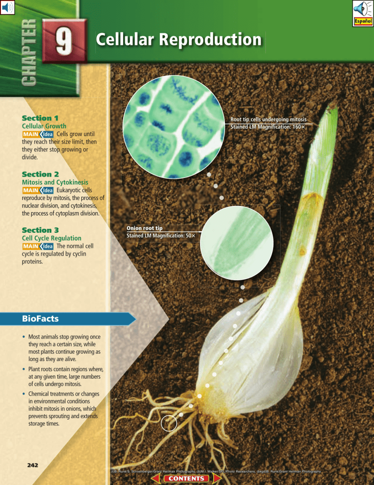
Cellular Reproduction Section 1 Root tip cells undergoing mitosis Stained LM Magnification: 160ⴛ Cellular Growth )DEA Cells grow until they reach their size limit, then they either stop growing or divide. Section 2 Mitosis and Cytokinesis )DEA Eukaryotic cells reproduce by mitosis, the process of nuclear division, and cytokinesis, the process of cytoplasm division. Section 3 Cell Cycle Regulation )DEA The normal cell cycle is regulated by cyclin proteins. Onion root tip Stained LM Magnification: 50ⴛ BioFacts • Most animals stop growing once they reach a certain size, while most plants continue growing as long as they are alive. • Plant roots contain regions where, at any given time, large numbers of cells undergo mitosis. • Chemical treatments or changes in environmental conditions inhibit mitosis in onions, which prevents sprouting and extends storage times. 242 (t)B. Runk/S. Schoenberger/Grant Heilman Photography, (b)M.I. Walker/SPL/Photo Researchers , (bkgd)B. Runk/Grant Heilman Photography Start-Up Activities LAUNCH Lab From where do healthy cells come? All living things are composed of cells. The only way an organism can grow or heal itself is by cellular reproduction. Healthy cells perform vital life functions, and they reproduce to form more cells. In this lab you will investigate the appearance of different cell types. Procedure 1. Read and complete the lab safety form. 2. Observe prepared slides of human cells under high magnification using a light microscope. 3. Observe onion root tip cells under the microscope. 4. Observe other cells on the prepared slides your teacher will give you. 5. Draw diagrams of the sample cells you observed. Identify and label any of the structures you recognize. Analysis 1. Compare and contrast the different cells you observed. 2. Hypothesize why the cells you observed had different appearances and structures. How could you identify diseased cells? Mitosis and Cytokinesis Make this Foldable to help you understand how cells reproduce by a process called mitosis, resulting in two genetically identical cells. STEP 1 Stack three sheets of notebook paper approximately 1.5 cm apart vertically as illustrated. STEP 2 Roll up the bottom edges and fold to form six tabs. STEP 3 Staple along the folded edge to secure all sheets. Rotate the Foldable and, with the stapled end at the top, label the tabs as illustrated. ÌÃÃÊ* >Ãià >`Ê ÞÌiÃà Visit biologygmh.com to: ▶ study the entire chapter online ▶ explore the Concepts in Motion, Microscopy Links, Virtual Labs, and links to virtual dissections ▶ access Web links for more information, projects, and activities ▶ review content online with the Interactive Tutor and take Self-Check Quizzes *À« >Ãi iÌ>« >Ãi >« >Ãi /i« >Ãi ÞÌiÃà &/,$!",%3 Use this Foldable with Section 9.2. As you study the section, record what you learn about each of the four phases of mitosis. In the tab labeled Cytokinesis, write a brief description of cytokinesis, the division of cytoplasm. Section Chapter 1 • XXXXXXXXXXXXXXXXXX 9 • Cellular Reproduction 243 Section 9.1 Objectives ◗ Explain why cells are relatively small. ◗ Summarize the primary stages of the cell cycle. ◗ Describe the stages of interphase. Review Vocabulary selective permeability: process in which a membrane allows some substances to pass through while keeping others out Cellular Growth )DEA Cells grow until they reach their size limit, then they either stop growing or divide. Real-World Reading Link If you’ve ever played a doubles match in tennis, you probably felt that you and your partner could effectively cover your half of the court. However, if the court were much larger, perhaps you could no longer reach your shots. For the best game, the tennis court must be kept at regulation size. Cell size also must be limited to ensure that the needs of the cell are met. New Vocabulary Cell Size Limitations cell cycle interphase mitosis cytokinesis chromosome chromatin Most cells are less than 100 µm (100 × 10 –6 m) in diameter, which is smaller than the period at the end of this sentence. Why are most cells so small? This section investigates several factors that influence cell size. Ratio of surface area to volume The key factor that limits the size of a cell is the ratio of its surface area to its volume. The surface area of the cell refers to the area covered by the plasma membrane. Recall from Chapter 7 that the plasma membrane is the structure through which all nutrients and waste products must pass. The volume refers to the space taken by the inner contents of the cell, including the organelles in the cytoplasm and the nucleus. #ONNECTION TO -ATH To illustrate the ratio of surface area to volume, consider the small cube in Figure 9.1, which has sides of one micrometer (µm) in length. This is approximately the size of a bacterial cell. To calculate the surface area of the cube, multiply length times width times the number of sides (1 µm × 1 µm × 6 sides), which equals 6 µm2. To calculate the volume of the cell, multiply length times width times height (1 µm × 1 µm × 1 µm), which equals 1 µm3. The ratio of surface area to volume is 6:1. ¥ ¥ ¥ ■ Figure 9.1 Note how the ratio of surface area to volume changes as the size of the cell increases, and note the amount of contents the nucleus must control as cell size increases. Infer How does the amount of surface area change as the cell’s volume increases? 244 Chapter 9 • Cellular Reproduction LM Magnification: 150⫻ If the cubic cell grows to 2 µm per side, as represented in Figure 9.1, the surface area becomes 24 µm2 and the volume is 8 µm3. The ratio of surface area to volume is now 3:1, which is less than it was when the cell was smaller. If the cell continues to grow, the ratio of surface area to volume will continue to decrease, as shown by the third cube in Figure 9.1. As the cell grows, its volume increases much more rapidly than the surface area. This means that the cell might have difficulty supplying nutrients and expelling enough waste products. By remaining small, cells have a higher ratio of surface area to volume and can sustain themselves more easily. Reading Check Explain why a high ratio of surface area to volume benefits a cell. Transport of substances Another task that can be managed more easily in a small cell than in a large cell is the movement of substances. Recall that the plasma membrane controls cellular transport because it is selectively permeable. Once inside the cell, substances move by diffusion or by motor proteins pulling them along the cytoskeleton. Diffusion over large distances is slow and inefficient because it relies on random movement of molecules and ions. Similarly, the cytoskeleton transportation network, shown in Figure 9.2, becomes less efficient for a cell if the distance to travel becomes too large. Therefore, cells remain small to maximize the ability of diffusion and motor proteins to transport nutrients and waste products. Small cells maintain more efficient transport systems. Figure 9.2 In order for the cytoskeleton to be an efficient transportation railway, the distances substances must travel within a cell must be limited. ■ Investigate Cell Size Could a cell grow large enough to engulf your school? What would happen if the size of an elephant were doubled? At the organism level, an elephant cannot grow significantly larger, because its legs would not support the increase in mass. Do the same principles and limitations apply at the cellular level? Do the math! Procedure 1. Read and complete the lab safety form. 2. Prepare a data table for surface area and volume data calculated for five hypothetical cells. Assume the cell is a cube. (Dimensions given are for one face of a cube.) Cell 1: 0.00002 m (the average diameter of most eukaryotic cells) Cell 2: 0.001 m (the diameter of a squid’s giant nerve cell) Cell 3: 2.5 cm Cell 4: 30 cm Cell 5: 15 m 3. Calculate the surface area for each cell using the formula: length ⫻ width ⫻ number of sides (6). 4. Calculate the volume for each cell using the formula: length ⫻ width ⫻ height. Analysis 1. Cause and Effect Based on your calculations, confirm why cells don’t become very large. 2. Infer Are large organisms, such as redwood trees and elephants, large because they contain extra large cells or just more standard-sized cells? Explain. Section 1 • Cellular Growth 245 Dr. Gopal Murti/Visuals Unlimited ■ Figure 9.3 The cell cycle involves three stages—interphase, mitosis, and cytokinesis. Interphase is divided into three substages. Hypothesize Why does cytokinesis represent the smallest amount of time a cell spends in the cell cycle? Study Tip Diagram Draw your own version of the cell cycle, including the terms interphase, mitosis, and cytokinesis as you read the text. Cellular communications The need for signaling proteins to move throughout the cell also limits cell size. In other words, cell size affects the ability of the cell to communicate instructions for cellular functions. If the cell becomes too large, it becomes almost impossible for cellular communications, many of which involve movement of substances and signals to various organelles, to take place efficiently. For example, the signals that trigger protein synthesis might not reach the ribosome fast enough for protein synthesis to occur to sustain the cell. The Cell Cycle VOCABULARY WORD ORIGIN Cytokinesis cyto– prefix; from the Greek word kytos, meaning hollow vessel –kinesis from the Greek word kinetikos, meaning putting in motion. 246 Chapter 9 • Cellular Reproduction Once a cell reaches its size limit, something must happen—either it will stop growing or it will divide. Most cells will eventually divide. Cell division not only prevents the cell from becoming too large, but it also is the way the cell reproduces so that you grow and heal certain injuries. Cells reproduce by a cycle of growing and dividing called the cell cycle. Each time a cell goes through one complete cycle, it becomes two cells. When the cell cycle is repeated continuously, the result is a continuous production of new cells. A general overview of the cell cycle is presented in Figure 9.3. There are three main stages of the cell cycle. Interphase is the stage during which the cell grows, carries out cellular functions, and replicates, or makes copies of its DNA in preparation for the next stage of the cycle. Interphase is divided into three substages, as indicated by the segment arrows in Figure 9.3. Mitosis (mi TOH sus) is the stage of the cell cycle during which the cell’s nucleus and nuclear material divide. Mitosis is divided into four substages. Near the end of mitosis, a process called cytokinesis begins. Cytokinesis (si toh kih NEE sis) is the method by which a cell’s cytoplasm divides, creating a new cell. You will read more about mitosis and cytokinesis in Section 9.2. The duration of the cell cycle varies, depending on the cell that is dividing. Some eukaryotic cells might complete the cycle in as few as eight minutes, while other cells might take up to one year. For most normal, actively dividing animal cells, the cell cycle takes approximately 12–24 hours. When you consider all that takes place during the cell cycle, you might find it amazing that most of your cells complete the cell cycle in about a day. The stages of interphase During interphase, the cell grows, develops into a mature, functioning cell, duplicates its DNA, and prepares for division. Interphase is divided into three stages, as shown in Figure 9.3: G1, S, and G2, also called Gap 1, synthesis, and Gap 2. The first stage of interphase, G1, is the period immediately after a cell divides. During G1, a cell is growing, carrying out normal cell functions, and preparing to replicate DNA. Some cells, such as muscle and nerve cells, exit the cell cycle at this point and do not divide again. The second stage of interphase, S, is the period when a cell copies its DNA in preparation for cell division. Chromosomes (KROH muh sohmz) are the structures that contain the genetic material that is passed from generation to generation of cells. Chromatin (KROH muh tun) is the relaxed form of DNA in the cell’s nucleus. As shown in Figure 9.4, when a specific dye is applied to a cell in interphase, the nucleus stains with a speckled appearance. This speckled appearance is due to individual strands of chromatin that are not visible under a light microscope without the dye. The G2 stage follows the S stage and is the period when the cell prepares for the division of its nucleus. A protein that makes microtubules for cell division is synthesized at this time. During G2, the cell also takes inventory and makes sure it is ready to continue with mitosis. When these activities are completed, the cell begins the next stage of the cell cycle—mitosis. Mitosis and cytokinesis The stages of mitosis and cytokinesis follow interphase. In mitosis, the cell’s nuclear material divides and separates into opposite ends of the cell. In cytokinesis, the cell divides into two daughter cells with identical nuclei. These important stages of the cell cycle are described in Section 9.2. Stained LM Magnification: 400⫻ Figure 9.4 The grainy appearance of this nucleus from a rat liver cell is due to chromatin, the relaxed material that condenses to form chromosomes. ■ Prokaryotic cell division The cell cycle is the method by which eukaryotic cells reproduce themselves. Prokaryotic cells, which you have learned are simpler cells, reproduce by a method called binary fission. You will learn more about binary fission in Chapter 18. Section 9.1 Assessment Section Summary Understand Main Ideas ◗ The ratio of surface area to volume describes the size of the plasma membrane relative to the volume of the cell. 1. ◗ Cell size is limited by the cell’s ability to transport materials and communicate instructions from the nucleus. ◗ The cell cycle is the process of cellular reproduction. ◗ A cell spends the majority of its lifetime in interphase. Relate cell size to cell functions, and explain why cell size is limited. Think Scientifically )DEA 5. Hypothesize 6. "IOLOGY If a cube representing a cell is 5 µm on a side, calculate the surface area-to-volume ratio, and explain why this is or is not a good size for a cell. 2. Summarize the primary stages of the cell cycle. 3. Describe what happens to DNA during the S stage of interphase. 4. Make a diagram of the stages of the cell cycle and describe what happens in each. Self-Check Quiz biologygmh.com what the result would be if a large cell managed to divide, despite the fact that it had grown beyond an optimum size. Section 1 • Cellular Growth 247 Michael Abbey/Visuals Unlimited Section 9. 2 Section Objectives ◗ Describe the events of each stage of mitosis. ◗ Explain the process of cytokinesis. Review Vocabulary life cycle: the sequence of growth and development stages that an organism goes through during its life Mitosis and Cytokinesis )DEA Eukaryotic cells reproduce by mitosis, the process of nuclear division, and cytokinesis, the process of cytoplasm division. Real-World Reading Link Many familiar events are cyclic in nature. The course of a day, the changing of seasons year after year, and the passing of comets in space are some examples of cyclic events. Cells also have a cycle of growth and reproduction. New Vocabulary prophase sister chromatid centromere spindle apparatus metaphase anaphase telophase Mitosis You learned in the last section that cells cycle through interphase, mitosis, and cytokinesis. During mitosis, the cell’s replicated genetic material separates and the cell prepares to split into two cells. The key activity of mitosis is the accurate separation of the cell’s replicated DNA. This enables the cell’s genetic information to pass into the new cells intact, resulting in two daughter cells that are genetically identical. In multicellular organisms, the process of mitosis increases the number of cells as a young organism grows to its adult size. Organisms also use mitosis to replace damaged cells. Recall the last time you accidently got cut. Under the scab, the existing skin cells divided by mitosis and cytokinesis to create new skin cells that filled the gap in the skin caused by the injury. The Stages of Mitosis ■ Figure 9.5 Chromosomes in prophase are actually sister chromatids that are attached at the centromere. Like interphase, mitosis is divided into stages: prophase, metaphase, anaphase, and telophase. Prophase The first stage of mitosis—the stage of mitosis during which a dividing cell spends the most time—is called prophase. In this stage, the cell’s chromatin tightens, or condenses, into chromosomes. In prophase, the chromosomes are shaped like an X, as shown in Figure 9.5. At this point, each chromosome is a single structure that contains the genetic material that was replicated in interphase. Each half of this X is called a sister chromatid. Sister chromatids are structures that contain identical copies of DNA. The structure at the center of the chromosome where the sister chromatids are attached is called the centromere. This structure is important because it ensures that a complete copy of the replicated DNA will become part of the daughter cells at the end of the cell cycle. Locate prophase in the cell cycle illustrated in Figure 9.6, and note the position of the sister chromatids. As you continue to read about the stages of mitosis, refer back to Figure 9.6 to follow the chromatids through the cell cycle. Reading Check Compare the key activity of interphase with the key Color-Enhanced SEM magnification: 6875⫻ 248 Chapter 9 • Cellular Reproduction Andrew Syred/Photo Researchers activity of mitosis. Visualizing the Cell Cycle Figure 9.6 The cell cycle begins with interphase. Mitosis follows, occurring in four stages—prophase, metaphase, anaphase, and telophase. Mitosis is followed by cytokinesis, then the cell cycle repeats with each new cell. LM Magnification: 118⫻ LM Magnification: 118⫻ LM Magnification: 118⫻ LM Magnification: 118⫻ LM Magnification: 118⫻ LM Magnification: 118⫻ Interactive Figure To see an animation of the cell cycle, visit biologygmh.com. Section 2 • Mitosis and Cytokinesis 249 (cw from top)Thomas Deerinck/Visuals Unlimited, (2)Thomas Deerinck/Visuals Unlimited, (3)Thomas Deerinck/Visuals Unlimited, (4)Thomas Deerinck/Visuals Unlimited, (5)Thomas Deerinck/Visuals Unlimited, (6)Thomas Deerinck/Visuals Unlimited LM Magnification: 100⫻ Sister chromatids Centrioles Spindle fibers Aster ■ Figure 9.7 In animal cells, the spindle apparatus is made of spindle fibers, centrioles, and aster fibers. Incorporate information from this section into your Foldable. ■ Figure 9.8 In metaphase, the chromosomes align along the equator of the cell. Compare a cell’s equator to Earth’s equator. As prophase continues, the nucleolus seems to disappear. Microtubule structures called spindle fibers form in the cytoplasm. In animal cells and most protist cells, another pair of microtubule structures, called centrioles, migrates to the ends, or poles, of the cell. Coming out of the centrioles are yet another type of microtubule called aster fibers, which have a starlike appearance. The whole structure, including the spindle fibers, centrioles, and aster fibers, is called the spindle apparatus and is shown in Figure 9.7. The spindle apparatus is important in moving and organizing the chromosomes before cell division. Centrioles are not part of the spindle apparatus in plant cells—only spindle fibers are present. Near the end of prophase, the nuclear envelope seems to disappear. The spindle fibers attach to the sister chromatids of each chromosome on both sides of the centromere and then attach to opposite poles of the cell. This arrangement ensures that each new cell receives one complete copy of the DNA. Metaphase During the second stage of mitosis, metaphase, the sister chromatids are pulled by motor proteins along the spindle apparatus toward the center of the cell and line up in the middle, or equator, of the cell, as shown in Figure 9.8. Metaphase is one of the shortest stages of mitosis, but when completed successfully, it ensures that the new cells have accurate copies of the chromosomes. Photomicrograph Magnification: 450⫻ Duplicated chromosomes at equator Asters radiating from centrosome Spindle fibers connecting to centromere 250 Chapter 9 • Cellular Reproduction (t)Dr. Conley L. Rieder and Dr. Alexey Khodjakov/Visuals Unlimited, (b)Carolina Biological Supply Co./PhotoTake NYC LM Magnification: 450⫻ Anaphase The chromatids are pulled apart during anaphase, the third stage of mitosis. In anaphase, the microtubules of the spindle apparatus begin to shorten. This shortening pulls at the centromere of each sister chromatid, causing the sister chromatids to separate into two identical chromosomes. All of the sister chromatids separate simultaneously, although the exact mechanism that controls this is unknown. At the end of anaphase, the microtubules, with the help of motor proteins, move the chromosomes toward the poles of the cell. Figure 9.9 By the end of telophase, the cell has completed the work of duplicating the genetic material and dividing it into two “packages,” but the cell has not completely divided. ■ Telophase The last stage of mitosis is called telophase. Telophase is the stage of mitosis during which the chromosomes arrive at the poles of the cell and begin to relax, or decondense. As shown in Figure 9.9, two new nuclear membranes begin to form and the nucleoli reappear. The spindle apparatus disassembles and some of the microtubules are recycled by the cell to build various parts of the cytoskeleton. Although the four stages of mitosis are now complete and the nuclear material is divided, the process of cell division is not yet complete. Data Analysis lab 9.1 Based on Real Data* Predict the Results What happens to the microtubules? Scientists performed experiments tracking chromosomes along microtubules during mitosis. They hypothesized that the microtubules are broken down, releasing microtubule subunits as the chromosomes are moved toward the poles of the cell. The microtubules were labeled with a yellow fluorescent dye, and using a laser, the microtubules were marked midway between the poles and the chromosomes by eliminating the fluorescence in the targeting region as shown in the diagram. Data and Observations Fluorescent-labeled microtubules Think Critically 1. Explain What was the purpose of the fluorescent dye? 2. Predict Draw a diagram of how the cell might appear later in anaphase. *Data obtained from: Maddox, P., et al. 2003. Direct observation of microtubule dynamics at kinetochores in Xenopus extract spindles: implications for spindle mechanics. The Journal of Cell Biology 162: 377-382. Maddox, et al. 2004. Controlled ablations of microtubules using picosecond laser. Biophysics Journal 87: 4203-4212. Laser-marked microtubules Section 2 • Mitosis and Cytokinesis Michael Abbey/Photo Researchers 251 Color-Enhanced SEM Magnification: 125⫻ Stained LM Magnification: 1000⫻ Cell plate Furrow Animal cell ■ Plant cells Figure 9.10 Cytokinesis Left: In animal cells, cytokinesis begins with a furrow that pinches the cell and eventually splits the two cells apart. Right: Plant cells build a cell plate that divides the cell into the two daughter cells. Section 9. 2 Toward the end of mitosis, the cell begins another process called cytokinesis that will divide the cytoplasm. This results in two cells, each with identical nuclei. During the later phases of mitosis, microtubules are formed that will be involved in cytokinesis. In animal cells, cytokinesis is accomplished by using microfilaments to constrict, or pinch, the cytoplasm, as shown in Figure 9.10. Recall from Chapter 7 that plant cells have a rigid cell wall covering their plasma membrane. Instead of pinching in half, a new structure, called a cell plate, forms between the two daughter nuclei, as illustrated in Figure 9.10. Cell walls then form on either side of the cell plate. Once this new wall is complete, there are two genetically identical cells. Prokaryotic cells, which divide by binary fission, finish cell division in a different way. When prokaryotic DNA is duplicated, both copies attach to the plasma membrane. As the plasma membrane grows, the attached DNA molecules are pulled apart. The cell completes fission, producing two new prokaryotic cells. Assessment Section Summary Understand Main Ideas ◗ Mitosis is the process by which the duplicated DNA is divided. 1. ◗ The stages of mitosis include prophase, metaphase, anaphase, and telophase. ◗ Cytokinesis is the process of cytoplasm division that results in genetically identical daughter cells. Explain why mitosis alone does not produce daughter cells. Think Scientifically )DEA 6. Hypothesize 7. "IOLOGY If a plant cell completes the cell cycle in 24 hours, how many cells will be produced in a week? 2. Describe the events of each stage of mitosis. 3. Diagram and label a chromosome in prophase. 4. Identify the stage of mitosis in which a cell spends the most time. 5. Contrast cytokinesis in a plant cell and an animal cell. 252 Chapter 9 • Cellular Reproduction (l)RMF/Visuals Unlimited, (r)B. Runk/S. Schoenberger/Grant Heilman Photography what would happen if a drug that stopped microtubule movement but did not affect cytokinesis was applied to a cell. Self-Check Quiz biologygmh.com Section 9. 3 Objectives ◗ Summarize the role of cyclin proteins in controlling the cell cycle. ◗ Explain how cancer relates to the cell cycle. ◗ Describe the role of apoptosis. ◗ Summarize the two types of stem cells and their potential uses. Review Vocabulary nucleotide: subunit that makes up DNA and RNA molecules New Vocabulary cyclin cyclin-dependent kinase cancer carcinogen apoptosis stem cell Cell Cycle Regulation )DEA The normal cell cycle is regulated by cyclin proteins. Real-World Reading Link No matter how many new homes a builder builds, even if building the same design, the crew always relies on blueprint instructions. Similarly, cells have specific instructions for completing the cell cycle. Normal Cell Cycle The timing and rate of cell division are important to the health of an organism. The rate of cell division varies depending on the type of cell. A mechanism involving proteins and enzymes controls the cell cycle. The role of cyclins To start a car, it takes a combination of a key turning in the ignition to signal the engine to start. Similarly, the cell cycle in eukaryotic cells is driven by a combination of two substances that signal the cellular reproduction processes. Proteins called cyclins bind to enzymes called cyclin-dependent kinases (CDKs) in the stages of interphase and mitosis to start the various activities that take place in the cell cycle. Different cyclin/CDK combinations control different activities at different stages in the cell cycle. Figure 9.11 illustrates where some of the important combinations are active. In the G1 stage of interphase, the combination of cyclin with CDK signals the start of the cell cycle. Different cyclin/CDK combinations signal other activities, including DNA replication, protein synthesis, and nuclear division throughout the cell cycle. The same cyclin/CDK combination also signals the end of the cell cycle. Figure 9.11 Signaling molecules made of a cyclin bound to a CDK kick off the cell cycle and drive it through mitosis. Checkpoints monitor the cell cycle for errors and can stop the cycle if an error occurs. ■ - # ' 3 ' Section 3 • Cell Cycle Regulation 253 Careers In biology Pharmaceutical QC Technician Just as the cell cycle has built-in quality control checkpoints, so do biological product manufacturing processes. A QC technician in a pharmaceutical manufacturing company uses various science and math skills to monitor processes and ensure product quality. For more information on biology careers, visit biologygmh.com. Quality control checkpoints Recall the process of starting a car. Many manufacturers use a unique microchip in the key to ensure that only a specific key will start each car. This is a checkpoint against theft. The cell cycle also has built-in checkpoints that monitor the cycle and can stop it if something goes wrong. For example, a checkpoint near the end of the G1 stage monitors for DNA damage and can stop the cycle before entering the S stage of interphase. There are other quality control checkpoints during the S stage and after DNA replication in the G2 stage. Spindle checkpoints also have been identified in mitosis. If a failure of the spindle fibers is detected, the cycle can be stopped before cytokinesis. Figure 9.11 shows the location of key checkpoints in the cell cycle. Abnormal Cell Cycle: Cancer #ONNECTION TO (EALTH Although the cell cycle has a system of quality control checkpoints, it is a complex process that sometimes fails. When cells do not respond to the normal cell cycle control mechanisms, a condition called cancer can result. Cancer is the uncontrolled growth and division of cells—a failure in the regulation of the cell cycle. When unchecked, cancer cells can kill an organism by crowding out normal cells, resulting in the loss of tissue function. Cancer cells spend less time in interphase than do normal cells, which means cancer cells grow and divide unrestrained as long as they are supplied with essential nutrients. Figure 9.12 shows how cancer cells can intrude on normal cells. Causes of cancer Cancer does not just occur in a weak organism. In fact, cancer occurs in many healthy, active, and young organisms. The changes that occur in the regulation of cell growth and division of cancer cells are due to mutations or changes in the segments of DNA that control the production of proteins, including proteins that regulate the cell cycle. Often, the genetic change or damage that occurs is repaired by various repair systems. But if the repair systems fail, cancer can result. Various environmental factors can affect the occurrence of cancer cells. Substances and agents that are known to cause cancer are called carcinogens (kar SIH nuh junz). Cancer cells Normal cells Figure 9.12 A medical professional can identify cancer cells because they often have an abnormal, irregular shape compared to normal cells. If left unchecked, a cancerous tumor can grow to the point where it can kill its host organism. ■ 254 Chapter 9 • Cellular Reproduction Although not all cancers can be prevented, avoiding known carcinogens can help reduce the risk of cancer. A governmental agency called the Food and Drug Administration (FDA) works to make sure that the things you eat and drink are safe. The FDA also requires labels and warnings for products that might be carcinogens. Industrial laws help protect people from exposure to cancer-causing chemicals, such as asbestos, in the workplace. For example, asbestos has been removed from many old buildings to protect people living and working inside them. Avoiding tobacco of all kinds, even secondhand smoke and smokeless tobacco, can reduce the risk of cancer. Some radiation, such as ultraviolet radiation from the Sun, is impossible to avoid completely. There is a connection between the amount of ultraviolet radiation to which a person is exposed and the risk of developing skin cancer. Therefore, sunscreen is recommended for everyone who is exposed to the Sun. Other forms of radiation, such as X rays, are used for medical purposes, such as to look at a broken bone or check for tooth cavities. To protect against exposure, you might have worn a heavy lead apron when an X ray was taken. Cancer genetics More than one change in DNA is required to change an abnormal cell into a cancer cell. Over time, it is possible that there might be many changes in DNA. This might explain why the risk of cancer increases with age. The fact that multiple changes must occur also might explain why cancer runs in some families. An individual who inherits one or more changes from a parent is at a higher risk for developing cancer than someone who does not inherit these changes. LAUNCH Lab Review Based on what you’ve read about the abnormal cell cycle and its results, how would you now answer the analysis questions? VOCABULARY SCIENCE USAGE V. COMMON USAGE Inheritance Science usage: the passing of genetic traits from parent to offspring via DNA. A person’s body structure and facial appearance are the result of genetic inheritance. Common usage: assets acquired from a deceased person that can be given to surviving family members. The house was Jim’s inheritance from his uncle. Compare Sunscreens Do sunscreens really block sunlight? Sunscreens contain a variety of different compounds that absorb UVB from sunlight. UVB is linked to mutations in DNA that can lead to skin cancer. Find out how effective at blocking sunlight various sunscreens are. Procedure 1. Read and complete the lab safety form. 2. Choose one of the sunscreen products provided by your teacher. Record the active ingredients and the Sun protection factor (SPF) on a data sheet. 3. Obtain two sheets of plastic wrap. On one sheet use a permanent marker to draw two widely spaced circles. Place a drop of sunscreen in the middle of one circle and a drop of zinc oxide in the middle of the other. 4. Lay the second sheet on top of both circles. Spread the drops by pressing with a book. 5. Take a covered piece of Sun-sensitive paper and your two pieces of plastic wrap to a sunny area. Quickly uncover the paper, lay the two pieces of plastic wrap on top, and place in the sunlight. 6. After the paper is fully exposed (1–5 minutes), remove it from the sunlight and develop according to instructions. Analysis 1. Think Critically Why did you compare the sunscreens to zinc oxide? 2. Draw Conclusions After examining the developed Sun-sensitive papers from your class, which sunscreens do you think would be most likely to prevent DNA mutations? Section 3 • Cell Cycle Regulation 255 Apoptosis VOCABULARY ACADEMIC VOCABULARY Mature: To have reached full natural growth or development. After mitosis, the two new cells must mature before they divide. Not every cell is destined to survive. When an embryo divides, some cells go through a process called apoptosis (a pup TOH sus), or programmed cell death. Cells going through apoptosis actually shrink and shrivel in a controlled process. All animal cells appear to have a “death program” that can be activated. One example of apoptosis occurs during the development of the human hand and foot. When the hands and feet begin to develop, cells occupy the spaces between the fingers and toes. Normally, this tissue undergoes apoptosis, with the cells shriveling and dying at the appropriate time so that the webbing is not present in the mature organism. An example of apoptosis in plants is the localized death of cells that results in leaves falling from trees during autumn. Apoptosis also occurs in cells that are damaged beyond repair, including cells with DNA damage that could lead to cancer. Apoptosis can help to protect organisms from developing cancerous growths. Stem Cells The majority of cells in a multicellular organism are designed for a specialized function. Some cells might be part of your skin, and other cells might be part of your heart. In 1998, scientists discovered a way to isolate a unique type of cell in humans called the stem cell. Stem cells are unspecialized cells that can develop into specialized cells when under the right conditions, as illustrated in Figure 9.13. Stem cells can remain in an organism for many years while undergoing cell division. There are two basic types of stem cells: embryonic stem cells and adult stem cells. Embryonic stem cells After a sperm fertilizes an egg, the resulting mass of cells divides repeatedly until there are about 100–150 cells. These cells have not become specialized and are called embryonic stem cells. If separated, each of these cells has the capability of developing into a wide variety of specialized cells. If the embryo continues to divide, the cells specialize into various tissues, organs, and organ systems. Embryonic stem cell research is controversial because of ethical concerns about the source of the cells. ■ Figure 9.13 Because stem cells are not locked into becoming one particular type of cell, they might be the key to curing many medical conditions and genetic defects. 256 Chapter 9 • Cellular Reproduction P. Sorrentino-Eurelios/PhotoTake NYC ■ Figure 9.14 Research with adult stem cells has led to advances in treatments for numerous injuries and diseases. Adult stem cells The second type of stem cells, adult stem cells, is found in various tissues in the body and might be used to maintain and repair the same kind of tissue in which they are found. The term “adult stem cells” might be somewhat misleading because even a newborn has adult stem cells. Like embryonic stem cells, certain kinds of adult stem cells also might be able to develop into different kinds of cells, providing new treatments for many diseases and conditions. In 1999, researchers at Harvard Medical School used nervous system stem cells to restore lost brain tissue in mice. In 2000, a team of researchers at the University of Florida used pancreatic stem cells to restore pancreas function in a mouse with diabetes. Research with adult stem cells, like that shown in Figure 9.14, is much less controversial because the adult stem cells can be obtained with the consent of their donor. Section 9. 3 Assessment Section Summary Understand Main Ideas ◗ The cell cycle of eukaryotic cells is regulated by cyclins. 1. ◗ Checkpoints occur during most of the stages of the cell cycle to ensure that the cell divides accurately. 2. Explain how the cancer cell cycle is different from a normal cell cycle. ◗ Cancer is the uncontrolled growth and division of cells. 4. Contrast apoptosis and cancer. ◗ Apoptosis is a programmed cell death. ◗ Stem cells are unspecialized cells that can develop into specialized cells with the proper signals. Describe how cyclins control the cell cycle. Think Scientifically )DEA 7. Hypothesize 8. "IOLOGY Write a statement for a dental brochure describing the safety of X rays as a diagnostic tool. what might happen if apoptosis did not occur in cells that have significant DNA damage. 3. Identify three carcinogens. 5. Describe a possible application for stem cells. 6. Explain the difference between embryonic stem cells and adult stem cells. Self-Check Quiz biologygmh.com Section 3 • Cell Cycle Regulation 257 Stem Cells: Paralysis Cured? CNS stem cells Bone marrow stem cells A race car driver is paralyzed in a crash. A teen is paralyzed after diving into shallow water. Until recently, these individuals would have little hope of regaining the full use of their bodies, but new research on adult stem cells shows promise for reversing paralysis. How can stem cells be used? Scientists are trying to find ways to grow adult stem cells in cell cultures and manipulate them to generate specific cell types. For example, stem cells might be used to repair cardiac tissue after a heart attack, to restore vision in diseased or injured eyes, to treat diseases such as diabetes, or to repair spinal cells to reverse paralysis. Late actor and paralysis victim Christopher Reeve was a strong proponent for stem cell research because he believed there is much potential in science to improve the condition of life for others who suffer from paralysis. Stem cells and paralysis In Portugal, Dr. Carlos Lima and his team of researchers found that tissue taken from the nasal cavity is a rich source of adult stem cells. These stem cells become nerve cells when transplanted into the site of a spinal cord injury. The new nerve cells replace the cells that were damaged. More than forty patients with paralysis due to accidents have undergone the Portuguese procedure. All patients have regained some sensation in paralyzed body areas. Most have regained some motor control. With intensive physical therapy, about ten percent of the patients now can walk with the aid of supportive devices, such as walkers and braces. This is promising news to the many individuals facing illnesses or injuries that have robbed them of the full use of their bodies. 258 Chapter 9 • Cellular Reproduction Fat cells Cardiac muscle cells Epithelial cells Blood cells Nerve cells Skeletal muscle cells Stem cells from bone marrow or the central nervous system can be manipulated to generate many cell types that can be transplanted to treat illness or repair damage. Stem cells and the future Scientists are eager to do the research necessary to make adult stem cell treatments a regular part of health care. Paralysis might not have to be permanent: stem cells could provide the cure. "IOLOGY Pamphlet Create a pamphlet depicting the benefits of adult stem cell research. Conduct additional research on adult stem cell research at biologygmh.com in order to include the research methodology, treatment, examples, cell physiology, and history of adult stem cell research. Be sure to illustrate your pamphlet. DOES SUNLIGHT AFFECT MITOSIS IN YEAST? Background: Ultraviolet (UV) radiation is a component of sunlight that can damage DNA and interrupt the cell cycle. Question: Can sunscreens prevent damage to UV-sensitive yeast? Materials sterile pipettes (10) aluminum foil test-tube rack sterile spreaders or sterile cotton swabs (10) dilution of UV-sensitive yeast yeast extract dextrose (YED) agar plates (10) sunscreens with various amounts of SPF Safety Precautions Procedure 1. Read and complete the lab safety form. 2. Obtain a test tube containing a diluted broth culture of the UV-sensitive yeast. 3. Formulate a hypothesis, then choose a sunscreen and predict how it will affect the yeast when exposed to sunlight. 4. Label ten YED agar plates with your group name. Label two plates as control. The control plates will not be placed in the sunlight. Label four of the experimental plates as “no sunscreen” and four as “sunscreen.” 5. Spread a 0.1 mL sample of the yeast dilution on all ten YED agar plates. Wrap the control plates in foil and give them to your teacher for incubation. 6. With direction from your teacher, decide how long to expose each of the experimental plates and label each plate accordingly. Prepare a table in which to collect your data. 7. Wrap the “no sunscreen” plates in foil. Apply sunscreen to the lids of the four sunscreen plates and wrap them in foil. 8. Remove only enough aluminum foil from each of the experimental plates to expose the dish lids. Expose the plates for the planned times. Re-cover the plates after exposure and give them to your teacher for incubation. 9. After incubation, count and record the number of yeast colonies on each plate. 10. Cleanup and Disposal Wash and return all reusable materials. Dispose of the YED plates as instructed by your teacher. Disinfect your work area. Wash your hands thoroughly with soap and water. Analyze and Conclude 1. Estimate Assume that each yeast colony on a YED plate grew from one yeast cell in the dilution. Use the number of yeast colonies on your control plate to determine the percent of yeast that survived on each exposed plate. 2. Graph Data Draw a graph with the percent survival on the y-axis and the exposure time on the x-axis. Use a different color to graph the data from the plates with and without sunscreen. 3. Evaluate Was your hypothesis supported by your data? Explain. 4. Error Analysis Describe several possible sources of error. Apply your Skill Brainstorm ideas about how UV-sensitive yeast could be used as a biological monitor to detect increases in the amounts of UV light reaching Earth’s surface. To learn more about mitosis in yeast, visit Biolabs at biologygmh.com. BioLab 259 Download quizzes, key terms, and flash cards from biologygmh.com. FOLDABLES Research and sequence key events occurring in the area of stem cell research since 1998. Include information on the discoveries of embryonic and adult stem cells and political and ethical debates over the use of embryonic stem cells in research. Vocabulary Key Concepts Section 9.1 Cellular Growth • • • • • • cell cycle (p. 246) chromatin (p. 247) chromosome (p. 247) cytokinesis (p. 246) interphase (p. 246) mitosis (p. 246) • • • • )DEA Cells grow until they reach their size limit, then they either stop growing or divide. The ratio of surface area to volume describes the size of the plasma membrane relative to the volume of the cell. Cell size is limited by the cell’s ability to transport materials and communicate instructions from the nucleus. The cell cycle is the process of cellular reproduction. A cell spends the majority of its lifetime in interphase. Section 9.2 Mitosis and Cytokinesis • • • • • • • anaphase (p. 251) centromere (p. 248) metaphase (p. 250) prophase (p. 248) sister chromatid (p. 248) spindle apparatus (p. 250) telophase (p. 251) )DEA Eukaryotic cells reproduce by mitosis, the process of nuclear division, and cytokinesis, the process of cytoplasm division. • Mitosis is the process by which the duplicated DNA is divided. • The stages of mitosis include prophase, metaphase, anaphase, and telophase. • Cytokinesis is the process of cytoplasm division that results in genetically identical daughter cells. Section 9.3 Cell Cycle Regulation • • • • • • apoptosis (p. 256) cancer (p. 254) carcinogen (p. 254) cyclin (p. 253) cyclin-dependent kinase (p. 253) stem cell (p. 256) )DEA The normal cell cycle is regulated by cyclin proteins. • The cell cycle of eukaryotic cells is regulated by cyclins. • Checkpoints occur during most of the stages of the cell cycle to ensure that the cell divides accurately. • Cancer is the uncontrolled growth and division of cells. • Apoptosis is a programmed cell death. • Stem cells are unspecialized cells that can develop into specialized cells with the proper signals. 260 Chapter 9 X • Study Guide Vocabulary PuzzleMaker biologygmh.com Vocabulary PuzzleMaker biologygmh.com Section 9.1 Vocabulary Review Match the correct vocabulary term from the Study Guide page to the following definitions. 1. the period in which the cell is not dividing 2. the process of nuclear division 3. the sequence of events in the life of a eukaryotic cell Understand Key Concepts 4. Which is not a reason why cells remain small? A. Cells remain small to enable communication. B. Large cells have difficulty diffusing nutrients rapidly enough. C. As cells grow, their ratio of surface area to volume increases. D. Transportation of wastes becomes a problem for large cells. Use the hypothetical cell shown below to answer question 5. 8. As a cell’s volume increases, what happens to the proportional amount of surface area? A. increases B. decreases C. stays the same D. reaches its limit Constructed Response 9. Short Answer Why are cellular transport and cellular communication factors that limit cell size? 10. Short Answer Summarize the relationship between surface area and volume as a cell grows. 11. Short Answer What types of activities are going on in a cell during interphase? Think Critically 12. Criticize this statement: Interphase is a “resting period” for the cell before it begins mitosis. 13. Explain the relationship of DNA, a chromosome, and chromatin. Section 9.2 Vocabulary Review Complete the concept map using vocabulary terms from the Study Guide page. Cell Cycle 5. What is the ratio of surface area to volume? A. 2:1 C. 4:1 B. 3:1 D. 6:1 interphase mitosis 14. 6. Of the surface area-to-volume ratio, what does the surface area represent in a cell? A. nucleus B. plasma membrane C. mitochondria D. cytoplasm 15. 7. Which describes the activities of a cell that include cellular growth and cell division? A. chromatin C. mitosis B. cytoplasm D. cell cycle 19. Starting with one cell that underwent six divisions, how many cells would result? A. 13 C. 48 B. 32 D. 64 Chapter Test biologygmh.com 18. 16. 17. Understand Key Concepts Chapter 9 • Assessment 261 The following graph shows a cell over the course of its cell cycle. Use this graph to answer questions 20 and 21. 25. Short Answer Describe the events that occur in telophase. Think Critically 26. Evaluate While looking through a microscope, you see a cell plate forming. This cell is most likely what type of cell? 27. 20. What stage occurred in the area labeled A? A. prophase C. S stage D. G2 stage B. G1 stage 21. What process occurred in the area labeled B? A. interphase C. mitosis B. cytokinesis D. metabolism "IOLOGY A biologist examines a series of cells and counts 90 cells in interphase, 13 cells in prophase, 12 cells in metaphase, 3 cells in anaphase, and 2 cells in telophase. If a complete cycle for this type of cell requires 24 hours, what is the average duration of mitosis? Section 9.3 Vocabulary Review 22. The cancer drug vinblastine interferes with synthesis of microtubules. In mitosis, this would interfere with what? A. spindle formation B. DNA replication C. carbohydrate synthesis D. disappearance of the nuclear envelope 28. Stem cells undergo uncontrolled, unrestrained growth and division because their genes have been changed. Constructed Response 29. Cancer is a cell response to DNA damage that results in cell death. 23. Short Answer During the cell cycle, when would a chromosome consist of two identical sister chromatids? 24. Short Answer In the following image of a section of onion root tip, identify a cell in each of the following stages: interphase, prophase, metaphase, anaphase, and telophase. Stained LM Magnification: 130⫻ The sentences below include term(s) that have been used incorrectly. Replace the incorrect term(s) with vocabulary terms from the Study Guide page to make the sentences true. 30. Cyclins are substances that cause cancer. Understand Key Concepts 31. What is the role of cyclins in a cell? A. to control the movement of microtubules B. to signal for the cell to divide C. to stimulate the breakdown of the nuclear membrane D. to cause the nucleolus to disappear 32. What substances form the cyclin-cyclin dependent kinase combinations that control the stages in the cell cycle? A. fats and proteins B. carbohydrates and proteins C. proteins and enzymes D. fats and enzymes 262 Chapter 9 • Assessment Biodisc/Visuals Unlimited Chapter Test biologygmh.com 33. Which is not a characteristic of cancer cells? A. uncontrolled cell division B. lack of cell cyclins C. cancer cells crowd out normal tissue D. contain only one genetic change 34. Which describes apoptosis? A. occurs in all cells B. is a programmed cell death C. disrupts the normal development of an organism D. is a response to hormones 35. Why have some stem cell researchers experienced roadblocks in their studies? A. Stem cells cannot be found. B. There are ethical concerns about obtaining stem cells. C. There are no known uses for stem cells. D. Stem cells do not become specialized cells. Additional Assessment 41. "IOLOGY Write a skit using props and people to demonstrate mitosis. 42. Research chemicals that are carcinogens and write about how these chemicals damage DNA. Document-Based Questions Dr. Chang and co-workers evaluated the risk of pancreatic cancer by studying its occurrence in a population group. Their data included age at diagnosis. The graph below shows cancer diagnosis rates for AfricanAmerican men and women. Data obtained from: Chang, K. J. et al. 2005. Risk of pancreatic adenocarcinoma. Cancer 103: 349-357. Constructed Response Refer to the diagram to answer question 36. 36. Short Answer Explain the relationship between cancer cells and the cell cycle. 37. Short Answer Distinguish between mitosis and apoptosis. Think Critically 38. Describe how stem cells might be used to help a patient who has a damaged spinal cord. 39. Predict why too-frequent or too-infrequent apoptosis could endanger health. 40. Apply Hundreds of millions of dollars are spent annually in the U.S. on the research and treatment of cancer, with much less being spent on cancer prevention. Compose a plan that would help Americans increase cancer prevention. Chapter Test biologygmh.com 43. Summarize the relationship between the occurrence of cancer and age. 44. Considering what you know about cancer and the cell cycle, explain why incidences of cancer increase with age. 45. Compare the ages of men and women who are diagnosed with cancer. Cumulative Review 46. Discuss the importance of enzymes in living organisms. Include the concept of catalysis in your response. (Chapter 6) 47. Describe the basic structure of the plasma membrane. (Chapter 7) Chapter 9 • Assessment 263 Standardized Test Practice Cumulative Multiple Choice 1. Carbon (C) has four electrons in its outer energy level, and fluorine (F) has seven. Which compound would carbon and fluorine most likely form? A. CF2 B. CF3 C. CF4 D. CF5 Use the diagram below to answer question 6. Use the diagram below to answer questions 2 and 3. 6. What are the structures projecting from the cells in the diagram? A. cilia B. flagella C. microfilaments D. villi 2. Which stage of mitosis is shown in this diagram? A. anaphase B. interphase C. metaphase D. telophase 3. To which structure does the arrow in the diagram point? A. centromere B. chromosome C. nucleolus D. spindle 4. Which stage of photosynthesis requires water to complete the chemical reaction? A. action of ATP synthase on ADP B. conversion of GAP molecules into RuBP C. conversion of NADP+ to NADPH D. transfer of chemical energy to form GAP molecules 5. Which carbon-containing compound is the product of glycolysis? A. acetyl CoA B. glucose C. lactic acid D. pyruvate 264 Chapter 9 • Assessment 7. Which cellular process stores energy? A. the breaking of lipid chains B. the conversion of ADP to ATP C. the synthesization of proteins from RNA codons D. the transportation of ions across the membrane 8. Which contributes to the selective permeability of cell membranes? A. carbohydrates B. ions C. minerals D. proteins 9. If data from repeated experiments support a hypothesis, which would happen next? A. A conclusion would be established. B. The data would become a law. C. The hypothesis would be rejected. D. The hypothesis would be revised. 10. Which type of heterotroph is a mouse? A. carnivore B. detrivore C. herbivore D. omnivore Standardized Test Practice biologygmh.com Extended Response Short Answer Use the diagram below to answer questions 11–13. B Use the diagram below to answer questions 18 and 19. 8 <' <& H 18. Analyze the diagram and describe the importance of the spindle fibers to chromatids during prophase. 19. Describe the function of the centromere and predict what might happen if cells did NOT have centromeres. 11. In the past, interphase often was called the “resting” phase of the cell cycle. Explain why this is inaccurate. Essay Question 12. Explain what the cell does at the checkpoint indicated by the stoplight in the diagram. The same organelles are found in many different types of cells in an animal’s body. However, there are differences in the number of organelles present, depending on the function of the different cells. For instance, the cells that require a great amount of energy to carry out their work would contain more mitochondria. 13. Use the diagram to compare the relative rates at which mitosis and cytokinesis occur. 14. Hypothesize how an organism could be both a heterotroph and an autotroph. 15. Suppose you had ink, pebbles, and table salt. Describe what kind of mixture each one of these would make if mixed with water. Explain your answers. Using the information in the paragraph above, answer the following question in essay format. 16. Name two enzymes involved in photosynthesis and describe their roles. 17. Infer how the ratio of surface area to volume changes as a cell grows larger. 20. How do you think two types of animal cells would differ in terms of the kinds of organelles they contain? Write a hypothesis about the cellular differences between two types of animal cells and then design an experiment to test your hypothesis. NEED EXTRA HELP? If You Missed Question . . . 1 Review Section . . . 6.1 2 3 4 5 6 7 8 9 10 11 12 13 14 15 16 17 18 19 20 9.2 9.2 8.2 8.3 7.3 8.1 7.2 1.3 2.1 9.1 9.1 8.2 8.1 6.3 8.2 9.1 9.2 9.2 7.3 Standardized Test Practice biologygmh.com Chapter 9 • Assessment 265
