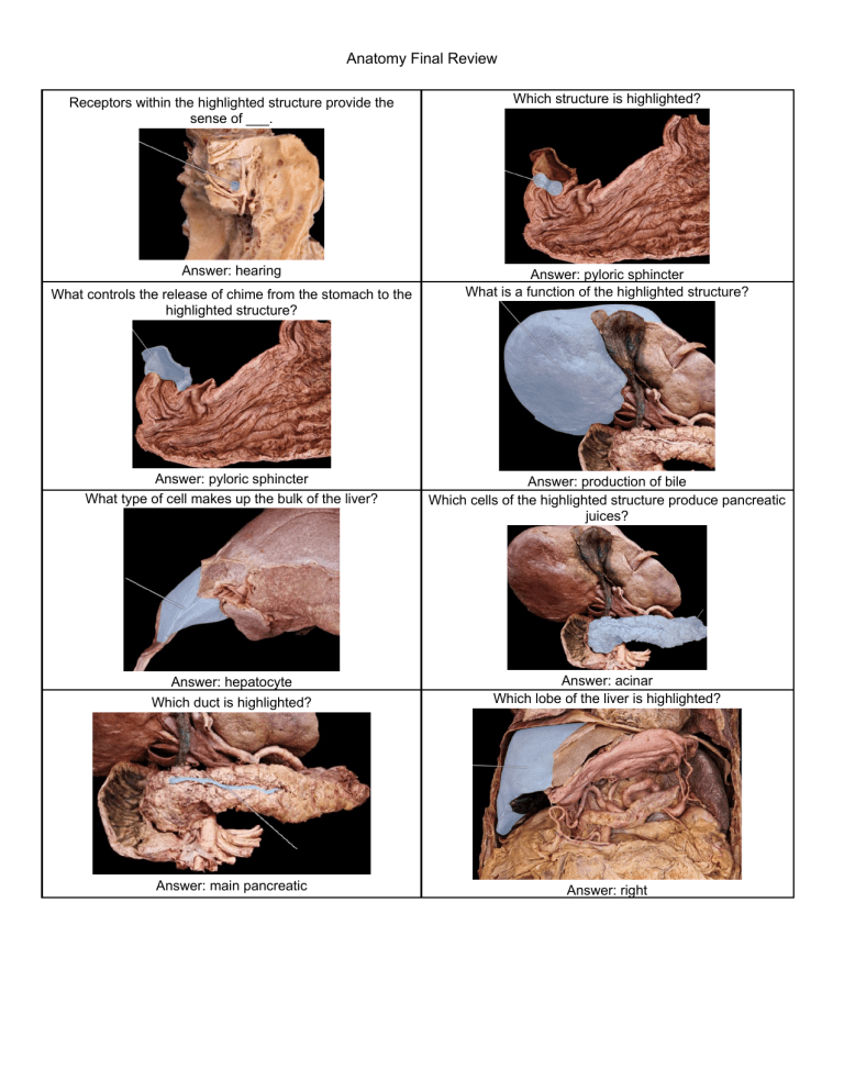
Anatomy Final Review Receptors within the highlighted structure provide the sense of ___. Which structure is highlighted? Answer: hearing Answer: pyloric sphincter What is a function of the highlighted structure? What controls the release of chime from the stomach to the highlighted structure? Answer: pyloric sphincter What type of cell makes up the bulk of the liver? Answer: production of bile Which cells of the highlighted structure produce pancreatic juices? Answer: hepatocyte Which duct is highlighted? Answer: acinar Which lobe of the liver is highlighted? Answer: main pancreatic Answer: right Which region of the colon is highlighted? Which region of the small intestine empties into the highlighted structure? Answer: ascending Answer: ileum Which structure is highlighted? Which region is highlighted? Answer: body What structure is highlighted? Answer: descending colon Which structure is highlighted? Answer: hard palate Which structure is highlighted? Answer: cystic duct Which structure is highlighted? Answer: gallbladder Which structure is highlighted? Answer: hepatic artery proper Which structure is highlighted? Answer: oropharynx Answer: hepatic portal vein Which structure is highlighted? The highlighted structures contain __ to allow for taste. Answer: chemoreceptors Answer: parotid duct The maculaelocated within the highlighted structure contain the receptors that monitor___. What is the action of the highlighted structure? Answer: static equilibrium and linear acceleration Which muscle is highlighted? Answer: elevates eye and turns it laterally Which nerve is highlighted? Answer: inferior rectus Which nerve is highlighted? Answer: vestibular Which structure is highlighted? Answer: vestibulocochlear Which structure is highlighted? Answer: auricle Which structure is highlighted? Answer: cornea Which structure is highlighted? Answer: incus Which structure is highlighted? Answer: olfactory tract Which structure is highlighted? Answer: pharyngotympanic tube Which structure is highlighted? Answer: retina Which structure is highlighted? Answer: sclera Which structure is highlighted? Answer: semicircular canals Which structure is highlighted? Answer: tympanic membrane Which structure is highlighted? Answer: inferior nasal concha Which structures are highlighted? Answer: primary gustatory cortex What is the name of the concave depression in the central retina for gathering light? Answer: optic tracts Answer: fovea Lack of thyroid hormone in a newborn infant leads to what type of retardation? The highlighted organ secretes which of the following hormones in response to growth hormones? Answer: cretinism The hormone secreted by the highlighted organ acts to ___. Answer: IGF-1 Answer: decrease blood pressure Which class of hormone released by the highlighted structure increases blood glucose? What structure allows the highlighted structure to have direct communication with the anterior pituitary? Answer: hypophyseal portal system Which gland is highlighted? Answer: glucocorticoid Which gland is highlighted? Answer: pituitary gland Which hormones does the highlighted organ secrete in response to hypoxia? Answer: thyroid Answer: erythropoietin Which of the following hormones is released by the highlighted structure? Which structure is highlighted? Answer: progesterone Which structure is highlighted? Answer: epididymis Answer: adrenal gland Which structure is highlighted? Which structure is highlighted? Answer: ovary Which structure is highlighted? Answer: pancreas Which structure is highlighted? Answer: pineal gland Which structure is highlighted? Answer: right atrium Which structure of the thyroid is highlighted? Answer: tuber cinereum Which structure of the thyroid is highlighted? Answer: isthmus Answer: left lobe What type of cells stain brown and are found in abundance in the posterior pituitary? Answer: ??? What is the name of the strong basophilic region between the pars anterior and pars posterior which is responsible for producing pro-opio-melano-cortins? Answer: pituitary pars intermedia The highlighted cell __. Answer: initiates inflammatory response How do the highlighted valve cusps function during ventricular contraction? Answer: they are held closed by the chordae tendineae to prevent blood from flowing back into the right atrium What does (AP), (PP) and (S) represent? Answer: AP- anterior pituitary PP- posterior pituitary S- infundibular stem ? What is the name of these critically important pale stained collection of cells responsible for the production of glucagon, insulin, and somatostatin? Answer: Islet of Langerhans Which cells is highlighted? Answer: lymphocyte The function of the highlighted structure is to prevent backflow of the blood into the __. Answer: right atrium What type of connective tissue is the highlighted structure composed of? Which artery is highlighted? Answer: dense irregular Which artery is highlighted? Answer: anterior interventricular Which artery is highlighted? Answer: aortic arch Which artery is highlighted? Answer: circumflex Which structure is highlighted? Answer: left marginal Which structure is highlighted? Answer: pericardium Which structure is highlighted? Answer: visceral pericardium covering the heart Which structure is highlighted? Answer: left auricle Which valve is highlighted? Answer: right coronary artery Answer: bicuspid Which vein is highlighted? Which vessel is highlighted? Answer: great cardiac Which vessel is highlighted? Answer: ascending aorta Which vessel is highlighted? Answer: inferior vena cava Which vessel is highlighted? Answer: right marginal artery Which vessels are highlighted? Answer: superior vena cava The highlighted artery supplies blood to which of the following organs? Answer: pulmonary vein The primary function of the highlighted artery is to supply blood to the ___. Answer: spleen Answer: brain Where is the highlighted vessel bifurcates there are structure that monitor CO2 and O2 concentration in the blood. What are the structures called? Which artery is highlighted? Answer: carotid bodies Which artery is highlighted? Answer: common iliac Which vessel is highlighted? Answer: external iliac What kind of tissue is this? Answer: superior vena cava What is the purpose of the highlighted vessels? Answer: cardiac muscle tissue Answer: to drain fluid from the tissues of the lower limb back towards vessels near the heart Which lymph nodes are highlighted? Which lymph nodes are highlighted? Answer: axillary Which structure is highlighted? Answer: tracheobronchial Which structure is highlighted? Answer: cisterna chyli Answer: lingual tonsil Which structure is highlighted? Which structure is highlighted? Answer: palatine tonsil Answer: spleen In this gland, what is the lamellated area at called__. Which structure is highlighted? Answer: thoracic duct What is the name of the outer densely stained area, and the pale lighted stained area? Answer: outer = cortex Inner = medulla What type of cells are represented at the apical surface? Answer: simple columnar epithelium Answer: Hassal’s corpuscles located in the medulla of the thymus What tissue type does this micrograph represent? Answer: non-keratinized stratified squamous epithelial tissue The highlighted vessels are __ blood to the heart. Answer: veins that carry oxygenated The ligaments running through the highlighted structures consist of mainly which type of fiber? What is an action of the highlighted muscles? Answer: elastic Answer: depresses ribcage Which cartilage is highlighted? What type of epithelium lines the highlighted structure? Answer: ciliated pseudostratified columnar Which cartilage is highlighted? Answer: tracheal Answer: thyroid Which cartilages are highlighted? Which muscles are highlighted? Answer: arytenoid Which structure is highlighted? Answer: external intercostals Which structure is highlighted? Answer: left inferior lobar bronchus Which structure is highlighted? Answer: cricothyroid muscle Answer: oblique fissure of left lung Which structure is highlighted? Which structure is highlighted? Answer: inferior nasal concha Which structure is highlighted? Answer: left main bronchus Which structure is highlighted? Answer: nasopharynx Which structure is highlighted? Answer: right inferior lobar bronchus Which structure is highlighted? Answer: right pulmonary artery Which structure of the lung is highlighted? Answer: vocal fold What type of tissue is represented at (E)? What are the clear large, clear cells scattered throughout this area? Answer: superior lobe of left lung What does this SEM 2000 represent? Answer: (E)- pseudostratified ciliated columnar epithelium ; large, clear cells are thin walled venules What does this slide present? Answer: primary bronchus Answer: the picture shoes the ureter What is the functional unit of this organ? Which structure is highlighted? Answer: the function of the kidneys is to filter, balance, remove waste from the blood and body fluids, and form urine. Which structure is highlighted? Which structure is highlighted? Answer: membranous urethra Which structure is highlighted? Answer: minor calyces Which structure is highlighted? Answer: renal pelvis Which structure is highlighted? Answer: renal vein Which structure is highlighted? Answer: ureter Which structure is highlighted? Answer: urethra What does the (W) represent in this micrograph? Answer: urinary bladder Answer: (W) - Answer: cortex What essential cells to the production of life is contained in these follicles? What is the name of these structure where spermatogenesis takes place? Answer: Which structure is highlighted? Answer: Which structure is highlighted? Answer: ductus deferens Which structure is highlighted? Answer: glans penis Which structure is highlighted? Answer: right uterine tube Answer: scrotum Which structure is highlighted? What structure is highlighted? Answer: spongey urethra Which structures are highlighted? Answer: tunica albuginea ☺ Answer: corpus cavernosa


