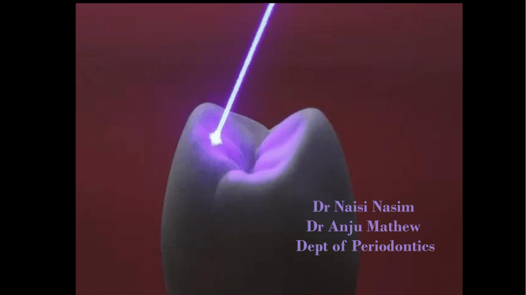
Dr Naisi Nasim Dr Anju Mathew Dept of Periodontics INTRODUCTION SOFT TISSUE APPLICATIONS GINGIVAL SOFT TISSUE PROCEDURES FRENECTOMY • Frenectomy procedures with a laser are predictably successful so long as the following steps are incorporated: 1. Creation of a periosteal fenestration at the base of the frenectomy to prevent reattachment of fibers 2. Removal of all impeding muscle fibers Depth of penetration for diode and Nd:YAG lasers is much higher (500 μm) than for erbium or CO2 lasers (5 to 40 μm), so settings must be monitored closely to prevent thermal damage to the underlying periosteum and bone. (Themes U 2018) Procedure Diode laser (810 nm). An initiated tip of 200 μm was used with an average power of 2 W in a continuous mode. Applied in a contact mode with focused beam for excision of the tissue . The tip of the laser was moved from the apex of the frenum to the base in a brushing stroke cutting the frenum. The ablated tissue was continuously mopped using wet gauze piece. No suturing ……..Antibiotics, analgesics and warm saline rinses to facilitate faster healing. FRENECTOMY DEPIGMENTATION • Recognized as one of the most effective, comfortable, and reliable techniques. • Based on the principle of selective photothermolysis • Laser light must be at a wavelength that is specific and well absorbed by the particular chromophore being treated. • Diode laser is 810 nm, allows high levels of energy to be absorbed by soft tissues, water, and chromophores, such as melanin and oxyhemoglobin. Anderson RR et al,1983 DE PIGMENTATION GINGIVECTOMY The gingivectomy can be used when suprabony pockets are present and access to osseous structures is not necessarily important. The procedure assists in decreasing gingival tissue in cases of enlargement and in altering fibrotic gingiva. Gingivectomy is contraindicated when (1) access to osseous structure is critical or (2) gingival attachment is inadequate (minimal) or absent. Local anesthesia 2% lidocaine was administrated in the buccal vestibule of the maxillary incisors. Measurements were made with a periodontal probe following the maximum reduction measurements and bleeding points made with an explorer. A 980-nm diode soft tissue laser was set to continuous mode with 0.5-watt amplitude. The laser was initiated by touching the tip to occlusal paper at 45 degrees and running it along the paper until smoke was released. Following initiation, the laser was changed to pulsating mode and 0.6-watt amplitude. GINGIVECTOMY GINGIVOPLASTY SOFT TISSUE CROWN LENGTHENING CROWN LENGTHENING Operculectomy LA Angled handpiece …held perpendicular to the target lesion Avoid contact between the laser beam and the tooth enamel. If operculum covers part of the tooth, an adaptive tool (for example, a wax spatula) needs to be inserted between the tissue and the tooth to prevent possible damage/ shield the tooth during the procedure) Vestibuloplasty LA 808 nm wave length diode laser (SUNNYTM, MSI, Bangalore) with 400 μm surgical tip was used (1 to 1.5W in a continuous mode using an initiated tip.) Initiated at the MGJ …horizontal stoke …parallel to the bone slowly relieving the muscle fibers till the desired depth . Tension …retracting the patient’s lip to enable the laser assisted excision of the muscle fibers. After a sufficient vestibular depth ….lip was once again pulled …residual muscle fibers if any excised with laser tip. FLAP PROCEDURES Practitioners perform periodontal flap procedures either exclusively or adjunctively with a laser. Once the flap is reflected, lasers again can be used for sulcular debridement and deepithelialization on the inside of the flap. If root debridement will be done with a laser, it is strongly suggested that only an erbium laser be used, because of possible thermal damage with diode and Nd:YAG lasers. CO2 lasers may be used according to the protocol of Crespi et al. to increase fibroblast attachment to the root surface. When osseous surgery is necessary after flap reflection, it can be performed with erbium lasers or conventional instrumentation such as a high/slow-speed handpiece, diamond/carbide burs, and manual devices (e.g., chisel). PHOTOBIOMODULATION (PBM) Therapeutic low intensity laser and light emitting diode (LED) irradiation is termed photobiomodulation (PBM) and this type of treatment has been suggested as an adjunctive measure in the management of periodontitis. PBM has been found to have the capacity to modify the cytokine cascade away from a reactive pro inflammatory locally destructive process to an anti-inflammatory cycle. LASER ASSISTED NEW ATTACHMENT PROCEDURE The innovators, Gregg and McCarthy formulated a set of criterion for LANAP which received Food and Drug Administration (FDA) clearance in 2004. Patients needing standard periodontal treatment with pocket depth (PD) ≥4 mm are indicated for LANAP. LANAP, when compared to conventional periodontal surgery, provided some elusive advantages like, Minimally invasive with better patient compliance Decreased postoperative pain and morbidity Less likely to develop hypersensitivity Less prone to recession Faster healing Natural teeth as well as implant both show regeneration of the surrounding tissues PROCEDURE 1. The patient is profoundly anesthetized initially with a local anesthetic to properly access the extent of intrabony flaw with a probe. 2. An optic fiber tip measuring 0.3-0.4 µ is placed parallel to the root surface, to carry away the epithelium lining of the pocket in coronal to apical motion to reflect the gingival flap. 3. Calcified plaque adherent to the root surface is removed. 4. Selective photothermolysis removes unhealthy, infected and inflamed epithelium of the pocket sparing the intact connective tissue separation of the layers of tissues at rete pegs and ridges level. REMOVAL OF POCKET EPITHELIUM Bleaching

