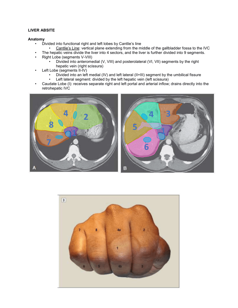
LIVER ABSITE Anatomy • Divided into functional right and left lobes by Cantlie’s line • Cantlie’s Line: vertical plane extending from the middle of the gallbladder fossa to the IVC • The hepatic veins divide the liver into 4 sectors, and the liver is further divided into 9 segments. • Right Lobe (segments V-VIII) • Divided into anteromedial (V, VIII) and posterolateral (VI, VII) segments by the right hepatic vein (right scissura) • Left Lobe (segments II-IV) • Divided into an left medial (IV) and left lateral (II+III) segment by the umbilical fissure • Left lateral segment: divided by the left hepatic vein (left scissura) • Caudate Lobe (I): receives separate right and left portal and arterial inflow; drains directly into the retrohepatic IVC 8 7 4 a 4 b 2 1 5 6 3 1 Left Liver Right Liver Extended Right Hepatectomy Extended Left Hepatectomy Superior (II) Inferior (III) Posterior (VI, VII) Anterior (V, VIII) Left Medial (IV) Left Lateral (II, III) Liver Abscesses • Pyogenic • Most common; GNRs (#1 E. coli, #2, K. pneumoniae) • Direct spread from biliary tree (#1) • CT: peripheral enhancing, central hypoattenuating, multi-loculated (vs. amebic) • FNA with WBCs +/- organisms • Tx: percutaneous drainage + Abx • Amebic • Epi: male predominant; hx of travel (Central/South America) • Pathogen: E. histolytica (fecal-oral); +Abs • CT: well-defined hypoattenuating lesion with peripheral wall of enhancing edema • Central localized hepatic necrosis (no organisms on FNA) • Tx: metronidazole; drainage if poor response/superinfection • Echinococcal • Epi: endemic to Mediterranean, East Africa, Middle East, South America, Asia • Pathogen: E. granulosus; fecal-oral; eosinophilia, +serology, +Casoni • CT: hypoattenuating lesion, septae/daughter cysts (75%), +/- peripheral calcifications (50%) • Pericyst, ectocyst, endocyst (germinal layer -- gives rise to daughter cysts) • Tx: albendazole, PAIR/surgical resection Benign Liver Lesions • Hemangioma • Most common benign tumor; F > M (3:1) • CT: late contrast puddling is pathognomonic • PERIPHERAL enhancement on ARTERIAL phase • CENTRIPETAL filling on PORTAL VENOUS phase • Retention of contrast on washout or delayed phase • Tx: angioembolization for bleeding; enucleation if symptomatic • Hepatic Adenoma • Female predominant; hormonally sensitive (OCPs, steroids) • Risk of bleeding and malignant transformation • CT: well-marginated, isointense on NC CT; HYPERINTENSE on ARTERIAL phase; rapid washout; +/- heterogenous due to hemorrhage • No Eovist (Gd) [MRI] or sulfur colloid [NM scan] retention (no Kupffer cells) • Tx: discontinue offending agent; resection if >5cm, symptomatic • Focal Nodular Hyperplasia (FNH) • No risk of bleeding or malignant transformation • CT: homogenous mass with CENTRAL scar; HYPERINTENSE on ARTERIAL phase • Central Scar: hypoattenuating on arterial phase; hyperattenuating on delay • Eovist (Gd) and sulfur colloid uptake (+ Kupffer cells) • Tx: observation Malignant Liver Lesions • Hepatocellular Carcinoma • Most common primary hepatic malignancy • HBV infection #1 cause worldwide; 80% in setting of cirrhosis • Screening: AFP + ultrasound every 6 months for AT RISK PATIENTS • CT: primarily arterial inflow • Heterogenous; Early arterial enhancement and washout of contrast on delayed phase • Tx: resection/ablation (if localized and adequate remnant), OLT (Milan criteria), sorafenib (metastatic/unresectable) • Metastatic Liver Tumors • More common than primary liver tumor (20:1) • CT: Hypoattenuating on NC CT compared to liver; hypovascular lesions with minimal arterial enhancement Liver Lesion Hepatic Hemangioma Non-contrast Hypointense to liver Focal Nodular Hyperplasia Isointense to the liver Homogenous lesion Hepatic Adenoma Isointense to the liver Heterogenous (bleed, necrosis, fat) HCC Isointense/hypointense to liver Metastases Hypointense Arterial Peripheral enhancement Rapid homogenous enhancement w/ hypoattenuating central scar Rapid enhancement Homogenous rapid enhancement Hyperintense (NET) Hypointense (CRC) Portal Centripetal filling Venous Retention of contrast Rapid washout Isointense w/ delayed enhancement of central scar Rapid washout Isointense to liver Rapid washout Isointense Central filling (CRC) Washout Liver Failure and Portal Hypertension • • Portal HTN • Dx: hepatic venous pressure gradient (HVPG): pressure gradient between the wedge hepatic venous pressure (WHVP) and the free hepatic venous pressures • Estimate of pressure gradient between PV and IVC • Portal HTN: HVPG greater than or equal to 5mmHg; clinically significant when >10mmHg Liver Failure • Causes: Pre-hepatic, Intrahepatic, Post-hepatic • Intrahepatic: pre-sinusoidal, sinusoidal, post-sinusoidal • Child’s Pugh Score: • Components: total bilirubin, PT/INR, albumin, ascites, encephalopathy • MELD Score: • Components: total bilirubin, PT/INR, creatinine


