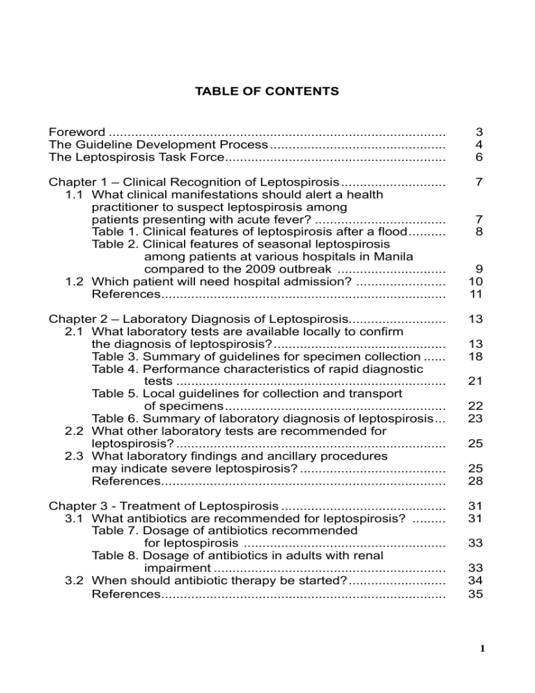
Leptospirosis CPG 2010 TABLE OF CONTENTS Foreword .......................................................................................... The Guideline Development Process ............................................... The Leptospirosis Task Force........................................................... 3 4 6 Chapter 1 – Clinical Recognition of Leptospirosis ............................ 1.1 What clinical manifestations should alert a health practitioner to suspect leptospirosis among patients presenting with acute fever? ................................... Table 1. Clinical features of leptospirosis after a flood .......... Table 2. Clinical features of seasonal leptospirosis among patients at various hospitals in Manila compared to the 2009 outbreak ............................. 1.2 Which patient will need hospital admission? ........................ References............................................................................ 7 Chapter 2 – Laboratory Diagnosis of Leptospirosis.......................... 2.1 What laboratory tests are available locally to confirm the diagnosis of leptospirosis? .............................................. Table 3. Summary of guidelines for specimen collection ...... Table 4. Performance characteristics of rapid diagnostic tests ........................................................................ Table 5. Local guidelines for collection and transport of specimens ........................................................... Table 6. Summary of laboratory diagnosis of leptospirosis ... 2.2 What other laboratory tests are recommended for leptospirosis? ........................................................................ 2.3 What laboratory findings and ancillary procedures may indicate severe leptospirosis? ....................................... References............................................................................ Chapter 3 - Treatment of Leptospirosis ............................................ 3.1 What antibiotics are recommended for leptospirosis? ......... Table 7. Dosage of antibiotics recommended for leptospirosis ...................................................... Table 8. Dosage of antibiotics in adults with renal impairment .............................................................. 3.2 When should antibiotic therapy be started? .......................... References............................................................................ 7 8 9 10 11 13 13 18 21 22 23 25 25 28 31 31 33 33 34 35 1 Leptospirosis CPG 2010 Chapter 4 – Antibiotic Prophylaxis for leptospirosis.......................... 4.1 What is the recommended pre-exposure prophylaxis? ........ 4.2 What is the recommended post-exposure prophylaxis? ....... Figure 1. Algorithm for post-exposure prophylaxis................ Pharmacology of Doxycycline ............................................... References............................................................................ 36 36 38 40 41 43 Chapter 5 – Leptospirosis-Associated Acute Kidney Injury .............. 5.1 Diagnosis of AKI due to leptospirosis.................................... 5.2 Management of AKI .............................................................. Figure 2. Algorithm for the management of oliguria .............. References............................................................................ 45 45 46 49 50 Chapter 6 – Pulmonary Complications of Leptospirosis .................. 6.1 Clinical diagnosis of pulmonary complications ..................... Table 9. Characteristics of patients with and without pulmonary involvement............................... Table 10. American-European consensus conference criteria for ARDS ................................ 6.2 Diagnostic studies ................................................................. References............................................................................ 6.3 Management of pulmonary complications ............................ Figure 3. Algorithm for the diagnosis and management of pulmonary complications .................................... References............................................................................ 52 52 Appendix Leptospirosis Data Collection Form ...................................... 2 53 55 56 57 59 61 62 64 Leptospirosis CPG 2010 FOREWORD Leptospirosis is an endemic zoonosis in the Philippines with an average of 680 leptospirosis cases and 40 deaths from the disease reported every year and a prevalence of 10/100,000. It is seasonal with a peak incidence during the rainy months of July to October. Clinical studies in the 60s and 70s and seroepidemiological surveys have documented the presence of leptospiral serovars in the country. Specifically, antibodies to various leptospiral serovars have been reported in urban domestic rats, rural field rats, water buffaloes, cattle, pigs, dogs and monkeys in the Philippnes. It has been more than a year now since Typhoon Ketsana, locally known as “Ondoy” and Typhoon Parma or “Peping” ravaged the country in succession. Leptospirosis reemerged as an aftermath of the heavy rainfall which led to massive flooding in the cities of Metro Manila and in the provinces of Luzon. Within only six hours, 455mm of rain fell on the area, an amount equivalent to a typical month’s rainfall in the monsoon season according to the Philippine Atmospheric, Geophysical and Astronomical Services Administration (PAGASA). Several communities were under water due to clogged drainage systems brought about by poor garbage disposal systems. The Center for Health Development offices of the Department of Health reported that “Ondoy” affected 12 regions in the country and afflicted more than 800,000 families. The most affected regions were the CALABARZON, the National Capital Region (NCR) and Central Luzon, putting these areas under a state of calamity. As soon as the leptospirosis outbreak was recognized in Metro Manila in October 2009, interim clinical practice guidelines were drafted by the Philippine Society for Microbiology and Infectious Diseases, the Philippine Society of Nephrology and the Council for Critical Care and Vascular Pulmonary Diseases of the Philippine College of Chest Physicians. Consensus meetings were held to formulate recommendations on the diagnosis and management of suspected leptospirosis cases and its complications. These interim guidelines were released by the Philippine College of Physicians and the Department of Health to guide the health care practitioners in the affected areas in the diagnosis, treatment and prevention of leptospirosis and its complications. As of November 2009, the National Epidemiology Center of the Department of Health reported 2,299 presumptive cases of leptospirosis with 178 deaths in 15 hospitals in Metro Manila over a period of two months. In the Luzon region, 1090 cases and 71 deaths in Regions I, III, IV-A and Cordillera Autonomous Region were recorded. Overall case fatality rate was 7.4%. This was way beyond the yearly endemic threshold of leptospirosis cases in the country. It is anticipated that leptospirosis will continue to re-emerge in the country as a result of rapid urbanization, deforestation, poor sanitation and increased incidence of typhoons brought about by climate changes. Thus, the Leptospirosis Task Force composed of members of the PSMID, PSN and PCCP have decided to update and finalize the 2009 interim guidelines released during the outbreak. This updated guideline is intended to guide health care practitioners in the early recognition, prompt management and prevention of leptospirosis and its complications in primary, secondary and tertiary health facilities. The guideline also aims to heighten the awareness and index of suspicion of clinicians not just during an outbreak but also during the rainy months and in cases associated with travel, recreational sports and occupational exposures. 3 Leptospirosis CPG 2010 The Guideline Development Process Phase 1: Preparation of the evidence-based draft The members of the Leptospirosis Task Force agreed to review the evidence on the following topics: 1. 2. 3. 4. 5. Clinical diagnosis of leptospirosis Laboratory diagnosis of leptospirosis Treatment of Leptospirosis Prevention of leptospirosis Diagnosis and management of complications a. Acute kidney injury b. Pulmonary hemorrhage The task force members then searched the MEDLINE database up to September 2010 and the Cochrane Library Issue 2010 for relevant literature. The group also searched the websites of the Philippine College of Physician and Philippine Society for Microbiology and Infectious Diseases for local literature. The HERDIN database was also searched and experts in the field were contacted for published and unpublished local literature. Phase 2: Preparation of the intermediate and penultimate drafts The PSMID was assigned to prepare the summaries of evidence on the diagnosis, treatment and prevention of leptospirosis. The PSN was assigned to prepare the draft for the diagnosis and management of leptospirosis-associated acute kidney injury, while the PCCP Council on Critical Care and Pulmonary Vascular diseases prepared the draft for the diagnosis and management of pulmonary complications associated with leptospirosis. These evidence-based drafts were then presented and discussed in subsequent meetings of the task force. The task force formulated recommendations on diagnosis, treatment and prevention based on the level of quality of the evidence, applicability and availability of health resources. We used the following system for grading the recommendations and quality of the evidence: 4 Leptospirosis CPG 2010 Grading System for the Strength of the Recommendations and Quality of Evidence GRADE Strength of recommendation DEFINITION A Good evidence to support a recommendation for or against use Moderate evidence to support a recommendation for or against use Poor evidence to support a recommendation for or against use B C Quality of evidence Level 1 Level 2 Level 3 Evidence from at least one properly randomized trial or a well-conducted systematic review Evidence from at least one well-designed trial, without randomization; from cohort or case-control analytic studies (preferably from >1 center); from multiple time series; or from dramatic results of uncontrolled experiments Evidence from opinions of experts, based on clinical experience, descriptive studies, or reports from expert committees, clinical trials or systematic reviewswith high risk for bias based on methodologic quality In grading the recommendations, the task force considered not just the overall quality of the evidence but also the consistency of the evidence as well as issues on applicability and availability of the diagnostic, therapeutic or preventive intervention at different levels of health care facilities. The tradeoffs between benefit and harm were also weighed, e.g. risk of progression to severity, risk exposure assessment, development of complications, and severity of adverse effects versus the benefits of treatment or prophylaxis, including cost-effectiveness. 5 Leptospirosis CPG 2010 The Leptospirosis Task Force Philippine Society for Microbiology and Infectious Diseases Chair: Co-Chair: Members: Manolito L. Chua, MD Marissa M. Alejandria, MD Rhona G. Bergantin, MD Raul P. Destura, MD Mario M. Panaligan, MD Cecilia S. Montalban, MD Minette O. Rosario, MD Paul P. Salandanan, MD Rontgene M. Solante, MD Maria Fe R. Tayzon, MD Dionisio M. Tiu, MD Philippine Society of Nephrology Irmingarda Gueco, MD Susan Anonuevo, MD Roberto Tanchanco, MD Jose Marcia, MD Melvin Marcial, MD Philippine College of Chest Physicians Council on Critical Care and Pulmonary Vascular Diseases Aileen Guzman-Banzon, MD, FPCCP Joseph Hope G. Cal, MD, FPCCP Teresita S. de Guia, MD, FPCCP 6 Leptospirosis CPG 2010 1. CLINICAL RECOGNITION OF LEPTOSPIROSIS 1.1 What clinical manifestations should alert a health practitioner to suspect leptospirosis among patients presenting with acute fever? Any individual presenting with acute febrile illness of at least 2 days AND either residing in a flooded area or has high-risk exposure (defined as wading in floods and contaminated water, contact with animal fluids, swimming in flood water or ingestion of contaminated water with or without cuts or wounds) AND presenting with at least two of the following symptoms: myalgia, calf tenderness, conjunctival suffusion, chills, abdominal pain, headache, jaundice, or oliguria should be considered a suspected leptospirosis case. [Grade A] Leptospirosis occurs throughout the world but is highest in the tropics. It is one of the most common zoonoses with human infection occurring commonly through superficial cuts and open wounds after exposure to a contaminated environment (e.g. flood), direct contact with infected animals or following rodent bites.1,2 The spectrum of presentation of leptospirosis is protean and varies from a mild and inapparent form to a severe one involving multiorgan system.1 Clinicians should therefore have a high index of suspicion among patients with febrile illness and high risk exposures because mortality may be as high as 15%.1,3 A review of the clinical presentation of 353 cases of laboratory confirmed leptospirosis in Hawaii from 1974 to 1998 showed that the most common presentation included fever, myalgia and headache.4 Leptospirosis is endemic in the Philippines and the number of cases peak during the rainy months of June to August. Outbreaks have been associated with wading in flood waters. A review of patients hospitalized for suspected leptospirosis in the 70’s showed abrupt fever, myalgia, headache, abdominal pain, meningismus, conjunctival suffusion and gastrocnemius or calf tenderness to be the common symptoms.5,6,7 In the eighties and nineties, other symptoms observed included oliguria/anuria, diarrhea, thrombocytopenia and bleeding diatheses.8-13 Usually, an average of 680 leptospirosis cases and 40 deaths from the disease are reported every year in the Philippines. However, in October 2009 a leptospirosis outbreak was declared by the Department of Health two weeks after the heavy rainfall typhoon Ketsana last September 26, 2009. As of 13 November 2009 a total of 2,292 suspected cases of leptospirosis were recorded with 178 deaths (8%) in 15 hospitals in Metro Manila.14 The clinical features of 257 confirmed 7 Leptospirosis CPG 2010 cases (Philippine General Hospital = 90, National Kidney and Transplant Institute =77, The Medical City = 52, University of Santo Tomas Hospital = 11, Manila Doctors Hospital = 7, Ospital ng Maynila = 6, Cardinal Santos Medical Center = 5, East Avenue Medical Center = 5, Makati Medical Center = 4) diagnosed during the outbreak are compared in Table 1 with reports of leptospirosis outbreaks in Brazil,15 India16 and Korea17; and in Table 2 with seasonal leptospirosis in the Philippines. No significant differences were seen in the clinical features of leptospirosis after an outbreak with that of seasonal leptospirosis.18 Table 1. Clinical features of leptospirosis after a flood18 Signs & symptoms (%) Number of patients Korea 1987 8 Mumbai, India 2005 Philippines 2009 93 confirmed 193 confirmed 237 probable 257 confirmed cases & probable cases cases 97 93.8 100 98.4 88 93.8 39.7 78.6 70 74.6 NR 55.6 58 28.5 24.5 59.1 40 NR 4.2 52.1 36 NR 7.6 39.3 16 92.7 81.4 38.1 33.2 37.6 56.8 15 23.9 NR 75.1 40 15.0 7.6 (8.6) Fever Myalgia Headache Conjunctival suffusion Abdominal pain Diarrhea Jaundice Oliguria NR Renal failure Respiratory symptoms (pulmonary hemorrhage) Thrombocytopenia CNS manifestations - altered sensorium, meningitis/ meningismus NR-not reported Salvador, Brazil 1996 18 6 NR 24.9 14.3 2.1 14.8 5.1 92 92 NR 62 91 NR 74 NR 80 NR NR 24 17 confirmed 100 71 58.8 41 71 6 29 29 17.6 5.9 NR 35 Number of cases Fever Myalgia Headache Conjunctival Suffusion Abdominal pain Diarrhea Jaundice Oliguria/anuria Renal failure Pulmonary Hemorrhage Thrombocytopenia CNS manifestations NR=not reported PGH 19786 34 Confirmed UST 1967-715 Symptoms & signs (%) NR 18 20 60 60 NR 3.4 70 100 40 65.5 60 13 confirmed 16 clinical UST 1974-787 NR 12.5 33.6 70.2 43.3 74 5 69.2 97.1 82.9 64.4 79.8 104 clinical & probable UST 1979-938 50 20.9 NR 77 68 2.1 NR 100 95.3 88.5 95.8 191 presumptive cases PGH 1985-919 61 17 61 68 83 93 18.6 61 100 68 81.3 76 59 confirmed cases PGH 1995-9610 NR 2.7 NR 56.5 37.9 89.8 13.0 NR 95.2 92.5 74.1 85 147 presumptive cases PGH 1990-9711 NR 7.6 30.7 73 69.0 30.7 NR 42 100 19.2 50.0 61.5 26 presumptive cases QMMC 199912 30 NR NR 61 66 89.1 2.4 26.5 99 87 NR 99 83 presumptive cases JRRMH 2000-0113 14.8 5.1 39.3 38.1 56.8 75.1 8.6 52.1 98.4 78.6 55.6 59.1 257 confirmed cases 2009 outbreak14 Table 2. Clinical features of seasonal leptospirosis admitted at various hospitals in Metro Manila compared with the 2009 outbreak.18 Leptospirosis CPG 2010 9 Leptospirosis CPG 2010 1.2 Which patient will need hospital admission? Any suspected case of leptospirosis presenting with acute febrile illness and various manifestations BUT with stable vital signs, anicteric sclerae, with good urine output, and no evidence of meningismus / meningeal irritation, sepsis / septic shock, difficulty of breathing nor jaundice and can take oral medications is considered MILD LEPTOSPIROSIS and can be managed on an OUT-PATIENT SETTING. [Grad e A] Any suspected case of leptospirosis presenting with acute febrile illness associated with unstable vital signs, jaundice/icteric sclerae, abdominal pain, nausea, vomiting and diarrhea, oliguria/anuria, meningismus / meningeal irritation, sepsis / septic shock, altered mental states or difficulty of breathing and hemoptysis is considered MODERATE – SEVERE LEPTOSPIROSIS and BEST managed in a HEALTHCARE / HOSPITAL SETTING. [Grade A] The incubation period of leptospirosis may range from 2 to 28 days. Signs and symptoms are highly variable. Asymptomatic seroconversion is the most common result of infection. The mildest presentation of leptospirosis is fever, headache, and myalgia, accompanied by other nonspecific findings such as nausea and vomiting, diarrhea, nonproductive cough, and maculopapular rash. Conjunctival suffusion (red eyes without exudate) and severe calf pain may be characteristic of acute leptospirosis, but are not specific. Mild leptospirosis may resolve spontaneously without requiring antimicrobial therapy. Severe manifestations of leptospirosis include any combination of jaundice, renal failure, hemorrhage (most commonly pulmonary), myocarditis, and hypotension refractory to fluid resuscitation. Other complications include aseptic meningitis and ocular involvement including uveitis. As originally described in the 19th century, Weil’s disease is characterized by a triad of fever, jaundice, and splenomegaly. Current usage of the term “Weil’s disease” refers to fever, jaundice, and renal failure and is often considered synonymous with severe leptospirosis.19 Clinical features associated with increased risk for mortality include altered mental status, respiratory insufficiency (rales, infiltrates), hemoptysis, oliguric hyperkalemic acute renal failure, and cardiac involvement (myocarditis, complete or incomplete heart block, atrial fibrillation). In a retrospective study of 68 patients with leptospirosis in a teaching hospital of Pointe-a-Pitre in French West Indies, prognostic factors independently associated with mortality were: dyspnea (OR 10 Leptospirosis CPG 2010 11.7, 95% CI 2.8 to 48.5), oliguria (OR 9.0, 95%CI 2.1 to 37.9), WBC >12,900/cu mm (OR 2.5, 95% CI 1.8 to 3.5), repolarization abnormalities on EKG (OR 5.9, 95% CI 1.4 to 24.8) and alveolar infiltrates on chest radiograph (OR 7.3, 95%CI 1.7 to 31,7).20 In a more recent case-control study of 89 mortalities and 281 discharged confirmed leptospirosis patients in Brazil, predictors of mortality included age > 40 years (OR 2.2, 95%CI 1.1 to 4.3), development of oliguria (OR 3.0, 95%CI 1.2 to 9.0), platelet count <70,000/uL (OR 2.2, 95%CI 1.2 to 4.7), creatinine > 3mg/dl (OR2.3, 95%CI 1.1 to 5.3) and pulmonary involvement (OR 6.0, 95%CI 3 to 12).21 In the urban epidemic of 326 cases of severe leptospirosis in Salvador, Brazil in 1996, altered mental status was the strongest independent predictor of death (OR 9.12, 95% CI 4.28 to 20.3). Other significant predictors identified were age > 37 years, renal insufficiency and respiratory insufficiency.15 Locally, severe jaundice, acute renal failure and bleeding diatheses have consistently been described among those who died.5,8,9-12 Concomitant comorbid illnesses and advanced age contributed to poor prognosis.8 In a review of 83 presumptive leptospirosis patients at Jose Reyes Memorial Hospital, leukocytosis (WBC >10,000) thrombocytopenia (platelet count <100,000/uL), evidence of bleeding, oliguria, length of time from onset of disease up to time of consult (>6 days) were significantly associated with poor outcome.13 REFERENCES 1. Levett PN. Usefulness of serologic analysis as a predictor of the infecting serovar in patients with severe leptospirosis. Clinical Infectious Diseases 2003;36:447-52. 2. Gilks CF, Lambert HP, Broughton ES, Baker CC. Failure of penicillin prophylaxis in laboratory-acquired leptospirosis. Postgraduate Medical Journal 1988;64:236-8 3. Haake DA, Dundoo M, Cader R, Kubak BM, Hartskeerl RA, Sejvar JJ, et al. Leptospirosis, water Sports and chemoprophylaxis. Clinical Infectious Diseases 2002;34:e40-3. 4. Katz AR, Ansdell VE, Effler PV, Middleton CR, Sasaki DM. Assessment of the clinical presentation and treatment of 353 Cases of laboratory-confirmed leptospirosis in Hawaii, 1974-1988. Clinical Infectious Diseases 2001;33:1834-41. 5. Alora B, Nambayan A, Perez J, Famatiga E and Tan Alora A. Leptospirosis in Santo Tomas University Hospital analysis of 17 cases, 1967-71. Phil J Microbiol Infect Dis 1973; 2:11-22. 6. Mendoza M,Tan S,Torres D, Tupasi T. Human leptospirosis: clinical and laboratory diagnosis. J Phil Med Assn 1979; 5:219-24 7. Manaloto CR, Alora AT, Alora BD. Leptospirosis: an analysis of 29 cases (Jan 1974Dec 1975). Phil J Microbiol Infect Dis 1980; 9:75-81 8. Marcial M, Dy E, Tan-Alora A. Leptospirosis revisited at the Santo Tomas University Hospital. Phil J Microbiol Infect Dis 1994; 24:21-33 9. Edmilao, M.,Lim, A, Abalos, M. Acute renal failure and mortality predictor factors in leptospirosis: a retrospective analysis. Phil J Intern Med 1995;33:189-99. 11 Leptospirosis CPG 2010 10. Casiple, LC. Thrombocytopenia and bleeding in leptospirosis. Phil J Microbiol Infect Dis 1998; 27:18-22 11. Cordero,C, Valdez J. The impact of the algorithm for the diagnosis and treatment of cases of acute renal failure secondary to leptospirosis at the UP-PGHMC. Phil J Intern Med 2000; 249-264 12. Reyes M, Peña A. Clinical and laboratory profile of leptospirosis: an analysis of twenty-six cases at Quirino Memorial Medical Center admitted in August 1999. Phil J Microbiol Infect Dis 2001;30:18-21 13. Orpilla-Bautista I and Panaligan,M. Predictors of mortality among patients with leptospirosis admitted at the JRRMMC. Phil J Microbiol Infect Dis 2002; 31:145-9 14. Department of Health National Epidemiology Center Statistics 15. Ko AI, Reis G, Ribeiro Dourado CM, Johnson WD, Riley LW and the Salvador Leptospirosis Study Group. Urban epidemic of severe leptospirosis in Brazil. Lancet 1999;354:820-5 16. Mathur M, De A, Turbadkar D. Leptospirosis outbreak in 2005: L.T.M.G. hospital experience. Indian Journal of Medical Microbiology 2009; 27-2.153-155. 17. Park YK, Park SK, Rhee YK, Kang SK. Leptospirosis in the Chonbuk province of Korea in 1987. Korean J Intern Med 1990; 5:34-43. 18. Roxas EA, Penamora A, Ginete JK, Leyritana K, Roman AD, Galura A, et al. Leptospirosis outbreak after a heavy rainfall typhoon in the Philippines: Clinical features and outcome. Unpublished 19. Ricaldi JN, Vinetz JM. Leptospirosis in the tropics and in travelers. Curr Infect Dis Rep 2006;8:51-8 20. Dupont H, Dupont-Perdrizet D, Perie JL, Zehner-Hansen S, Jarrige B, Daijardin JB. Leptospirosis: prognostic factors associated with mortality. Clinical infectious Diseases 1997;25:720-4. 21. Spichler AS, Villaca PJ, Athanazio DA, Albuquerque JOM, Buzar M, Castro B. Predictors of lethality in severe leptospirosis in urban Brazil. Am J Trop Med Hyg 2008; 79:911-4 12 Leptospirosis CPG 2010 2. LABORATORY DIAGNOSIS OF LEPTOSPIROSIS 2.1 What are the locally available laboratory tests that can be used to confirm the diagnosis of leptospirosis? Generally, it is not necessary to confirm the diagnosis or wait for the result of the tests before starting treatment. The clinical assessment and epidemiologic history are more important. Early recognition and treatment is MORE important to prevent complications of the severe disease and mortality. However, if definitive or confirmatory diagnosis is warranted in suspected cases and for epidemiological and public health reasons, these are the locally available diagnostic tests for leptospirosis. A. Direct Detection Method 1. Culture and isolation remains the GOLD standard BUT is time-consuming, labor-intensive, requires 6 to 8 weeks for the result, needs darkfield microscopy and has low diagnostic yield. It can identify the serovar but is insensitive. 2. Polymerase Chain Reaction (PCR) has the advantage of early confirmation of the diagnosis especially during the acute leptospiremic phase (first week of illness) before the appearance of antibodies.1 Its utilization in the clinical setting is currently not generally available because of the cost-limiting nature of the test and the need for trained personnel. B. Indirect Detection Methods 1. Microagglutination Test (MAT) - a four-fold rise of the titer from acute to convalescent sera is confirmatory of the diagnosis.1,2 It is highly sensitive and specific BUT time-consuming and hazardous to perform because of the risk of exposure to the live antigen. Cross-reactions may occur with syphilis, viral hepatitis, HIV, relapsing fever, Lyme’s disease, legionellosis and autoimmune diseases.1 In endemic areas like the Philippines, a single titer of at least 1:1600 in symptomatic patients is indicative of leptospirosis.24 2. Specific IgM Rapid Diagnostic Tests like LeptoDipstick®, Leptospira IgM ELISA (PanBio), MCAT and Dridot® are serologic tests in a single test format for the quick detection of Leptospira genus-specific IgM antibodies in human sera. The sensitivity rates are between 63%-72% and specificity rates between 93%-96% when tested in illnesses of less than 7 days. If serum samples are taken beyond 7 days, sensitivity improves to > 90%. Therefore, false negative results can be a problem if the tests are performed during the early stage of the illness.5-9 A second sample should be obtained for suspected cases with initial negative or doubtful results. 3. Nonspecific Rapid Diagnostic Tests like LAATS (Leptospira AntigenAntibody Agglutination Test (Leptospira Serology Bio-Rad) detects Leptospira antibody in human serum through agglutination reaction which may persist for years. This is used as a screening test but is NOT sensitive. A positive result should be confirmed with MAT. 13 Leptospirosis CPG 2010 Early diagnosis of leptospirosis is essential since the disease has protean manifestations and can present with a rapid and fatal course. It may manifest with relatively mild flu-like symptoms or as Weil’s disease, characterized by renal failure, liver impairment, and lung haemorrhages. Weil’s disease has a high fatality rate.1 Early detection will facilitate a more directed intervention and could prevent complications. Unfortunately, diagnostic tests are not always available or accessible, especially in resource-constrained areas. The World Health Organization (WHO) has developed a scoring system for making a presumptive diagnosis of leptospirosis. However, validation of this scoring system in a small cross-sectional study at the Philippine General Hospital with microscopic agglutination test (MAT) as the gold standard, revealed low sensitivity of 33% and a specificity of 65%.3 The predictive values and accuracy rate were also low. Laboratory confirmation is equally important for epidemiological and public health reasons – to determine which serovar caused the infection, the likely source of infection and the potential reservoir - to guide control strategies. Direct Detection Methods Microscopic demonstration Leptospires may be visualized in clinical specimens by dark-field microscopy or by immunofluorescence or light microscopy after appropriate staining. Approximately 104 leptospires/ml are necessary for one cell per field to be visible by dark-ground microscopy (DGM). The quantitative buffy coat method has a sensitivity of approximately 103 leptospires/ml. Microscopy of blood is of value only during the first 7-10 days of the acute illness during leptospiremia. Dark-field microscopic examination of body fluids such as blood, urine, CSF, and dialysate fluid has been used but is both insensitive and lacks specificity. False positive and false negative results are easily made even in experienced hands. Immunofluorescence staining of bovine urine, water, and soil and immunoperoxidase staining of blood and urine have been applied to increase the sensitivity of direct microscopic examination. Histopathological stains and immunohistochemical methods have been applied for the detection of leptospires in tissues. Leptospires were first visualized by silver staining, and the WarthinStarry stain is widely used for histologic examination.9 14 Leptospirosis CPG 2010 Culture isolation of leptospires Leptospiremia occurs during the first stage of the disease, beginning before the onset of symptoms, and ends by the first week of the illness. Thus blood cultures should be taken as soon as possible after the patient’s presentation and before antibiotics. Leptospires survive in conventional blood culture media for a number of days. CSF and dialysate fluid can also be cultured during the first week of illness. Urine can be cultured from the second week of symptomatic illness. The duration of urinary excretion varies but may last for several weeks. Isolation of leptospires from clinical samples gives a definitive diagnosis and also aids in identifying the prevalent serovar.9 Culture methods are very tedious, complicated, expensive, technically demanding, time consuming, requiring prolonged incubation (minimum 1 month before declaring a sample negative) and may not be successful (low sensitivity). The organism also has a relatively long doubling time (6 to 8 h or more). Additionally they are highly infectious organisms requiring ‘Biosafety level II’ facilities. Identification of leptospiral isolates Isolated leptospires are identified either by serological methods or by recently developed molecular techniques. The use of panels of monoclonal antibodies allows laboratories, which can perform the microscopic agglutination test to identify isolates with relative rapidity. Molecular methods have become more widely used for identification and subtyping of isolates. A limitation of PCR-based diagnosis of leptospirosis is the inability of most PCR assays to identify the infecting serovar. While this is not significant for individual patient management, the identity of the serovar has significant epidemiological and public health value. PCR has also been used to distinguish pathogenic from nonpathogenic serovars.9 A limiting factor in all methods which analyze chromosomal DNA is the requirement for large quantities of purified DNA. Profiles are affected markedly by the primer used, the quantity and quality of the DNA template, and the electrophoresis conditions. The greatest value of arbitrary primer techniques lie in their ability to differentiate between isolates when the range of potential serovars is limited, allowing rapid identification of freshly isolated strains.9 Susceptibility testing Leptospires are susceptible to beta-lactams, macrolides, tetracyclines, fluoroquinolones and streptomycin. Development of rapid, standardized methods for susceptibility testing, however, is hampered by the long incubation time required, the use of media containing serum and the difficulty in quantifying growth accurately. 15 Leptospirosis CPG 2010 Indirect Detection Methods Antigen detection Detection of leptospiral antigens in clinical material offer greater sensitivity. Radioimmunoassay (RIA) can detect 104 to 105 leptospires/ml and an enzymelinked immunosorbent assay (ELISA) method can detect 105 leptospires/ml.9 Antibody detection Most cases of leptospirosis are diagnosed by serology. Serological methods can be divided into genus specific and serogroup specific tests. Antibodies are detectable by the 6th to 10th day of disease and generally reach peak levels within 3 to 4 weeks. Antibody levels then gradually recede but may remain detectable for years. Patients with leptospirosis may produce antibodies that cross react with several serovars, particularly during the initial phase of the disease. After the acute phase, cross-reactive antibodies gradually disappear as the immune response “matures”, usually in the course of weeks or months, while serogroupand serovar-specific antibodies often persist for years. Thus, the genus-specific antibodies usually remain detectable for months, the serovar-specific antibodies for years. Serovar-specific antibodies are protective and a patient is immune to reinfection with the same serovar as long as the titer of specific antibodies is high enough.10 Microscopic agglutination test (MAT) The reference method for serological diagnosis of leptospirosis is the MAT, wherein patient sera are reacted with live antigen suspensions of leptospiral serovars and then read by dark-field microscopy. The end point is the highest dilution of serum at which 50% agglutination occurs. Because of the difficulty in detecting when 50% of the leptospires are agglutinated, the end point is determined by the presence of approximately 50% free, unagglutinated leptospires compared to the control suspension. Considerable effort is required to reduce the subjective effect of observer variation, even within laboratories. Different laboratories use different cut-off titers ranging from 1:100 to 1:800 for diagnosis and may result in overdiagnosis and overestimation of disease burden.9 Thus it is important to establish baseline titers in the community. A high degree of cross-reaction that occurs between different serogroups, especially in acute-phase samples complicates the interpretation of results. Paradoxical reactions wherein the highest titers are detected to a serogroup unrelated to the infecting one, are also common. The broad cross-reactivity in 16 Leptospirosis CPG 2010 the acute phase, followed by relative serogroup specificity in convalescent-phase samples, results from the detection in the MAT of both IgM and IgG antibodies and the presence of several common antigens among leptospires. Thus MAT is a complex test to control, perform, and interpret. Live cultures of all serovars required for use as antigens need to be maintained, whether the test is performed with live or formalin-killed antigens. Other drawbacks of MAT include the continuous risk of cross-contamination of the antigen cultures, requiring periodic verification of each serovar. MAT titers are affected by the culture medium in which the antigens are grown. Moreover, the repeated weekly subculture of large numbers of strains presents hazards for laboratory workers. 17 Leptospirosis CPG 2010 Table 3. Summary of guidelines on specimen collection for the diagnosis of leptospirosis10 SAMPLE TYPE Blood with heparin (to prevent clotting) IDEAL TIME FOR COLLECTION First 10 days Clotted blood or serum for Collected twice at an interval of several days serology COMMENTS Blood culture more than 10 days after disease onset is not worth while as leptospires have mostly disappeared from the blood and antibodies will have become detectable in the serum allowing serodiagnosis. One or two drops of blood are inoculated into 10 ml of semisolid medium containing 5-fluorouracil at bedside. For the greatest recovery rate, multiple cultures should be performed, but this is rarely possible. Inoculation of media with dilutions of blood samples may increase recovery. Samples for culture should be stored and transported at ambient temperatures, since low temperatures are detrimental to pathogenic leptospires. The testing of paired sera is necessary to detect a rise in titers between the two samples or seroconversion to confirm the diagnosis of leptospirosis. A negative serological result in the early phase of the disease does not exclude leptospirosis. Urine for culture Inoculated into an appropriate culture medium not more than 2 hours after voiding Leptospires die quickly in urine. Survival of leptospires in acid urine may be increased by making it neutral. Urine should be processed immediately not more than 2 hours after voiding by centrifugation, followed by resuspending the sediment in phosphate buffered saline (to neutralize the pH) and inoculating into semisolid medium containing 5-fluorouracil. Cultures are incubated at 28 to 30°C and examined weekly by dark-field microscopy for up to 13 weeks before being discarded. Contaminated cultures may be passed through a 0.2-μm or 0.45-μm filter before subculture into fresh medium.9 Cerebrospinal fluid and dialysate for culture First week of illness Leptospires may be observed by dark-field microscopy and isolated by culture by inoculating 0.5 ml cerebrospinal fluid into 5 ml semi-solid culture medium during the first weeks of illness. Postmortem samples As soon as possible after death The specimens collected will depend on the resources available and cultural restrictions. Postmortem samples should be collected aseptically and as soon as possible after death; they should also be inoculated into culture medium as soon as possible. The samples should be stored and transported at +4 °C. Source: World Health Organization. Human leptospirosis: guidance for diagnosis, surveillance and control. 2003 18 Leptospirosis CPG 2010 Rapid Diagnostic Tests Because of the complexity of the MAT, rapid screening tests for leptospiral antibodies in acute infection have been developed. Screening tests use broadreacting genus-specific antigens to detect the patient’s immune response to the infecting leptospires. Since the prevalence of leptospiral serogroups varies geographically, antigenic characteristics of the pathogen causing infection may vary from one location to another. The sensitivity of the screening test in any given setting therefore, depends on the ability of test antigens to detect antibodies produced against the site-specific leptospiral serovars. Thus, laboratories should validate the performance of screening tests in the setting where they will be used. The number of antibody positive subjects in a population depends on two factors: disease prevalence and clinical criteria used to select the study population. IgM EIA, microcapsule agglutination test (MCAT), LEPTO Dipstick, macroscopic slide agglutination test (Macroscopic SAT), LEPTO Lateral flow, indirect haemagglutination assay (IHA) and LEPTO Dri Dot are some of the rapid diagnostic tests used for screening. LEPTO Dipstick and LEPTO Lateral flow are IgM immunoassays whereas LEPTO Dri Dot is a latex agglutination test. The principle of MCAT is similar to that of latex agglutination assay. Generally, the sensitivity of these tests is usually low during the first week of illness, then increases to a peak by 10-12 days or during the second week of the disease. IgM antibodies become detectable during the first week of illness allowing the diagnosis to be confirmed and treatment initiated while it is likely to be most effective. Antibody levels are generally low or absent during the first 3 days of illness. IgM detection has been shown to be more sensitive than MAT when the first specimen is taken early in the acute phase of the illness. However most of the commercially available ELISA kits use non-pathogenic L.biflexa patoc 1 strain as an antigen. Despite limitations in sensitivity during the early phase of illness, these rapid assays have high specificities and can be more feasibly implemented than the MAT. Although the assays used different formats and antigen preparations, they demonstrated similar sensitivities and specificities as shown in Table 4. The type of assay format, whether ELISA or rapid detection based, was not associated with a significant increase in performance. The selection of an assay will therefore depend more on availability, cost, and the feasibility of implementing the test for point-of-care diagnosis. It is important to remember that screening assays do not discriminate among infections due to different infecting serogroups. Though the assays are more sensitive than MAT it is less specific. Additional diagnostic methods, such as culture isolation and the MAT, will continue to be required in order to monitor changes in circulating serogroups that may occur during surveillance or an outbreak. 19 Leptospirosis CPG 2010 Prevalent serovars in the Philippines A survey conducted in Metro Manila and Laguna in 2006-07 found that 92% of 106 rat serum samples were positive for anti- Leptospira antibodies and the most common infecting serovars were Manilae, Hebdomadis, and Losbanos using the microscopic agglutination test. Based on pulsed-field gel electrophoresis and gyrase B gene sequence analyses, four groups of rat kidney isolates were found: L. interrogans serovar Manilae, serovar Losbanos, and serogroup Grippotyphosa, and L. borgpetersenii serogroup Javanica. Most isolates were lethal after experimental infection of golden Syrian hamsters. These four Leptospira serovars and serogroups are circulating among rats, and animals may be one of the possible transmission sources of leptospirosis in the Philippines.15 Earlier surveys on animals have isolated serovars Autumnalis, Grippotyphosa from dogs and Pyrogenes, Pomona from pigs.16 Antibodies to serovars Pyrogenes, Pomona, Grippotyphosa, Tarassovi, Sejroe and Poi were detected among carabaos.16,17 Monkeys were positive for Bataviae, Hyos, Australis and Grippotyphosa.18 Among humans, the seroepidemiological survey done in 1990-2001 reported that the prevalent serovars in Metro Manila, Cavite and Bulacan were Manilae, Lospbanos, Poi and Tarassovi using MAT.19 Clinical studies in the 60s and 70s found 15 patients in Sto Tomas University Hospital to be positive for serovars Manilae, Bataviae, Pyrogenes, Grippotyphosa and Pomona using MAT20, while blood culture isolates from PGH patients belonged to the pyrogenes group serotype Manilae.21 In children, the major serovars detected through MAT from patients in PGH were Pyrogenes, Manilae, Icterohemorrhagiae, Pomona, Javanica and Grippotyphosa.22 20 Leptospirosis CPG 2010 Table 4. Performance characteristics of rapid diagnostic tests at different stages of illness compared with MAT as gold standard Tests and duration of illness Number of specimens IgM ELISA Pan Bio Hawaii11 Brazil12 Acute phase Convalescent phase LeptoTek Dri Dot Brazil12 Acute phase Convalescent phase LATEX agglutination Hawaii11 Barbados13 IgM indirect fluorescent antibody 11 assay - Hawaii Indirect hemagglutination assay Hawaii11 US, Hawaii, Thailand14 Acute phase Convalescent phase IgM DOT ELISA 11 Hawaii US, Hawaii, Thailand14 Acute phase Convalescent phase Lepto DIPSTICK Hawaii11 Multicenter international5 Acute phase Convalescent phase Microcapsule agglutination test Philippines7 Single Paired 379 80 72 50 Sensitivity % 35 (25-46) Specificity % 98 (96-99) 88 (78-94) PPV % NPV % 86 (70-95) 83 (79-87) 67 (54-77) 92 (80-97) 80 72 50 50 (38-62) 84 (70-92) 95 (87-98) 373 186 379 90 (81-95) 52 (31-72) 38 (28-49) 10 (7-15) 98 (95-100) 85 (80-90) 23 (19-28) 88 43 (32-55) 77 (60-88) 98 82 (77-86) 376 26 (18-37) 100 (98-100) 96 (94-97) 96 (77-100) 82 (77-86) 148 128 38 (31-47) 67 (58-75) 379 51 (40-62) 95 (92-97) 99 (98-100) 76 (63-86) 87 (82-90) 148 128 50 (42- 58) 84 (77-90) 379 534 28 (19-39) 96 (93-98) 69 (52-83) 82 (77-86) 60 (35-81) 87 (69-100) 94 (87- 99) 93 (84 -100) 67 93 76 76 85 88 54 84 150 21 22 BioRad® macroscopic agglutination test Microscopic agglutination test (MAT for leptospirosis) Lepto (IgM) card kit/Dridot® PCR for Leptospira Culture for leptospira Laboratory test Serum Urine 2nd to 4th week > 1 week of illness Urine Blood or serum preferably collected twice at an interval of 10 days Whole blood, serum or plasma > 1 week of illness > 1 week of illness Blood, CSF within 7 days of illness Urine 2nd week to 4th week of illness Blood, CSF within 7 days of illness Best time to collect the specimen Blood in EDTA (purple top) Whole blood or serum (red top) CSF Urine CSF Blood in EDTA (purple top) Citrated blood (green top) Specimen to be collected With ice With ice if serum Room temperature if newly collected blood With ice Chilled or with cold packs Urine - within 1 hr (protect from excessive heat or cold) Blood, CSF – room temperature Transport requirements Daily Cutoff time: 3 pm Daily Thursday Daily except Saturday Sunday and holidays Daily except Saturday Sunday and holidays Running days 2 minutes 4 hours Thursday 3 pm 24-48 hours 6 weeks Turnaround time St Luke’s Medical Center Pathology Laboratory The Medical City Pathology Laboratory PGH-MRL receiving counter 2nd floor, ER complex 2. Research Institute for Tropical Medicine (RITM) Microbiology Dept 9002 Research Drive, Filinvest Corporate City Alabang, Muntinlupa RITM Microbiology Dept 9002 Research Drive, Filinvest Corporate City Alabang, Muntinlupa 1. Philippine General Hospital (PGH) Medical Research Laboratory (MRL) receiving counter 2nd floor, ER complex Where to send the specimen Table 5. Local guidelines for collection and transport of specimens for leptospirosis Leptospirosis CPG 2010 Genus Specific Tests -IgM ELISA -IgG ELISA -Indirect Fluorescent Antibody Test (IFAT) Serogroup specific test Microscopic Agglutination Test (MAT) Serology a)ANTIBODY Detection paired sera required to confirm diagnosis. Fourfold or greater rise in titer between paired sera confirms diagnosis Culture -Silver staining -Warthin-starry stain -Immunohistochemistry -Quantitative buffy coat Microscopy -Dark Field (DFM) -Immunofluorescence Staining -Immunoperoxidase staining Tests Most widely used laboratory method for leptospira diagnosis, Time taken to perform Gold Standard High sensitivity Detection of group specific antibody possible nd Positive from 2 wk of illness onward Gives confirmed diagnosis Demostration of leptospires in tissue Convenient for visualizing leptospires in blood, urine, rarely in CSF Advantages Complex due to requirement of maintaining strains for the preparation of live antigen DFM- lacks sensitivity and specificity, 4 approximately 10 leptospires/ ml are necessary for one cell per field to be visible under DFM Cumbersome Disadvantages -/+ - Delayed Response Anamnestic response - - - + +/+/- Early Treatment Normal response Blood CSF Dialysate Urine: convalescent shedder Reservoir host +/- - +/(low titer) +/(high titer) + +/+/+ - - + (low titer) + (high titer) + + - +/- + (low titer) + (high titer) + + +/(declining titers) +/(declining titers) - + - +/(declining titer) + +/- +/- (declining titers) +/(declining titer) + +/- Correlation of clinical disease with investigations at different stages of disease Sample 1st wk 2nd wk 3rd wk 4th wk Month-years Years Table 6. Summary of laboratory tests for leptospirosis9 Leptospirosis CPG 2010 23 24 Successful method to detect Leptospira DNA in serum and even better in urine in the first week of infection, also used to detect Leptospira DNA in tissues for postmortem diagnosis, which is useful when conventional methods fail Greater specificity than dark field microscopy 4 Can detect up to 10 5 to 10 leptospires/ml assay varies from 30 seconds to 4 hours Inability of most PCR assays to identify the infecting serovar Sensitivity lower than PCR not extensively used for diagnosis Blood CSF Dialysate Fluid Urine: convalescent shedder Reservoir host +/+/+ + + +/+/- - Source: Ahmad SN, Shah S, Ahmad FMH. J Postgrad Med 2005; 51 (3):195-200 Molecular Diagnostic Dot –blotting In-situ hybridization PCR b) ANTIGEN Detection -Radioimmunoassay (RIA) -Enzyme-linked immunosorbent assay (ELISA) -Chemiluminiscent immunoassay - Staphylococcal coagglutination -IgM Dipstick -Macroscopic slide agglutination -Lateral Flow Assay -Indirect Hemagglutinin Assay -Microcapsule agglutination -Counterimmunoelectrophoresis -Complement Fixation + + + + + +/- + - Leptospirosis CPG 2010 Leptospirosis CPG 2010 2.2 What other laboratory examinations are recommended in leptospirosis? The following are non-specific laboratory tests that can support the diagnosis of leptospirosis and can be used to alert the health practitioner to monitor for the development of complications: 1. Complete blood count (CBC) with platelet count may show peripheral leukocytosis with neutrophilia. Thrombocytopenia is common. Platelet count of < 100,000/cu mm is a risk factor for bleeding and pulmonary hemorrhage. 2. Urinalysis shows proteinuria, pyuria, and often hematuria. Hyaline and granular casts may also be present during the first week of illness. Findings may sometimes be mistaken for UTI. 3. Serum creatinine can be initially normal and can elevate during the course of the illness. An increasing serum creatinine is indicative of impending acute kidney injury. 4. Serum creatine phosphokinase (CPK-MM) is elevated in patients with severe myalgia. 5. Liver function tests – Bilirubin, ALT, AST, and alkaline phosphatase may show slight elevation. Hyperbilirubinemia may take time to resolve. 6. Bleeding parameters (Prothrombin time, partial thromboplastin time PTT) may be prolonged. 2.3 What laboratory findings and ancillary procedures may indicate SEVERE leptospirosis? The following laboratory findings are markers of severe leptospirosis and should alert the practitioner to closely monitor the patient for progression into complications: 1. Complete blood count (CBC) with platelet count – leucocytosis (WBC>12,000 cells/cumm) with neutrophilia and thrombocytopenia (<100,000 cells/cu mm) 2. Serum creatinine > 3 mg/dL (or CrCl < 20 ml/min) and BUN > 23 mg/dL 3. Liver function tests - AST/ALT ratio > 4x, Bilirubin > 190 umol/L 4. Bleeding parameters - prolonged prothrombin time (PT) < 85% 5. Serum potassium > 4 mmol/L 6. Arterial blood gas (ABG) - severe metabolic acidosis(ph< 7.2, HCO3 < 10) and hypoxemia (PaO2 < 60 mmHg, SaO2 < 90%, PF ratio <250) 7. Chest radiograph demonstrating extensive alveolar infiltrates 8. Electrocardiogram showing signs of heart block, myocarditis, repolarization abnormalities 25 Leptospirosis CPG 2010 In anicteric leptospirosis, peripheral leukocyte counts range from 3,000 to 26,000/μL, with left shift. In severe leptospirosis, leukocytosis is often marked and thrombocytopenia is not uncommon. Mild thrombocytopenia occurs in 50% of patients and is associated with renal failure. A prospective study done in Indonesia revealed that 60% of 52 patients with leptospirosis had thrombocytopenia and was significantly associated with clinical bleeding (OR 4.6, 95% CI 1.3-16).23 The latter occurred more frequently in non-survivors but the difference was not significant (OR 2.1, 95% CI 0.5-9.0).23,24 On the other hand, in a review of 59 patients with confirmed leptospirosis at the Philippine General Hospital, 61% had thrombocytopenia with significantly higher mortality among those with thrombocytopenia. Myocarditis, pulmonary hemorrhage and ARDS also occurred more frequently in the thrombocytopenic group (p <0.05).25 In another review of 83 presumptive cases of leptospirosis at Jose Reyes Memorial Hospital, leucocytosis and thrombocytopenia were significantly associated with poor outcome.26 A case-control study of confirmed leptospirosis patients in Brazil also showed that thrombocytopenia (platelet count <70,000 u/l) was an independent predictor of mortality (OR 2.2, 95%CI 1.2 to 4.7).27 Leucocytosis (WBC > 12,900/cu mm) was identified as a prognostic factor associated with mortality (OR 2.5, 95% CI 1.8 to 3.5) in a study of 68 patients with leptospirosis in the French West Indies.28 The kidneys are consistently involved in leptospirosis. Urinary sediment changes observed include the presence of leukocytes, erythrocytes, and hyaline or granular casts especially in the 1st week of illness. Renal involvement ranges from mild proteinuria in anicteric leptospirosis to renal failure and azotemia in severe disease. In a study of 740 hospitalized adult patients with severe leptospirosis (14% case-fatality rate), patients’ serum creatinine levels increased significantly with age, and more than 90% of patients had serum creatinine >1.2 mg/dl.29 Multivariate analysis of factors associated with severity in leptospirosis showed that the only laboratory finding related to ICU admission was a creatinine level > 200 μmol/L (OR 6.69, CI 2.6-17.6).30 In a study done in Brazil, oliguria (urine output < 400 ml/day) and creatinine level of > 3 mg/dl predicted fatality, with case fatality rate for isolated renal forms of leptospirosis at 18%.27 Therefore, renal failure remains an important determinant of outcome.31-33 Patients with leptospirosis typically have elevated serum bilirubin and alkaline phosphatase levels, as well as mild increases in serum aminotransferases (up to 200 u/L). Hepatic cytolysis (alanine aminotransferase level >119 u/L or aspartate aminotransferase level >102 u/L) was found in 45% of case-patients in one study.33 This may occur in the absence of jaundice, and the increase in bilirubin may be disproportionately elevated in comparison to other liver function tests. Hyperbilirubinemia is not only related to hepatocellular dysfunction but also signifies impaired bilirubin excretion from renal failure, and bilirubin overproduction from tissue hemorrhage and intravascular hemolysis.34 26 Leptospirosis CPG 2010 In Weil’s syndrome, prothrombin time may be prolonged yet is remedied by administration of vitamin K. However, in one study, prolongation of prothrombin time was the only coagulation marker associated with mortality (OR 1.4, 95% CI 1.0 to 1.8).23 Fifty percent of patients with leptospirosis during the 1st week of illness have elevated levels of creatine phosphokinase, and this finding may help differentiate leptospirosis from viral hepatitis. In a study of 353 confirmed cases of leptospirosis (case fatality rate of 1.4%), pertinent laboratory results included evidence of urinary abnormalities, hepatic abnormalities, or both in 49-73% of patients.35 Pulmonary radiographic abnormalities are common in patients with severe leptospirosis. Such abnormalities frequently develop around 3-9 days after the onset of illness. Pulmonary involvement due to capillary damage varies between 20–70% and is a complication associated with mortality. The most common radiologic picture is that of a patchy alveolar pattern corresponding to scattered alveolar hemorrhage.36 Such abnormalities frequently affects the lower lobes in the periphery of the lung fields. Serum potassium >4.0 mmol/L (OR 19.9, 95% CI 1.2 to 342.8) was identified as an independent factor associated with mortality in a cohort of 42 patients with acute lung injury who were mechanically ventilated.37 The significant association between the CPK level and maximum creatinine level observed in these patients suggested that rhabdomyolysis may have contributed to the renal failure and higher potassium levels in the non survivors. In a review of 72 confirmed cases of leptospirosis in Turkey, serum potassium levels at admission (OR 4.2, 95%CI 1.4 to 13.1) and altered mental status (OR 8.9, 95% CI 1.6 to 50.7) were identified as independent risk factors for mortality.36 Cardiac manifestations have been described to be associated with severe leptospirosis. In one retrospective study in the French West Indies, repolarization abnormalities on EKG were independently associated with mortality (OR 5.9, 95% CI 1.4 to 24.8).28 Involvement of the heart is a common postmortem finding in patients who die of leptospirosis. Histologic evidence of myocarditis is seen in 50% to 100% of the hearts studied. Acute coronary arteritis and aortitis are also common autopsy findings. ECG findings that have been reported included intraventricular conduction delay and nonspecific T-wave changes. Rhythm disturbances such as atrial arrhythmias, premature ventricular contractions and relative bradycardia are commonly encountered in patients with leptospirosis.38 27 Leptospirosis CPG 2010 REFERENCES 1. Levett PN, Haake DA, Leptospira Species (Leptospirosis). In: Mandell GL, Douglas RG, Bennett JE, eds. Principles and Practice of Infectious Diseases. 17th ed. 2009; New York: Churchill Livingstone: 3059-66 2. Levett PN. Leptospirosis. Clin Microbiol Rev 2001;14:296-326. 3. Brato DG, Mendoza MT, Cordero CP. Validation of the World Health Organization (WHO) criteria using the microscopic agglutination test (MAT) as the gold standard in the diagnosis of leptospirosis. Phil J Microbiol Infect Dis 1998; 27: 125-8 4. Turner LH. Leptospirosis II: Serology. Trans Roy Soc Trop Med Hyg 1968; 62: 880-99 5. Smits HL, Anayina YV, Chereshsky A, Dancel L, Lai-A-Fat RFM, Chee HD, et al. International multicenter evaluation of the clinical utility of a dipstick assay for detection of Leptospira-specific immunoglobulin M antibodies in human serum specimens. J Clin Microbiol 1999; 37: 2904-9 6. Suputtamongkol Y, Sarawish S, Silpasakorn S, Potha U, Silpapojakul K, Naigowit P. Microcapsule agglutination test for the diagnosis of leptospirosis in Thailand. Annals Trop Med Parasitol 1998; 92: 797-801 7. Casiple L, Mendoza MT, Ang CF. The value of the single serum microcapsule agglutination test (MCAT) in detecting leptospirosis. Phil J Microbiol Infect Dis 1997; 26: 65-9 8. Smits HL, Chee HD, Eapen CK, Kuriakose M, Sugathan S, Gasem MH, et al. Latexbased, rapid and easy assay for human leptospirosis in a single test format. Trop Med Int Health 2001;6:114-8 9. Ahmad SN, Shah S, Ahmad FMH. Laboratory diagnosis of leptospirosis. J Postgrad Med 2005;51: 195-200. 10. World Health Organization. Human leptospirosis: guidance for diagnosis, surveillance and control. 2003 11. Effler PV, Bogard AK, Domen HY, Katz AR, Higa HY, Sasaki DM. Evaluation of Eight Rapid Screening Tests for Acute Leptospirosis in Hawaii. J Clin Microbiol 2002;40:1464-9. 12. McBride AJA, Santos BL,Queiroz A, Santos AC, Hattskeerl RA, Reis MG, Ko AI. Evaluation of four whole-cell Leptospira-based serological tests for diagnosis of urban leptospirosis. Clin Vacc Immunol 2007;14:1245-8. 13. Bajani MD, Ashford DA, Bragg SL, Woods CW, Aye T, Spiegel RA, et al. Evaluation of four commercially available rapid serologic tests fordiagnosis of leptospirosis. J Clin Microbiol 2003; 41:803-9 14. Hull-Jackson C, Glass MB, Ari MB, Bragg SL, Branch SL, Whittington CU, et al. Evaluation of a comercial latex agglutination assay for serological diagnosis of leptospirosis. J Clin Microbiol 2006; 44:1853-5 15. Villanueva SYAM, Ezoe H, Baterna RA, Yanagihara Y, Muto M, Koizumi N, et al. Serologic and molecular studies of Leptospira and leptospirosis among rats in the Philippines. Am J Trop Med Hyg 2010;82:889-98 16. Carlos ER, Kundin WD, Tsai CC, Irving GS, Watten RH, Batung-bakal C. Leptospirosis in the Philippines: I. Isolation studies. Acta Medica Philip 1970; 6(2):1. 17. Aragon,PR, Jacalne AV, Famatiga EG. Isolation of leptospira from rats, dogs and pigs, Acta Medica Philipp 1973; 94:1 . 28 Leptospirosis CPG 2010 18. Famatiga E, Topacio T, Vinculado F. Studies on leptospirosis in man and animals: isolation of leptospirosis from human cases. Acta Medica Philippina 1977; 9: 112116. 19. Yanagihara Y, Villanueva SYAM, Yoshida S, Okamoto Y, Masuzawa T. Current status of leptospirosis in Japan and Philippines. Comp Immunol Microbiol Infect Dis 2007;30:399-413 20. Alora B, Nambayan A, Perez J, Famatiga E, AIora A. Leptospirosis in Santo Tomas University Hospital: analysis of 17 cases (1967-1971). Phil J Microbiol Infect Dis 1973; 11(1):11-22. 21. Mendoza MT, Tan S, Torres D, Tupasi TE. Human leptospirosis: Clinical and laboratory diagnosis. J Phil Med Assn 1979;55:219-24 22. Santos-Ocampo PD, Chan AC, Pascual R, Dalmacio-Cruz A, Famatiga E, IlardeCelestino D. Leptospirosis in Filipino children. Phil J Pediatr 1977;26:41-56 23. Wagenaar JFP, Goris MGA, Partiningrum ML, Isbandrio B, Hartskeerl RA, Brandjes DPM, et al. Coagulation disorders in patients with severe leptospirosis are associated with severe bleeding and coagulopathy. Tropical Medicine and International Health 2010;15:152-9 24. Chierakul W, Tientadakul P, Suputtamongkol Y, Wuthiekanun V, Phimda K, Limpaiboon R, et al. Activation of the coagulation cascade in patients with leptospirosis. Clin Infect Dis 2008; 46: 254-60. 25. Casiple, LC. Thrombocytopenia and bleeding in leptospirosis.Phil J Microbiol Infect Dis 1998; 27:18-22 26. Orpilla-Bautista I, Panaligan,M. Predictors of mortality among patients with leptospirosis admitted at the JRRMMC. Phil J Microbiol Infect Dis 2002; 31:145-9 27. Spichler AS, Villaca PJ, Athanazio DA, Albuquerque JOM, Buzar M, Castro B. Predictors of lethality in severe leptospirosis in urban Brazil. Am J Trop Med Hyg 2008; 79:911-4 28. Dupont H, Dupont-Perdrizet D, Perie JL, Zehner-Hansen S, Jarrige B, Daijardin JB. Leptospirosis: prognostic factors associated with mortality. Clinical infectious Diseases 1997;25:720-4. 29. Lopes AA, Costa E, Costa YA, Sacramento E, de Oleveira Jr ARR, Lopes MR, et al. Comparative study of the in-hospital case-fatality rate of leptospirosis between pediatric and adult patients of different age groups. Rev Inst Med trop S. Paulo 2004; 46: 19-24. 30. Paganin F, Bourdin A, Dalban C, Courtin JP, Poubeau P, Borgherini G, et al. Leptospirosis in Reunion Island (Indian Ocean): analysis of factors associated with severity in 147 cases. Intensive Care Med 2007; 33:1959-66. 31. Daher EF, Silva Jr. GB, Karbage NNN, Carvalho Jr. PC, Kataoka RS, Silva EC, et al. Predictors of oliguric acute kidney injury in leptospirosis: a retrospective study on 196 consecutive patients. Nephron Clin Pract 2009; 112: c25-30. 32. Doudier B, Garcia S, Quennee V, Jarno P, Broqui P. Prognostic factors associated with severe leptospirosis. Clin Microbiol Infect 2006; 12: 299-300. 33. Herrmann-Storck C, Saint Louis M, Foucand T, Lamaury I, Deloumeaux J, Baranton G, et al. Severe leptospirosis in hospitalized patients, Guadeloupe. Emerging Infectious Diseases 2010; 16(2): 331-4. 34. Chang ML, Yang CW, Chen JC, Ho YP, Pan MJ, Lin CH, et al. Disproportional exaggerated aspartate transaminase is a useful prognostic parameter in late leptospirosis. World J Gastroenterol 2005; 11(35): 5553-56. 29 Leptospirosis CPG 2010 35. Katz AR, Ansdell VE, Effler PV, Middleton CR, and Sasaki DM. Assessment of clinical presentation and treatment of 353 cases of laboratory-confirmed leptospirosis in Hawaii, 1974-1998. Clinical Infectious Diseases 2001; 33: 1834-41. 36. Esen S, Sunbul M, Leblebicioglu H, Eroglu C, Turan D. Impact of clinical and laboratory findings on prognosis in leptospirosis. Swiss Med Weekly 2004; 134: 347-52. 37. Marotto PCF, Nascimento CMR, Neto JE, Marotto MS, Andrade L, Sztajnbok J, et al. Acute lung injury in leptospirosis: clinical and laboratory features, outcome and factors associated with mortality. Clin Infect Dis 1999; 29:1561-3 38. Dixon AC. The cardiovascular manifestations of leptospirosis. Western J Med 1991;154:331-4 30 Leptospirosis CPG 2010 3. TREATMENT OF LEPTOSPIROSIS 3.1. What antibiotics are recommended for patients suspected to have leptospirosis? For mild leptospirosis, doxycycline (hydrochloride, hyclate) is the drug of choice. Alternative drugs include amoxicillin and azithromycin dihydrate. [Grade B] For moderate-severe leptospirosis, penicillin G remains the drug of choice. Alternative drugs include parenteral ampicillin, 3rd generation cephalosporin (cefotaxime, ceftriaxone), and parenteral azithromycin dihydrate. [Grade A] Antibiotic therapy should be completed for 7 days, except for azithromycin dihydrate which could be given for 3 days. [Grade A] A Cochrane systematic review found three small randomized placebo controlled trials conducted in the 1980s on the effectiveness of antibiotics for leptospirosis.1 Two RCTs were on penicillin for severe leptospirosis which had conflicting results and one was on doxycycline for anicteric leptospirosis. Edwards’ trial on 79 patients with Weil’s disease conducted at Barbados found no significant difference in time to defervescence, return of biochemical parameters to normal, incidence of iritis, renal failure or mortality.2 On the other hand, Watt’s trial on 42 patients with severe leptospirosis conducted in the Philippines showed that duration of fever, serum creatinine rise and hospital stay was significantly shorter in the penicillin group.3 The trial on anicteric leptospirosis conducted in Panama on 29 patients showed a reduction in the duration of fever.4 All three trials showed significant reduction in leptospiruria after treatment (RR=0.09 95% CI 0.04, 0.22). Parenteral third-generation cephalosporins (e.g., ceftriaxone or cefotaxime), doxycycline, or azithromycin may also be used for the treatment of severe leptospirosis.5 A number of studies have been done recently comparing the effectiveness of antibiotics other than penicillin. Ceftriaxone and IV penicillin were shown to have comparable treatment outcomes in terms of duration of fever, duration of organ dysfunction, and mortality rate in severe leptospirosis in an open-label RCT of 173 patients conducted in Thailand.6 Another open-label RCT in Thailand found no significant differences among pencillin G, doxycycline and cefotaxime in 256 patients with confirmed leptospirosis with regards to mortality (5%), defervescence, and time to resolution of abnormal laboratory findings.7 In a multicenter randomized controlled trial of 296 patients with fever of less than 15 31 Leptospirosis CPG 2010 days, 23% of which were confirmed cases of leptospirosis, azithromycin was not inferior to doxycycline in terms of cure rate, fever clearance time, adverse drug events, and death.8 Although it may be argued that the efficacy of antibiotics in treating nonsevere leptospirosis is uncertain since acute mild leptospirosis usually resolves spontaneously; clinical indicators of progression from undifferentiated fever to severe disease are unknown. Rapid diagnostic tests are also not highly sensitive. Finally, fever is quite common among people in the tropical settings such that patients would not seek treatment until severe symptoms occur. A cost-benefit analysis of strategies for diagnosis and treatment of suspected leptospirosis showed that empiric doxycycline was the most efficient strategy, being both least costly and the one that resulted to the shortest duration of fever compared to the strategy of giving doxycyline only when a patient is tested positive for leptospirosis using MCAT, lateral flow assay or latex test or to the strategy of no patients tested or given antibiotic treatment.10 The limited sensitivity of the diagnostic tests, such as MCAT, latex test and lateral flow, implied that their use to guide treatment was not cost-effective. The most influential factor driving the results was the cost of treating patients with complications who did not receive adequate treatment because of misdiagnosis or a strategy of no antibiotic treatment.10 Jarisch-Herxheimer reactions have been reported in patients with leptospirosis treated with penicillin.11,12 It is postulated that the inflammatory process results from activation of the cytokine cascade during the degeneration of spirochetes.13 Patients receiving penicillin should be monitored for these reactions. Leptospires are susceptible to beta-lactams, macrolides, tetracyclines, fluoroquinolones and streptomycin. Using broth microdilution techniques, 13 human clinical Leptospira isolates representing at least 3 species (L interrogans, weilii, kirschneri) and 6 serovars (Bataviae, Grippotyphosa, Icterohaemorrhagiae, Pomona, Pyrogenes, Canicola) from Egypt, Thailand, Nicaragua and Hawaii were tested against 13 antimicrobials. Ampicillin, cefepime, azithromycin and clarithromycin had MICs below the lower limit of detection (0.016 ug/ml). Cefotaxime, ceftriaxone, imipenem-cilstatin, penicillin G, moxifloxacin, ciprofloxacin and levofloxacin had MIC90s between 0.030 and 0.125 ug/ml. Doxycycline and tetracycline had the highest MIC90s: 2 and 4 ug/ml, respectively.14 Tables 7 and 8 are the dosage recommendations for the different antibiotics with corresponding dose adjustments for patients with renal insufficiency.15 32 Leptospirosis CPG 2010 Table 7. Dosage of Antibiotics Recommended for Leptospirosis g Mild Leptospirosis Antibiotic Dosage Doxycycline (hydrochloride, hyclate) Amoxicillin Azithromycin dehydrate 100mg bid PO p p Moderate-Severe Leptospirosis Antibiotic Dosage First line agent Penicillin G 1.5 MU q6-8h Alternative agents 500mg q6h or 1g q8h PO Ampicillin IV 1 g initially, followed by 500 Azithromycin dihydrate mg OD for 2 more days PO Ceftriaxone Cefotaxime 0.5-1.0 gm q6h 500 mg OD for 5 days 1 gm q24h 1 gm q6h * Step-down therapy can be instituted once patient is clinically stable and able to tolerate oral medication. Any oral antibiotic under mild leptopspirosis can be selected. Table 8. Dosage of Antibiotics in Adults with Renal Impairment Antibiotic Amoxicillin Ampicillin Azithromycin Dehydrate Cefotaxime Ceftriaxone Doxycycline Penicillin G g Dose for Normal Renal Function 500mg q6h or 1g q8h 0.5-1gm q6h 500 mg OD 1 gm q6h 1 gm q24h 100mg BID 1.5 MU q6h p Adjustment for renal failure Estimated creatinine clearance (CrCl), ml/min 50-90 10-50 <10 Q8h Q8-12h Q24h Q6h Q6-12h Q12-24h No dose adjustment Q8-12h Q12-24h No dose adjustment No dose adjustment No dose adjustment Q24h 33 Leptospirosis CPG 2010 3.2 When should antibiotic therapy be started? Antibiotic therapy should be started as soon as the diagnosis of leptospirosis is suspected regardless of the phase of the disease or duration of symptoms. [Grade B] Classic leptospirosis include two phases: a septicemic (leptospiremic) phase that lasts about a week, usually characterized by fever of sudden onset, chills, severe myalgia, anorexia, conjunctival suffusion, nausea, vomiting, and prostration. After a 3- to 4-day period of relative improvement, illness may recur in the immune phase, when leptospires cannot be cultured from blood and antibiotic therapy does not appear useful. However, in an experimental study with leptospirosis-infected hamsters, decreased renal expression of major sodium transporters along the nephron was demonstrated. Early and late antimicrobial treatment with ampicillin was associated with minimal or no detection of leptospiral antigens, normal expression of major sodium transporters, and reduced levels of serum thiobarbituric acid, which detects a chemical which comprises lipopolysaccharide-like substance secreted by leptospires.16 The exact pathophysiology of different manifestations of severe leptospirosis in humans, and the effect of antimicrobial therapy remains to be elucidated. Thus, it is recommended that antibiotics be started as soon as the diagnosis of leptospirosis is suspected, regardless of the phase of the disease. A large case series of 353 laboratory confirmed leptospirosis in Hawaii from 1974-98 showed that duration of illness was significantly shortened by a mean of 4.5 days in patients treated with antibiotics before 7 days after symptom onset compared with those treated on or after the 7th day of symptoms (p=0.04). Initiation of treatment within 2 days of symptom onset was associated with a highly significant shortening of illness duration (p=0.006). No significant difference in length of illness was seen if antibiotic therapy was initiated after the 7th day of symptoms.17 The trial by Watt et al demonstrated clinical efficacy and reduction in mortality rates even with late administration of antibiotics in patients with severe disease.3 Cost-benefit analysis has also shown that a no antibiotic treatment strategy for suspected leptospirosis is not cost-effective as it would increase the cost of care resulting from complications in patients who were either misdiagnosed or who did not receive adequate treatment.10 34 Leptospirosis CPG 2010 REFERENCES 1. Guidugli F, Castro AA, Atallah ÁN. Antibiotics for leptospirosis. Cochrane Database of Systematic Reviews 2000, Issue 2. Art. No.: CD001306. DOI: 10.1002/14651858. CD001306. Available from URL: http://www.mrw.interscience.wiley.com/cochrane/ clsysrev/articles/CD001306/frame.html 2. Edwards CN, Nicholson GD, Hassell TA, Everard CO, Callender J. Penicillin therapy in icteric leptospirosis. Am J Trop Med Hyg. 1988 39(4):388-90. 3. Watt G, Padre LP, Tuazon ML, Calubaquib C, Santiago E, Ranoa CP, et al. Placebocontrolled trial of intravenous penicillin for severe and late leptospirosis. Lancet 1988;1(8583):433-5. 4. McClain JBL, Baliou WR, Harrison SM, Steinweg DL. Doxycycline therapy for leptospirosis. Ann Intern Med 1984;100:696-8 5. Stoddard R and Shadomy SV. Leptospirosis. CDC Traveler’s Health Yellow Book 2010 6. Panaphut T, Domrongkitchaiporn S, Vibhagool A, Thinkamrop B, Susaengrat W. Ceftriaxone compared with sodium penicillin G for treatment of severe leptospirosis. Clinical Infectious Diseases 2003;36:1507-13. 7. Suputtamonkol Y, Niwattayakul K, Suttinont C, Losuwanaluk K, Limpaiboon R, Chierakul K, et al. An open, randomized, controlled trial of penicillin, doxycycline, and cefotaxime for patients with severe leptospirosis. Clinical Infectious Diseases 2004;39:1417-24 8. Phimda K, Hoontrakul S, Suttinont C,Chareonwat S,Losuwanaluk K,Chueasuwanchai S, et al. Doxycycline versus azithromycin for treatment of leptospirosis and scrub typhus. Antimicrob Agents Chemother 2007;51(9):3259–63. 9. Ghouse M, AB Maulana AB, Mohamed Ali MD, Sarasa VD. A two-year study of the efficacy of azithromycin in the treatment of leptospirosis in humans. Indian Journal of Medical Microbiology 2006;24(4):345-6 10. Suputtamongkol Y, Pongtavornpinyo W, Lubell Y, Suttinont C, Hoontrakul S, Phimda K, et al. Strategies for diagnosis and treatment of suspected leptospirosis: a costbenefit analysis. PLOS Neglected Tropicl Diseases 2010;4:e610 11. Vaughan C, Cronin CC, Walsh EK, Whelton M. The Jarisch-Herxheimer reaction in leptospirosis. Postgrad Med J. 1994 (70)118-21 12. Friedland JS, Warrell DA. The Jarisch-Herxheimer reaction in leptospirosis: Possible pathogenesis and review. Rev Infect Dis. 1991;13:207-210 13. Levett PN, Haake DA, Leptospira Species (Leptospirosis). In: Mandell GL, Douglas RG, Bennett JE, eds. Principles and Practice of Infectious Diseases. 17th ed. 2009; New York: Churchill Livingstone: pp.3059-66 14. Ressner RA, Griffith ME, Beckius MI, Pimentel G, Scott Miller R, Mende K, et al. Antimicrobial susceptibility of geographically diverse clinical human isolates of Leptospira, Antimicrob Agents Chemother 2008;52: 2750-4 15. Gilbert DN, Moellering RC, Eliopoulos GM, Chambers HF, Saag MS. The Sanford Guide to Antimicrobial Therapy, 40th ed Sperryville, VA 2010:179-85. 16. Spichler A, Ko AI, Silva EF, De Brito T, Silva AM, Athanazio D, et al. Reversal of renal tubule transporter downregulation during severe leptospirosis with antimicrobial therapy. Am J Trop Med Hyg. 2007 77(6): 1111-9. 17. Katz AR, Ansdell VE, Effler PV, Middleton CR, Sasaki DM. Assessment of the clinical presentation and treatment of 353 cases of laboratory-confirmed leptospirosis in Hawaii, 1974-1998. Clin Infect Dis 2001; 33:1834-41 35 Leptospirosis CPG 2010 4. ANTIBIOTIC PROPHYLAXIS FOR LEPTOSPIROSIS 4.1 What is the recommended pre-exposure prophylaxis for leptospirosis? The most effective preventive measure is avoidance of high-risk exposure (i.e. wading in floods and contaminated water, contact with animal’s body fluid). If high risk exposure is unavoidable, appropriate personal protective measures include wearing boots, goggles, overalls, and rubber gloves. [Grade A] Pre-exposure antibioticprophylaxis is NOT ROUTINELY RECOMMENDED. However, in those individuals who intend to visit highly endemic areas AND are likely to get exposed (e.g. travelers, soldiers, those engaged in water-related recreational and occupational activities), pre-exposure prophylaxis may be considered for short-term exposures. [Grade B]. The recommended regimen for pre-exposure prophylaxis for non-pregnant, non-lactating adults is: Doxycycline (hydrochloride and hyclate) 200 mg once weekly, to begin 1 to 2 days before exposure and continued throughout the period of exposure [Grade B] Currently, there is NO recommended pre-exposure prophylaxis that is safe for pregnant and lactating women. In a Cochrane review of antibiotic prophylaxis for leptospirosis, only two randomized controlled trials were found comparing doxycycline with placebo as pre-exposure prophylaxis.1 The first trial with good methodologic quality and adequate sample size (940 soldiers) found that doxycycline given at 200 mg weekly over a 3-week period had a protective efficacy rate of 95% (RR=0.05, 95% CI 0.01 to 0.37) in preventing symptomatic leptospirosis infections among healthy male soldiers undergoing jungle training in an endemic area in Panama.2 The second trial conducted in a highly endemic area in North Andaman, India among 782 healthy individuals showed that doxycycline given at 100 mg BID weekly for 12 weeks did not reduce the incidence of laboratory-identified infection but it also significantly reduced the risk of developing symptomatic leptospirosis (RR=0.46, 95% CI 0.23 to 0.89).3 The most common adverse events in both trials consisted of vomiting especially after taking doxycycline without a meal. The risk of vomiting, however, 36 Leptospirosis CPG 2010 outweighs the benefit of reducing the clinical attack rate of leptospirosis. Gastrointestinal toxicity can be minimized by taking doxycycline with food, except for milk products, which cause drug interactions. Phototoxicity can be minimized by wearing protective sunscreens. Epidemiologic investigation of an outbreak of leptospirosis among “EcoChallenge” multisport race athletes in Borneo showed that doxycyline usage before and during the exposure was protective, although not significantly (RR=0.4, 95% CI 0.1 to 1.1) because of the small number of athletes who took doxycycline. Illness developed in 4 (20%) of the 20 athletes who reported taking doxycycline. The preventive efficacy attributable to doxycycline during the race was 55% (95%CI -0.05% to 95%).4 One clinical trial conducted in high transmission areas in Sri Lanka in October 2005 compared oral penicillin 500 mg BID given over a month during active farming season versus placebo as chemoprophylaxis against leptospirosis. Eight hundred active farmers were included but only 319 had good compliance. Of 5 subjects hospitalized with fever, 3 had serological evidence of leptospirosis, all of whom belonged to the placebo group indicating that chemoprophylaxis with penicillin may be effective.5 Compliance, however, is an important factor to consider as less than half of the study population were able to complete the regimen. No trials were found that compared doxycycline to other antibiotic regimens. There are also no trials that evaluated the efficacy and safety of chemoprophylaxis on a prolonged basis in situations wherein repeated exposure will occur over protracted periods. 37 Leptospirosis CPG 2010 4.2 What is the recommended post-exposure prophylaxis for leptospirosis? Doxycycline (hydrochloride and hyclate) is the recommended postexposure chemoprophylactic agent for leptospirosis. The duration of prophylaxis depends on the degree of exposure and the presence of wounds. Individuals should continue to monitor themselves for fever and other flu-like symptoms and should continue to wear personal protective measures since antibiotic prophylaxis is not 100% effective. The decision to give prophylaxis depends on the risk exposure assessment. 2.1. LOW-RISK EXPOSURE is defined as those individuals with a single history of wading in flood or contaminated water without wounds, cuts or open lesions of the skin. Doxycycline 200 mg single dose within 24 to 72 hours from exposure [Grade B] 2.2. MODERATE-RISK EXPOSURE is defined as those individuals with a single history of wading in flood or contaminated water and the presence of wounds, cuts, or open lesions of the skin, OR accidental ingestion of contaminated water. Doxycycline 200 mg once daily for 3-5 days to be started immediately within 24 to 72 hours from exposure6 [Grade C] 2.3. HIGH-RISK EXPOSURE is defined as those individuals with continuous exposure (those having more than a single exposure or several days such as those residing in flooded areas, rescuers and relief workers) of wading in flood or contaminated water with or without wounds, cuts or open lesions of the skin. Swimming in flooded waters especially in urban areas infested with domestic/sewer rats and ingestion of contaminated water are also considered high risk exposures. Doxycycline 200 mg once weekly until the end of exposure [Grade B] Only one trial on post-exposure prophylaxis was included in the Cochrane systematic review on antibiotics for preventing leptospirosis.1 This small trial compared single dose of doxycycline versus placebo given to residents of Sao Paulo, Brazil within 48 hours after a high-risk exposure to flood. Although a protective association of doxycycline for confirmed leptospirosis cases (RR = 2.3) 38 Leptospirosis CPG 2010 and seroconversion only (RR = 2.0) was found, the association was not statistically significant because of the small sample size of this study.7 Antibiotic prophylaxis in the prevention of leptospirosis is NOT 100% effective. Factors that may affect effectiveness include the timing of intake of prophylaxis, the quality of the drug, interaction with other drugs, the presence or absence of skin wounds, and degree and extent of exposure. Antibiotic prophylaxis for leptospirosis with doxycycline is recommended depending on the risk category of exposure. See Figure 1. In an outbreak investigation of leptospirosis among participants in an adventure race in Florida, the factors found to be associated with increased risk of leptospirosis included swallowing river water (OR 3.4; 95% CI 1.6-7.0), swallowing swamp water (OR 2.4; 95% CI 1.1-5.2), and being submerged in any water (OR, 2.3; 95% CI, 1.1-4.7).8 A study among US military personnel in Okinawa, Japan who were negotiating an underwater obstacle course found that the risk of leptospirosis was 18x higher among those who swallowed water during immersion.9 Another epidemiologic investigation of leptospirosis among athletes in the Eco-Challenge multisport race event showed that swimming in the flooded Segama River was an independent risk factor for leptospirosis (RR 2.0; 95% CI 1.3 to 3.1) with an attributable risk of 36%.4 Prolonged immersion in water and the presence of skin lesions as portals of entry contributed to the high attack rate.10 Cost-effectiveness analysis using data from the previously described two preexposure trials2,3, one post-exposure trial7 and data from an outbreak investigation among athletes4 showed that prophylaxis with doxycycline among high-risk individuals reduced hospitalization and mortality rates, resulting in significant cost savings for the United States and Barbados.11 Analytical cohort studies on outbreaks in Brazil and India showed that leptospirosis is more severe in urban areas infested with domestic/sewer rats compared to rural areas with field rats. A study in Peru found that environmental surface waters in slum communities contain high concentrations of pathogenic Leptospira serovars which are associated with acquiring severe disease forms.12 A case-control study in Salvador, Brazil identified residence in proximity of open sewers and sighting of rats in the household environment to be risk factors for acquiring severe leptospirosis.13 In India, risk factors identified for acquisition of leptospirosis after a flood were: contact of injured part with flood water (OR 6.69, 95% CI 3.05 to 14.64), walking barefoot (OR 4.95, 95% CI 2.22 to 11.06), constant 39 Leptospirosis CPG 2010 presence of rats in household (OR 4.95, 95% CI 1.53 to 16.05) and spending more than four days in cleaning (OR 2.64, 95% CI 1.18 to 5.89).14 The use of chemoprophylaxis REQUIRES PRIOR CONSULT WITH A PHYSICIAN. It should not be taken unless prescribed and fully explained by a physician, including common side-effects and contraindications. SINCE ANTIBIOTIC PROPHYLAXIS IS NOT 100% EFFECTIVE, INDIVIDUALS SHOULD CONTINUE TO MONITOR THEMSELVES FOR FEVER AND OTHER FLU-LIKE SYMPTOMS and should continue to wear personal protective measures. Figure 1. Post-exposure prophylaxis for leptospirosis HISTORY OF WADING IN FLOOD* Single exposure (-)wounds/cuts/ skin lesions Low Doxycycline (hydrochloride or hyclate) 2 capsules 100 mg single dose within 24 to 72 hours Continuous Exposure (+)wounds/cuts/ skin lesions (-)wounds/cuts/(+)wounds/cuts/ skin lesions Moderate Risk High Risk Doxycycline (hydrochloride or hyclate) 2 capsules 100 mg OD for 3-5 days to be started immediately within 24 to 72 hours from exposure Doxycycline (hydrochloride or hyclate) 2 capsules 100 mg once weekly until the end of exposure *MODIFIED from San Lazaro Hospital’s Guideline on Prophylaxis for Leptospirosis 2009 40 Leptospirosis CPG 2010 PHARMACOLOGY OF DOXYCYCLINE15-17 Contraindications • Do not use this medication if you are pregnant. It could cause harm to the unborn baby, including permanent discoloration of the teeth later in life. • Do not take this medication without telling your doctor if you are breastfeeding a baby. Doxycycline passes into breast milk and may affect bone and tooth development in a nursing baby. • Do not give doxycycline to a child younger than 8 years old. Doxycycline can cause permanent yellowing or graying of the teeth, and it can affect a child’s growth. • Do not use this medication if you are allergic to doxycycline, or to similar medicines such as demeclocycline, minocycline, or tetracycline. Precautions • Before taking doxycycline, tell your doctor if you have liver or kidney disease. You may not be able to take doxycycline, or you may need a dose adjustment or special tests during treatment • Doxycycline can make birth control pills less effective. Use a second method of birth control while you are taking doxycycline to keep from getting pregnant. • Avoid exposure to sunlight or artificial UV rays (sunlamps or tanning beds). Doxycycline can make your skin more sensitive to sunlight and sunburn may result. Use a sunscreen (minimum SPF 15) and wear protective clothing if you are out in the sun. • Do not take iron supplements, multivitamins, calcium supplements, antacids, or laxatives within 2 hours before or after taking doxycycline • Store this medication at room temperature away from moisture and heat • Throw away any unused doxycycline when it expires or when it is no longer needed. Do not take any doxycycline after the expiration date printed on the bottle. Expired doxycycline can cause a dangerous syndrome resulting in damage to the kidneys. 41 Leptospirosis CPG 2010 Drug Interactions • Drug interactions: antacids; minerals such as iron, zinc, calcium, magnesium, and over-the-counter vitamin and mineral supplements cholesterol-lowering medications such as cholestyramine or colestipol; isotretinoin; tretinoin; product that contains bismuth subsalicylate; warfarin ; penicillin antibiotic such as amoxicillin, penicillin, dicloxacillin, oxacillin (Bactocill), and others. Adverse Reactions • Get emergency medical help if you have any of these signs of an allergic reaction: hives; difficulty breathing; swelling of your face, lips, tongue, or throat. • Antibiotic medicines can cause diarrhea, which may be a sign of a new infection. If you have diarrhea that is watery or has blood in it, call your doctor. Do not use any medicine to stop the diarrhea unless your doctor has told you to. Other alarming side effects: • severe headache, dizziness, blurred vision; fever, chills, body aches, flu symptoms; severe blistering, peeling, and red skin rash; urinating less than usual or not at all; pale or yellowed skin, dark colored urine, fever, confusion or weakness; severe pain in your upper stomach spreading to your back, nausea and vomiting, fast heart rate; loss of appetite, jaundice (yellowing of the skin or eyes); or easy bruising or bleeding, unusual weakness. Less serious side effects may include: • swollen tongue, trouble swallowing; mild nausea, vomiting, diarrhea, or stomach upset; white patches or sores inside your mouth or on your lips; sores or swelling in your rectal or genital area; or vaginal itching or discharge. How to Reduce Doxycycline Side Effects • Ensure that your physician is aware of all medications and over-the-counter vitamin supplements or herbal remedies that you are taking. Antacids and certain vitamins and minerals are known to interfere with doxycycline absorption. • Take doxycycline with food or after a meal. If you have taken doxycycline on an empty stomach before and gotten away with it, the next time may be different. Doxycycline-induced nausea is quite unpleasant and more serious stomach irritation can occur. 42 Leptospirosis CPG 2010 • Don’t lie down for an hour following doxycycline intake to prevent one of the most formidable doxycycline side effects, esophageal damage. If reclined, the medication may dissolve in the esophagus instead of the stomach. If this doxycycline side effect occurs, a patient may gag on something as innocuous as sips of water. Treatment may take days for this condition to abate, depending on the extent of the irritation or damage. • Avoid, if possible, taking doxycycline along with other medications that are known to bother the stomach. Space out the medications accordingly and add more food intake if needed. Pain medications and NSAIDs (e.g., ibuprofen) combined with doxycycline may cause significant stomach distress. • Talk to your physician about the benefits of acid reducers, rather than antacids, as these may be helpful in reducing or eliminating some doxycycline side effects. • Discuss any recommended treatments for yeast infection should these occur. Some patients find lactobacillus helpful in preventing yeast infections. This healthy bacteria is found in some foods, including yogurt and cheese. It is also available as a supplement in capsule form. • Take doxycycline 100 mg capsule BID if 200 mg OD is not tolerated. REFERENCES 1. Brett-Major DM, Lipnick RJ. Antibiotic prophylaxis for leptospirosis. Cochrane Database of Systematic Reviews 2009, Issue 3. Art. No.: CD007342. DOI: 10.1002/14651858.CD007342.pub2. 2. Takafuji ET, Kirkpatrick JW, Miller RN, Karwacki JJ, Kelley PW, Gray MR, et al.An efficacy trial of doxycycline chemoprophylaxis against leptospirosis. New Engl J Med 1984;310(8): 497–500. 3. Sehgal SC, Sugunan AP, Murhekar MV, Sharma S, Vijayachari P. Randomized controlled trial of doxycycline prophylaxis against leptospirosis in an endemic area. Int J Antimicrob Agents 2000; 13 (4): 249-55. 4. Sejvar J, Bancroft E, Winthrop K, Bettinger J, Bajani M, Bragg S, et al. Leptospirosis in “Eco-Challenge” athletes in Malaysian Bornea, 2000. Emerg Infect Dis 2003;9: 702-7 5. Illangasekera VL, Kularatne SA, Kumarasiri PV, Pussepitiya D, Premaratne MD. Is oral penicillin an effective chemoprophylaxis against leptospirosis? A placebo controlled field study in the Kandy District, Sri Lanka. Southeast Asian J Trop Med Public Health 2008; 39(5):882-4 6. DOH- HEMS Manual on Treatment Protocols of Common Communicable Diseases and other Ailments During Emergencies and Disasters 43 Leptospirosis CPG 2010 7. Gonsalez CR, Casseb J, Monteiro FG, Paula-Neto JB, FernandezRB, Silva MV, et al. Use of doxycycline for leptospirosis after high-risk exposure in Sao Paulo, Brazil. Revista do Instituto de Medicina Tropical de Sao Paulo 1998; 40 (1): 59-61. 8. Stern EJ, Galloway R, Shadomy SV, Wannemuehler K, Atrubin D, Blackmore C et al. Outbreak of leptospirosis among Adventure Race participants in Florida, 2005. Clin Infect Dis 2010; 50: 843-9 9. Corwin A, Ryan A, Bloys W, Thomas R, Deniega B, Watts D. A waterborne outbreak of leptospirosis among United States military personnel in Okinawa, Japan. Int J Epidemiol 1990; 19:743-8 10. Haake DA, Dundoo M, Cader R, Kubak BM, Hartskeerl RA, Sejvar JJ, Ashford DA. Leptospirosis, water sports, and chemoprophylaxis. Clin Infect Dis 2002; 34:e40-3 11. Galloway RL, Levett PN, Tumeh JW, Flowers CR. Assessing cost-effectiveness of empiric and prophylactic therapy for managing leptospirosis outbreaks. Epidemiol Infect 2009; 137: 1323-32 12. Ganoza CA, Matthias MA, Collins-Richards D, Brouwer KC, Cunningham CB, et al. Determining risk for severe leptospirosis by molecular analysis of environmental furface waters for pathogenic Leptospira. PLoS Med 2006; 3: e308 13. Sarkar U, Nascimento SF, Barbosa R, Martins R, Nuevo H, Kalafanos I, et al. Population-based case-control investigation of risk factors forleptospirosis during an urban epidemic. Am J Trop Med Hyg 2002;66:605–610. 14. Bhardwaj P, Kosambiya JK, Desai VK. A case-control study to explore the risk factors for acquisition of leptospirosis in Surat city, after a flood. Indian J Med Sci 2008:62:431-8 15. h t t p : / / h e a l t h . y a h o o . c o m / i n f e c t i o u s d i s e a s e m e d i c a t i o n s / d o x y c y c l i n e / healthwised00037a1.html#d00037a1-drugs 16. http://www.drugs.com/pro/doxycycline.html 17. h t t p : / / w w w. e h o w. c o m / h o w _ 2 1 6 5 2 1 7 _ a v o i d - d o x y c y c l i n e - s i d e e f f e c t s . html?ref=fuel&utm_source=yahoo&utm_medium=ssp&utm_campaign=yssp_art 44 Leptospirosis CPG 2010 5. LEPTOSPIROSIS- ASSOCIATED ACUTE KIDNEY INJURY Acute kidney injury (AKI) is one of the major complications of leptospirosis. The incidence varies from 10 % to 60 %.1-5 Its presence is a marker of severity and is an indication for hospitalization as it may portend a poorer prognosis. 5.1 Diagnosis of Leptospirosis-associated Acute Kidney Injury (AKI) 1. What are the clinical features of AKI due to leptospirosis? The features may span from mild proteinuria to severe anuric acute renal failure.6 Commonly it may present as non-oliguric renal failure with mild hypokalemia. 7 Oliguria with hyperkalemia may reflect the severity of AKI and may connote poor prognosis. The underlying pathology in renal leptospirosis is a combination of acute tubular damage and tubule-interstitial nephritis.8 The presence of tubular dysfunction usually predisposes the patient to hypokalemia and polyuria.9 Acute kidney injury that is severe enough to manifest with oliguria and hyperkalemia portends a poorer prognosis. 2. What are the recommended laboratory tests for AKI? Creatinine, sodium, potassium, urinalysis and chest x-ray, urine or serum neutrophil gelatinase-associated lipocalin (NGAL), are recommended. Creatinine generally indicates the level of kidney function. Creatinine > 3mg / dL together with age > 40 years, oliguria, platelet count < 70,000 u / L and pulmonary involvement are predictors of mortality in severe leptospirosis.10 Hyponatremia and hypokalemia are common findings due to tubular dysfunction. Abnormal urine findings include pyuria, hematuria, proteinuria and crystalluria.11 Chest x-ray is indicated when ARDS or pulmonary hemorrhage is suspected. 45 Leptospirosis CPG 2010 Urine or serum NGAL (if available) will increase in ATN ahead of serum creatinine by at least two days12 and will help differentiate ATN from pre-renal azotemia.13 In acute kidney injury, serum creatinine rises rather late in the process, and so there has been an effort to identify biomarkers, such as urine or serum NGAL that indicate acute kidney injury much earlier. 3. What is oliguria and what are the predictors of oliguric AKI? Oliguria is defined as urine output < 0.5 mL/kg/hr or <400mL/day8 or a self report of decreased or no urine output within the last 12 hours In a retrospective study of 196 patients in Brazil with leptospirosis-associated AKI, predictors for developing oliguric AKI in leptospirosis on univariate analysis included age > 40 years, crackles, low arterial ph, hyponatremia, increased serum creatinine, elevated direct bilirubin and AST levels.11 On multivariate analysis, the independent predictors for developing oliguric AKI were presence of crackles (OR 5.3, 95%CI 1.78 to 13.35) and elevated direct bilirubin (OR 1.08, 95% CI 1.005 to 1.087).11 In a prospective study of acute renal failure secondary to leptospirosis at the Philippine General Hospital from 1992-95, predictors for oliguria identified on multivariate analysis of 187 patients were: elevated creatinine, low bicarbonate, thrombocytopenia and low alkaline phosphatase.14 An earlier retrospective study of 191 patients with leptospirosis-induced acute renal failure at the Philippine General Hospital from 1985-91 showed that high levels of SGOT, SGPT, creatinine, low levels of bicarbonate and maximum urine output of <1,996cc/24 hrs were associated with poor prognosis.15 4. What are the symptoms and signs of hypovolemia? a. Thirst b. Dry mucosal membranes and axillae c. Poor skin turgor especially over the sternum 5.2 Management of AKI 1. How should a leptospirosis patient with oliguria be managed? See Algorithm for Oliguria 46 Leptospirosis CPG 2010 2. What intravenous fluids should be used for hypovolemic patients? Plain NSS with K+ incorporation Since renal tubular dysfunction is common among patients with leptospiral AKI, hypokalemia is a recognized feature. Maintain serum K+ at 4meq/L. For dehydrated patients, give an initial bolus of 20 mL/kg of pNSS (without KCl incorporation), then reassess the fluid status after 15 minutes. If the patient remains dehydrated, then continue to hydrate the patient with close monitoring of fluid status. 3. What are the indications for acute renal replacement therapy or dialysis? Any one of the following is an indication for dialysis : [Grade A] a. Uremic symptoms – Nausea, vomiting, altered mental status, seizure, coma b. Serum creatinine > 3mg /dL c. Serum K > 5 meq /L in an oliguric patient d. ARDS, pulmonary hemorrhage e. pH < 7.2 f. Fluid overload g. Oliguria despite measures following the algorithm Presence of uremic symptoms is an absolute indication for dialysis. Lead time for insertion of vascular access and other procedure should however be provided when making plans for RRT. As previously mentioned, elevated serum creatinine, oliguria, low arterial pH and pulmonary involvement (ARDS / pulmonary hemorrhage) are predictors of lethality10, hence individually or collectively they should indicate early dialysis. Immediate renal replacement therapy is recommended for oliguric patients with potassium > 5 meq /L, since presence of hyperkalemia portends a higher mortality16-18 5. What dialysis modality should be used? Hemodialysis (including its variants) and hemofiltration are preferred over peritoneal dialysis. The latter is a valid option if hemodialysis is not readily available. [Grade A] 47 Leptospirosis CPG 2010 Hemodialysis and its variants offer a faster way of removal of toxins especially in hypercatabolic patients. For infection-associated AKI, hemofiltration was found to be superior to peritoneal dialysis; OR for deaths 5.1 (95% CI 1.6 to 16). The cost of hemofiltration was also found to be less than half that of peritoneal dialysis.19 A review of the leptospirosis AKI experience in Thailand showed that blood exchange (hemodialysis and hemofiltration) were associated with better results in terms of mortality (0 vs10 %), renal recovery time (8.3 days vs. 16.2 days) and reduction of serum bilirubin, urea, and creatinine as compared to peritoneal dialysis.20 6. How frequent should the dialysis be? Daily dialysis should be done for critically ill patients, especially in those with pulmonary involvement. [Grade B] A small observational study conducted in Sao Paulo, Brazil, which compared two time periods in 2002-03 with 2004-05 and included patients with suspected Weil’s disease admitted to the ICU with AKI and ARDS, showed that those who underwent prompt daily dialysis had lower mortality rate (16.7%) compared to those whose dialysis were delayed and given on alternate days (66.7%). Mean serum urea was also significantly lower in the daily dialysis group.21 Although the numbers are small, daily hemodialysis was found superior to alternate day treatment. A quasi-randomized trial conducted in the medical and surgical ICUs of Germany in 1993-98 among 160 acute renal failure patients also showed that daily dialysis showed better control of BUN, creatinine and lower mortality rates (28% vs 46%) compared to alternate-day dialysis.22 The Medical City experience during the 2009 outbreak showed that limiting the duration of dialysis to 2.5 hours to accommodate more patients did not result in a rise in mortality among dialyzed patients. 48 Leptospirosis CPG 2010 Figure 2. Algorithm for the Management of Oliguria in Leptospirosis Oliguria - <0.5 ml/kg/hr or <400ml/day or self-report of low or no urine output in 12 hrs Yes Mean Arterial Pressure <65 mm Hg Start Norepinephrine and titrate to keep MAP >65 mmHg No Assess Fluid Status Hypovolemic? Yes Fast drip Normal Saline Solution, 20 mL/kg/hr and reassess after 15 minutes Continue hydration till euvolemic Adjust IVF rate to suit patient needs No Urine Output >0.5 mL/kg/hr? Yes Furosemide 40 mg IV bolus or Bumetamide 1 mg IV No Urine Output >0.5 mL/kg/hr? Yes No Double dose of furosemide (or Bumetanide) hourly up to a maximum of 160 mg (or 4 mg) Urine Output >0.5 mL/kg/hr? Yes No Monitor hourly and adjust rate of IVF to maintain euvolemia Reassess kidney status Monitor hourly and adjust rate of IVF to maintain euvolemia Reassess kidney status Monitor hourly and adjust rate of IVF to maintain euvolemia Reassess kidney status Acute Renal Replacement Therapy 49 Leptospirosis CPG 2010 REFERENCES 1. Lomar AV, Veronesi R, Brito T, Diament D:Leptospiroses; in Veronesi R, Focaccia R (eds). Tratado de Infectologia, ed 2. São Paulo,Atheneu, 2002, pp 1007–1023. 2. Ko AI, Galvão Reis M, Ribeiro Dourado CM, et al. Urban epidemic of severe Leptospirosis in Brazil. Salvador Leptospirosis Study Group. Lancet 1999;354:820– 825 3. Edwards CN, Nicholson GD, Hassel TA, Everard COR, Callender J. Leptospirosis in Barbados: a clinical study. West Indian Med J 1990; 39: 27–34. 4. Marotto PCF, Marotto MS, Santos DL, Souza TNL, Seguro AC. Outcome of leptospirosis. Am J Trop Med Hyg 1997; 56: 307–310 5. Leblebiciouglu H, Sencan I, Sunbul M, AltintopI, Gunnaydin M, Weil’s disease: report of 12 cases. Scand J Infect Dis 1996; 28: 637–639. 6. Visith S, Kearkiat P, Nephropathy in leptospirosis (Symposium). J Postgrad Med 2005; 51: 184–188. 7. Seguro AC, Lomar AV, Rocha AS: Acute renalfailure in leptospirosis: nonoliguric and hypokalemic forms. Nephron 1990; 55: 146–151 8. Cerqueira TB, Athanazio DA, Spichler AS and Seguro AC: The Brazilian J Infect Dis 2008;12(3):248-252 9. Chih-Wei Yang, Mai-Szu Wu, Ming-Jeng Pan. Nephrol Dial Transplant 2001;16(Suppl 5):73-7 10. Spichler AS, Vilaça PJ, Athanazio DA, Albuquerque JO,Buzzar M, Castro B, et al. Predictors of lethality in severe leptospirosis in urban Brazil. Am J Trop Med Hyg 2008; 79(6): 911–914 11. Daher EF, Silva GB, Karbage NNN, Carvalho PC Jr, Kataoka RS, Silva EC, et al. Predictors of oliguric acute kidney injury in leptospirosis. Nephron Clin Pract 2009;112:c25-c30 12. Devarajan P: Neutrophil gelatinase-associated lipocalin—an emerging troponin for kidney injury. Nephrol Dial Transplant 2008; 23: 3737–3743 13. Nickolas TL, Matthew MS, ’Rourke JO, Yang J, Sise ME, Canetta PA, et al. Sensitivity and specificity of a single emergency department measurement of urinary neutrophil gelatinase-associated lipocalin for diagnosing acute kidney injury. Ann Intern Med 2008;148:810-819 14. Villela G, Edmilao MI, Cordero CP, Valdez J and the Leptospirosis Study Group. Predictors of oliguria and complications/mortality among acute renal failure leptospirosis cases admitted at the Philippine General Hospital. Phil J Intern Med 2000;38:2335-42 15. Edmilao, MI.,Lim, A, Abalos, M. Acute renal failure and mortality predictor factors in leptospirosis: a retrospective analysis. Phil J Intern Med 1995;33:189-99 16. Panaphut T, Domrongkitvhaiporn S, Thinkamrop B. Prognostic factors of death in leptospirosis: a prospective cohort study in Khon Kaen, Thailand. Int J Infect Dis 2002;6:52-9 17. Pappachan MJ, Mathew S, Aravindan KP, et al. Risk factors for mortality in patients with leptospirosis during an epidemic in northern Kerala. Natl Med J India 2004;17(5):240-2 50 Leptospirosis CPG 2010 18. Paganin F, Bourdin A, Dalban C, etal. Leptospirosis in Reunion Island (Indian Ocean): Analysis of factors associated with severity in 147 confirmed cases. Intensive Care Med 2007; 33(11):1959-66 19. Phu NH, Hien TT, Hoang Mai NT, Chau TTH, Chuong LV, LOC PP, et al. Hemofiltration and peritoneal dialysis in infection-associated acute renal failure in Vietnam.. N Engl J Med 2002; 347: 20. Wiwanitkit V. Comparison between blood exchange and classical therapy for acute renal failure in Weil’s disease: appraisal on Thai reports. Nephrology (Carlton) 2006;11(5):481. 21. Andrade L, Cleto S, Seguro A. Door to dialysis time and daily hemodialysis in patients with leptospirosis: Impact on mortality. Clin J Am Soc Nephrol 2007;2:739744 22. Schiffl H, Lang SM, Fisher R: Daily hemodialysis and the outcome of acute renal failure. N Eng J Med 2002; 346:305-310 51 Leptospirosis CPG 2010 6. PULMONARY COMPLICATIONS OF LEPTOSPIROSIS Pulmonary complications of leptospirosis have been infrequently described, but in recent years this scenario has changed. The incidence of pulmonary involvement varies, but ranges from 20-70%.1 There is a pronounced male predominance due to outdoor activity during heavy rainfall and flooding.2 However, with the increasing female social roles more cases are now reported in women. Pulmonary involvement in leptospirosis was reported to be present in urban but unknown in rural areas. Potential explanation could include: different frequencies of exposure to pathogenic leptospirosis in urban and rural populations, differing pathogenicity of serovars present in urban and in rural environments, the focal emergence of pulmonary tropism, or varying levels of infecting leptospira in environmental water sources of infections.5 6.1 Clinical Diagnosis of Pulmonary Complications of Leptospirosis 1. When would you suspect pulmonary complications of Leptospirosis? Tachypnea (Respiratory Rate > 30/min) is the first sign of pulmonary involvement in most cases. One should consider lung involvement with the onset of cough, hemoptysis or dyspnea in a patient with a clinical diagnosis of leptospirosis. Pulmonary symptoms usually appear between the 4th and 6th day of disease.8 Leptospirosis must be suspected when there is potential exposure to rats, especially in patients with high grade fever, myalgia, hepatitis and renal abnormalities. Cough (either dry or productive of blood stained sputum), frank hemoptysis and different grades of dyspnea are the most common symptoms suggestive of pulmonary involvement.6 Some patients may present with pleuritic chest pain. Pulmonary examination maybe normal or presence of crackles at the lung bases maybe noted in some cases. 2. What are the predictors for the development of pulmonary complications in leptospirosis? Significant risk factors for pulmonary complications are delayed antibiotic treatment and thrombocytopenia at the onset of the disease 52 Leptospirosis CPG 2010 Leptospira interrogens bataviae is the most common serotype seen in patients with pulmonary involvement.1 Patients with pulmonary complications had a longer duration of fever at presentation, delayed antibiotic treatment, platelet count < 100 x109 L, and most patients had serum creatinine > 177 μmol/L and bilirubin > 34.2 μmol/L.1 (Table 9) Table 9. Characteristics of patients with and without pulmonary involvement1 Pulmonary involvement (n=149) Age (median (years)) 43 (21-71 Gender Male: female 5:3 Median duration of fever at presentation (days [range]) 5[2-9] Delay in treatment after onset of fever (days [range]) 5[2-9] Bilirubin > 34.2 μmol/L 4/8 Creatinine >177 μmol/L 3/8 9 Platelet count <100x10 /L 5/8 Leptospira interrogans bataviae (%) 5/5 (100) No pulmonary involvement (n=8) p-value 37 (14-81) NS 110:39 3[1-14] 3[1-14] 17/78 23/124 14/119 73/78 (93.6) NS 0.043 0.048 NS NS <0.001 NS Martinez-Garcia and colleagues have shown that cigarette smoking is also a risk factor for the development of pulmonary involvement. Smoking can favor the development of pulmonary hemorrhage by increasing the permeability of lung capillaries through release of inflammatory mediators, damaging alveolar basement membrane and increasing the local inflammatory response.4 Others have reported that pulmonary complications are strongly associated with hypotension and renal failure.3 Furthermore, lung involvement implies a worse prognosis and the associated mortality is high.1 Independent factors associated with mortality in patients with pulmonary involvement are: hemodynamic disturbance, serum creatinine level > 265.2 μmol/L and serum potassium level > 4.0 mmol/L. Authors suggested identification of these factors associated with mortality early in the course of severe respiratory failure in leptospirosis.8 3. What are the pulmonary complications that can develop in leptospirosis? Pulmonary hemorrhage and Acute Respiratory Distress Syndrome (ARDS) are the two most common pulmonary complications of leptospirosis. They are usually associated with worse prognosis and high mortality. 53 Leptospirosis CPG 2010 Pulmonary Hemorrhage (Diffuse Alveolar Hemorrhage) Alveolar hemorrhage presents as dyspnea. Hemoptysis is the main pulmonary manifestation; it may vary from mild to severe, severity being related to mortality. Many case reports documented the frequent occurrence of hemoptysis and diffuse alveolar hemorrhage. Hemoptysis occurred in 50% of 168 cases in China between 1959 to 1960.12 In a 1987 epidemic in Korea, 37 of 93 patients (40%) with leptospirosis had hemoptysis, massive in all patients who died.11 Fourteen cases of pulmonary hemorrhage occurred among 75 patients (19%) in the Seychelles, all six deaths were from this cause.10 The pathogenesis is not clearly defined although vascular endothelial involvement has been demonstrated to occur through an immunologic mechanism in which the toxin acts as an antigen. The disruption of the vascular endothelium would lead to an increase in permeability, which would in turn give rise to alveolar bleeding.7 This would explain the hemoptysis and the radiographic findings of alveolar infiltrates. A few studies demonstrated the deposition of IgG, IgA and C3 along the alveolar basement membrane and proposed that an autoimmune process could be involved in the mechanism of severe pulmonary hemorrhage. These authors also demonstrated that CD4 and CD8 depletion will show more extensive areas of lung hemorrhage associated with vasculitis.8 Another controversial point in relation to pulmonary hemorrhage in leptospirosis is whether it could be associated with disseminated intravascular coagulation (DIC) or not. However, it is currently believed that the hemorrhagic phenomenon of leptospirosis is not a result of DIC.8 Although vasculitis is the most important cause of bleeding in leptospirosis, thrombocytopenia and coagulation disturbances individually or as a group are contributing factors that lead to and worsen the bleeding episodes.9 Acute Respiratory Distress Syndrome The American Thoracic Society (ATS) and European Respiratory Society (ERS) investigators published criteria for the diagnosis of ARDS which has since been widely adopted16 (Table 10). Recognizing the spectrum of severity of the disease the consensus panel recommended that a PaO2/FIO2 ratio of 300 or less regardless of PEEP would define an entity termed acute lung injury (ALI). ARDS is the most severe form of ALI and is diagnosed with PaO2/FIO2 of 200 or less regardless of PEEP level and in the absence of heart disease.17 Vieira et al reported that in patients with leptospirosis who developed ARDS the mean PaO2/ FiO2 was 171+ 73. 15 54 Leptospirosis CPG 2010 Table 10. American-European Consensus Conference Criteria for ARDS16 Timing /Oxygenation Chest Radiograph Pulmonary Artery Wedge Pressure ALI Acute PaO2/FIO2 ≤ 300mmHg Bilateral infiltrates ≤ 18mmHg if measured or no clinical evidence of left atrial hypertension ARDS Acute PaO2/FIO2 ≤ 200mmHg Bilateral infiltrates ≤ 18mmHg if measured or no clinical evidence of left atrial hypertension Pathologically, ARDS is characterized by impairment of the alveolar-capillary barrier. The etiology of ARDS in leptospirosis is unclear and the neutrophil mediated mechanism is less probable given the lack of inflammation noted in pathologic studies.15 Andrade et al reported that the leptospirosis infection leads to decreased expression of NHE3 and AQP2 water channels.13 The exact mechanism by which leptospirosis affects the expression of transporter membrane proteins has yet to be investigated. Leptospirosis has been shown to interfere with the NF-KB system and to induce apoptosis by triggering crosstalk between the JNK and NF-KB signaling pathways.13,14 Both mechanisms are likely to play a role in the dysregulation of these sodium transporters. The profound influence of leptospirosis on the sodium transport capacity of alveolar epithelial cells and impaired pulmonary fluid handling can impair pulmonary function, increasing the susceptibility of the lung to injury. The most frequent clinical and laboratory manifestations observed in leptospirosis with ARDS are: dyspnea, fever, myalgia, jaundice, hemoptysis, cough, elevated serum bilirubin and creatinine levels, anemia, leukocytosis, thrombocytopenia, elevated prothrombin time and acidosis. All patients had bilateral pulmonary infiltrates on chest x-ray. On physical examination, patients are tachypneic, crackles and rhonchi are heard over the chest and wheezing maybe present. Cyanosis maybe noted if alveolar flooding has seriously compromised gas exchange. In patients who developed acute respiratory failure in leptospirosis the mortality rate was 51%.15 Survivors had a significantly lower incidence of mental confusion and lower level of acidosis than non-survivors. On the other hand, nonsurvivors were significantly older than survivors. Treatment and its delay after the beginning of symptoms did not interfere in mortality.15 55 Leptospirosis CPG 2010 6.2 Diagnostic Studies for Leptospirosis Involving the Lungs • The severity of pulmonary involvement can be assessed by abnormalities on chest radiograph and arterial blood gas. • Radiographic findings commonly accompany pulmonary symptoms. All patients have bilateral pulmonary infiltration and maybe seen as early as the first 24 hours of the systemic stage of leptospirosis. • Hypoxemia and hypocarbia are common blood gas abnormalities. Elevated PCO2 is seen in severe cases. Continuous monitoring of oxygen saturation is recommended in the presence of pulmonary complications. 1. Radiographic Findings The reported frequency of abnormalities shown on chest radiograph in patients with leptospirosis varies from 11-67%.18 This wide range probably reflects natural differences in the virulence of the disease in different parts of the world, differences in individual reactivity and differences in selection of patients. Radiographic findings may be seen early in the systemic stage of the disease. Three radiographic patterns occur: (1) small “snowflake-like” nodular densities corresponding to areas of alveolar hemorrhage seen in 57% of patients. (2) large confluent consolidations seen in 16% and (3) diffuse, ill defined ground-glass pattern that may represent resolving hemorrhage and seen in 27% of cases.12,18 Serial radiographs show a tendency for the nodular pattern to be followed by confluent consolidation and/or ground glass density. Abnormalities are bilateral, non-lobar in most cases and have a marked tendency toward peripheral predominance. Accentuated markings of the lungs is also a common radiograph finding but other patterns like interstitial infiltration and pleural effusion are not commonly reported.6 2. Arterial Blood Gas Analysis The arterial PO2, PCO2 and pH are the most informative laboratory indicators of overall pulmonary function in acute respiratory failure. Arterial blood gas studies are not sensitive in the early stage of the disease but the ratio of the PaO2 and FiO2 can be a useful tool as defined by the American-European Consensus on ARDS. However, with alveolar flooding this seriously compromise gas exchange leading to hypoxemia. The hypoxemia is due to right-to-left shunting of blood from perfusion of non-ventilated fluid-filled alveoli. Ventilation-perfusion abnormalities 56 Leptospirosis CPG 2010 may also contribute to hypoxemia. In leptospirosis with pulmonary hemorrhage or ARDS, hypoxemia does not correlate with radiological severity and does not predict outcome.19 Arterial PCO2 is often low in ALI and ARDS especially in the early stage when tachypnea results in alveolar hyperventilation. An elevated PCO2 which is commonly seen in severe cases indicates hypoventilation. Causes of hypoventilation are: underlying lung disease, increased metabolic production of carbon dioxide, increase in wasted ventilation and mechanical impairment caused by weak or fatigued respiratory muscles working to ventilate stiff poorly compliant lungs.16 Elevated PCO2 does not necessarily indicate poor prognosis and often resolves with treatment by mechanical ventilation. 3. Pulse Oximetry Continuous monitoring of oxygen saturation is considered standard care in the intensive care unit. With pulse oximetry, blood oxygenation can easily be monitored continuously and non-invasively at the bedside. Generally, pulse oximeters are accurate when oxygenation is between 70% to 100%, but maybe inaccurate below that range.20 Pulse oximetry cannot replace ABG in the evaluation of severe hypoxemia and acidosis. REFERENCES 1. Thammakumpee K. Leptospirosis and its pulmonary complications. Respirology 2005;10:656-9 2. Shenoy VV, Nagar VS, Chowdhury AA, Bhalgat PS, Juvale NI. Pulmonary leptospirosis: an excellent response to bolus methylprednisolone. Postgrad Med J 2006;82:602-6 3. Niwattayakul K, et al. Hypotension, renal failure and pulmonary complications in Leptospirosis. Ren Failure 2002;24:297-305 4. Martinez-Garcia MA. Pulmonary involvement in leptospirosis. Eur J Clin. Microbial Infect Dis 2000;19:471-4 5. Segura E, Ganoza CA, Campos K, Ricaldi JN, Torres S, Silva H, et al. Clinical spectrum of pulmonary involvement in leptospirosis in a region of endemicity with quantification of letospirosis burden. Clin Infect Dis 2005;40 (3):343-51 6. Carvalho CR, Bethlem E. Pulmonary complications of leptospirosis. Clin Chest Med 2002;23:469-8 7. Marquez-Martin E, et al. Lung involvement in leptospirosis. Arch Bronconeumol 2006; 42 (4):202-4 8. Dolhnikoff M, Mauad T, Bethlem EP, Carvalho CRR. Pathology and pathophysiology of pulmonary manifestations in leptospirosis. Braz J Infect Dis 2007;11:142-8 57 Leptospirosis CPG 2010 9. Jayashree S, Moushumi S. Thrombocytopenia in leptospirosis and role of platelet transfusion. Asian J Transf Sci 2007;1:52-5 10. Yersin C, Bovet P, Merien F, et al. Human leptospirosis in the Seychelles (Indian Ocean): a population-based study. Am J Trop Med Hyg 1998;59:933-40 11. Park SK, Lee SH, Rhee YK, Kang SK. Leptospirosis in Chonbuk Province of Korea in 1987: a study of 93 patients. Am J Trop Med Hyg 1989;41:345-51 12. Andrew L, Lakshminarayanan S, Hirschmann JV. Leptospirosis presenting as diffuse alveolar hemorrhage: Case report and literature review. Chest 2003;123:639-643 13. Andrade L, Rodriques A, et al. Leptospirosis leads to dysregulation of sodium transporters in the kidney and lung. Am J Physiol Renal Physiol 2007;292:F586F592 14. Yang CW, Wu MS, et al. Leptospirosis outer membrane protein activates NF-KB and downstream genes expressed in medullary thick ascending limb cells. J Am Soc Nephrol 2000;11:2017-2026 15. Vieira SR, Brauner JS. Leptospirosis as a cause of acute respiratory failure: Clinical features and outcome in 35 critical care patients. Braz J Infect Dis. 2002;6(3):135139 16. Murray John, Nadel J. Textbook of Respiratory Medicine. Volume 2 5th ed. 2010 17. Bernard GR, ArtigasA, Brigham KL, et al. The American-European Consensus Conference on ARDS: Definitions, mechanisms, relevant outcomes and clinical trial coordination. Am J Respir Crit Care Med 1994; 149:818-24 18. Jung-Gi Im, Kyung Mo Yem, et al. Leptospirosis of the Lung: radiographic findings in 58 patients. AJR 1989;152:955-959 19. Ware LB, Matthay MA. The acute respiratory distress syndrome. N Engl J Med 2000; 343:606-1 20. Malley WJ. Clinical Blood Gases: Application and Non-invasive alternatives 1990:286-291 58 Leptospirosis CPG 2010 6.3 Management of Pulmonary Complications of Leptospirosis Site-of-care decisions 1. Which patients will need admission to the intensive care unit? • A management-oriented approach based on the patient’s clinical presentation/condition, status of any co-morbid condition, chest x-ray and arterial blood gas or oximetry findings should be utilized in the decision to determine the site of care for patients. • Patients who present with tachypnea, chest x-ray findings of localized or multilobar infiltrates or pleural effusion and who develop moderate to severe hypoxemia need to be admitted to the intensive care unit for closer monitoring and/or invasive ventilation. The physician’s decision to hospitalize a patient is generally based on the stability of the patient’s clinical condition, presence or absence of other active medical problems, and the risk of death and complications. Presence of pulmonary involvement during admission or in the course of hospital stay is a predictor of bad prognosis. Early identification of patients at high risk for death allows initiation of steroid therapy and admission to the intensive care setting where assisted ventilation and other support can be initiated if necessary. As tachypnea is often the first sign of pulmonary involvement, patients with a respiratory rate of >30 breaths/minute should be monitored closely as patients with pulmonary involvement have higher mortality.1 Presence of infiltrates whether localized or multilobar or pleural effusion on chest x-ray are also heralds of poorer prognosis in patients with leptospirosis.2 Four quadrant involvement on chest radiograph attributable to ALI is also an indicator of poor prognosis. The presence of hypoxemia with a PaO2/FiO2 ratio <200 may already portray the onset of acute lung injury/acute respiratory distress syndrome which is extremely fatal.3-5 These signs and symptoms herald presence or onset of pulmonary complications which is a predictor of poor prognosis and thus warrants closer monitoring in the intensive care unit where assisted ventilation and other support can be initiated. Treatment of Pulmonary Complications The onset of pulmonary symptoms is a harbinger of poor prognosis in patients with leptospirosis. Prompt action must be done in order to arrest further complications and death. The main objective is to maintain adequate ventilation. Although benefits of corticosteroids in acute lung injury and adult respiratory 59 Leptospirosis CPG 2010 distress syndrome has been extensively studied and accepted,6-7 evidence for use of corticosteroids in pulmonary leptospirosis is confined to occasional case reports or brief studies.8-10 1. What is the role of steroids in pulmonary complications in leptospirosis? • Bolus methyl prednisolone given within the first 12 hours of onset of respiratory involvement is life saving in pulmonary leptospirosis, more so in severe disease. It also reduces or delays the need for ventilator support. • Methylprednisolone should be given at a dose of 1gm IV/day for 3 days followed by oral Prednisolone 1 mg/kg/day for 7 days [Grade C] Many case reports, clinical series and descriptions of outbreaks have documented pulmonary involvement in leptospirosis.11,12 Direct involvement of the organism,13 inflammatory mediators14 and vasculitis have all been incriminated as the cause.15,16 Owing to the involvement of mediators and associated inflammatory process there is a potential role for steroids. The evidence for use of steroids in ARDS is conflicting17-19 but studies have reported it to be useful when used in early ARDS due to leptospirosis.20,21 The risk of corticosteroids include secondary infections, hyperglycemia, poor wound healing, psychosis, pancreatitis and prolonged muscle weakness with impaired functional status. 2. When should corticosteroids be administered? High dose glucocorticoid pulse therapy should be given as early as possible after the onset of dyspnea to all the patients with pulmonary involvement in leptospirosis. The recommended time period is within twelve hours from onset of respiratory symptoms. 3. How should corticosteroids be given? Methylprednisolone should be given as an IV bolus dose at 1gm/day for 3 days. This will be followed by oral Prednisolone at dose of 1mg/kg/day for 7 days. This recommendation is based on the case series by Shenoy et al21 where they showed that corticosteroids reduced mortality and changed outcome significantly when used early in the management of pulmonary leptospirosis within the first 12 hours after the onset of pulmonary manifestations. 60 Leptospirosis CPG 2010 4. When should assisted ventilation be initiated? As soon as a patient suspected to have leptospirosis manifests with pulmonary symptoms such as cough, dyspnea or hemoptysis, great care and attention must be afforded the patient. The patient’s oxygenation status must be determined by arterial blood gas studies if possible or pulse oximetry when ABG is not available. A chest x-ray must be done immediately and if there are bilateral infiltrates present and PaO2/FiO2 is <200 or SaO2 is less than 90%, then intubation should be strongly considered and patient is hooked to a mechanical ventilator. Recommended initial mechanical ventilator settings are as follows: assist/control mode, utilizing low tidal volume at 6ml/kg body weight, positive end expiratory pressure (PEEP) of 5cmH2O, FiO2 of 100% and respiratory rate of 25/ min. FiO2 and PEEP should be adjusted accordingly to maintain SaO2 ≥90%. If there are facilities available to measure plateau pressure, it should be maintained at ≤ 30cmH2O. Respiratory rate should also be adjusted according to the patient’s rate to avoid asynchrony. Figure 3. Algorithm for the Diagnosis and Management Leptospirosis with Pulmonary Complications of Suspected leptospirosis Cough, dyspnea, hemoptysis, RR>30 Yes Give methylpredinisolone 1 gm IV bolus Do chest x-ray, ABG, pulse oximetry No CXR – bilateral infiltrates (quantify quadrants), P/F ratio < 250 or SaO2 <90% at 6L/min O2 Continue observation Yes No Discontinue steroids Continue observation Intubate and hook to MV Initial MV setup: AC Mode, FiO2 100%, TV 6ml/kg BW PEEP 5 cmH2O (adjust accordingly) RR- adjust accordingly Continue methylprednisolone 1 gm IV/day for 2 days then Prednisolone 1 mg/kg/day for 7 days 61 Leptospirosis CPG 2010 REFERENCES 1. Dolhnikoff M, Mauad T, Bethlem EP, Carvalho CRR. Pathology and pathophysiology of pulmonary manifestations in leptospirosis. Braz J Infect Dis. 2007;11:1413-8670 2. Eko Budiono et al. Pulmonary involvement predicts mortality in severe leptospirosis patients. Acta Med Indones-Indones J Intern Med. 2009; 41(1):11-14 3. Farr RW Leptospirosis. Clin Infect Dis 1995;21:1-6. 4. Gonçalves AJR, Carvalho JEM., Silva JBG, et al. Hemoptysis and the adult respiratory distress syndrome as the causes of death in leptospirosis: changes in the clinical and anatomicopathological patterns [in Portuguese]. Rev Soc Bras Med Trop 1992;25:261-70 5. Marotto PCF, Nascimento CMR, Neto JE, Marotto MS, Andrade L, Sztajnbok J, et al. Acute lung injury in leptospirosis: clinical and laboratory features, outcome, and factors associated with mortality. Clin Infect Dis 1999;29:1561-3 6. Chadda K, Annane D. The use of corticosteroids in severe sepsis and acute respiratory distress syndrome. Ann Med 2002;34:582–9 7. Brower RG, Ware LB, Berthiaume Y, et al. Treatment of ARDS. Chest 2001;120:1347– 67 8. Courtin JP, Carre P, Poubeau P, et al. Diffuse alveolar hemorrhage and myositis in icterohemorrhagic leptospirosis. Rapid control by a single bolus of corticosteroids. Rev Mal Respir 1994;11:601–3 9. Udwadia FE. Multiple organ dysfunction syndrome due to tropical infections. Indian J Crit Care Med 2003;7:233–6 10. Trivedi SV, Chavda RK, Wadia PZ, et al. Role of glucocorticoid pulse therapy in pulmonary involvement in leptospirosis. J Assoc Physicians India 2001;49:901–3 11. O’Neil KM et al. Pulmonary manifestations of leptospirosis. Rev Infect Dis 1991;13:705-9 12. Martinez Garcia MA, de Diego Damia A, Menendez Villanueva R, López Hontagas JL. Pulmonary involvement in leptospirosis. Eur J Clin Microbiol Infect Dis 2000;19:471-4 13. Silva JJ, Dalston MO, Carvalho JE, Setubal S, Oliveira JM, Pereira MM. Clinicopathological and immunohistochemical features of the severe pulmonary form of leptospirosis. Rev Soc Bras Med Trop 2002;35:395-9 14. Nally JE, Chantranuwat C, Wu XY, Fishbein MC, Pereira MM, Da Silva JJ, et al . Alveolar septal deposition of immunoglobulin and complement parallels pulmonary hemorrhage in a guinea pig model of severe pulmonary leptospirosis. Am J Pathol 2004;164:1115-27 15. Miller NG, Allen JE, Wilson RB. The pathogenesis of hemorrhage in the lung of the hamster during acute leptospirosis. Med Microbiol Immunol 1974;160:269-78 16. De Brito T, Bohm GM, Yasuda PH. Vascular damage in acute experimental leptospirosis of the guinea-pig. J Pathol 1979;128:177-82 17. Steinberg KP, Hudson LD, Goodman RB, Hough CL, Lanken PN, Hyzy R, et al. Efficacy and safety of corticosteroids for persistent acute respiratory distress syndrome. N Engl J Med 2006;354:1671-84 62 Leptospirosis CPG 2010 18. Meduri GU et al. Methylprednisolone infusion in early severe ARDS. Chest 2007; 131:954-963 19. Meduri GU, Headley AS, Golden E, et al. Effect of prolonged methylprednisolone therapy in unresolving acute respiratory distress syndrome: a randomized controlled trial. JAMA 1998;280:159-165 20. Ittyachen AM, Lakshmanakumar VK, Eapen CK, Joseph MR. Methylprednisolone as adjuvant in treatment of acute respiratory distress syndrome owing to leptospirosis: A pilot study. Indian J Crit Care Med 2005;9:133-6 21. Shenoy VV, Nagar VS, Chowdhury AA, Bhalgat PS, Juvale NI. Pulmonary leptospirosis: An excellent response to bolus methylprednisolone. Postgrad Med J 2006;82:602-6 63 Leptospirosis CPG 2010 LEPTOSPIROSIS DATA COLLECTION FORM PATIENT INFORMATION Name: , Last Name Age: Address: Contact #: Hospital Case ID # First Name Sex: y Male y Female Hospital ID # Middle initial Occupation:__________________ Service y Privatey charity CLINICAL INFORMATION Date of consult at hospital where form is being filled up __/__/__ Mo day year Chief complaint_______________________Date of onset of symptoms__/__/__ Mo day year History of previous consult y Yes y No If YES Place________________ Diagnostics____________ History of antibiotic treatment (within 4 weeks prior to consult) y Yes y No Indication: y treatment y prophylaxis y others_______________ If yes : Drug Name_____________ Dosage:___________ Duration________ EXPOSURE HISTORY History of exposure (wading,swimming,submerged) in flood waters y None y Single y Multiple Date of first exposure__________________ Place of exposure _____________________ Contact with rodents, animal carcass, excreta, urine Yes No Other members of household diagnosed with leptospirosis y 0 y 1 y 2 y 3 y 4 y >4 SYMPTOMS: Fever Malaise Headache Chills Muscle/joint pains Diarrhea Abdominal pain Nausea/vomiting Jaundice yY yY yY yY yY yY yY yY yY yN yN yN yN yN yN yN yN yN Cough yY yN Hemoptysis yY yN Dyspnea yY yN Decreased urine output yY yN Hematuria/tea colored urine y Y y N Melena yY yN y Others______________________ ____________________________ PHYSICAL FINDINGS on consult: Vital signs: Temp_______, BP______, HR_______, RR________ Conjunctival suffusion yY yN Abdominal tenderness yY Meningismus yY yN Edema yY Presence of wound/skin lesions Icterisia/jaundice yY yN yY 64 yN yN yN Leptospirosis CPG 2010 Rales Wheezes breath sounds Irregular rhythm yY yY yY yY yN yN yN yN Hypertension COPD Asthma Malignancy yY yY yY yY yN yN yN yN Signs of dehydration yY yN Others CO MORBIDITIES Diabetes yY yN Existing CKD yY yN Liver disease yY yN Others_______________________ DIAGNOSTICS on Admission CBC Hgb___ Hct___ WBC___Neutrophil____Lymphocyte ____Platelet Ct_________ Creatinine ______mg/dL Serum K____ meq/L Serum Na _____meq/L Serum Ca____meq/L Serum Mg____meq/L Amylase____ u/L Lipase_____u/L Bilirubin _____ u/L SGPT_____ u/L SGOT_____ u/L CKMM ______ PT_____ PTT _____ ABG pH_____ PCO2_____ HCO3_____ PO2 CXR________________________________________________ Urinalysis color______ pH_______ spec gravity________ protein______ sugar______ WBC_____ RBC_____ others_____________________________________________ Leptospiral IgM determination Date extracted ___/___/____/ Result _________ Leptospiral PCR Date extracted ___/____/____ Result _________ Leptospirosis MAT Date extracted ___/____/_____ Acute serum __________ Date extracted___/____/_____ Convalescent serum __________ Culture Date ___/___/____BLOOD_________ Urine________ CSF___________ THERAPEUTICS Medicines given during admission (provide dose and duration) y Penicillin G _________________ y Ceftriaxone _______________ y Ampicillin__________________ y Amoxicillin ______________ y Doxycycline________________ y Azithromycin ______________ y Others______________ y Steroids y Hydrocortisone_____________ y Methylprednisolone_____________ y Prednisone _______________ y Furosemide (total dose_______/day) 65 Leptospirosis CPG 2010 y Noradrenaline _______________ y Dopamine __________________ y Others _____________________ HAD ANY OF THE FOLLOWING: y Renal replacement therapy y Hemodialysis No of sessions y1 y 2 y 3 y 4 y 5 y 6 y 7 y >7 y SLEDD y CRRT y Peritoneal Dialysis y Transfusion y Whole Blood y PRBC y FFP y platelet Concentrate y Intubation/Ventilatory support Ventilator daysy 0 y 1 y 2 y 3 y 4 y 5 y 6 y 7 y >7 y Pacemaker OUTCOME Complications y Renal failure y Pulmonary hemorrhage y Myocarditis y GI bleeding yMeningitis y Nosocomial pneumonia y Others___________________ ICU days y 0 y 1 y 2 y 3 y 4 y 5 y 6 y 7 y >7 Duration of hospital stay _______________ Date Discharged ______________ Date Died ____________ Cause of death y Pulmonary hemorrhage y ARDS y Multiorgan Dysfunction y Renal failure y Others__________________________ 66
