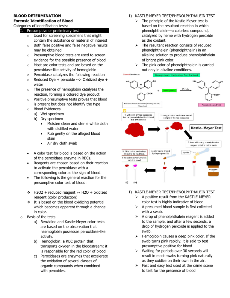
BLOOD DETERMINATION Forensic Identification of Blood Categories of identification tests: I. Presumptive or preliminary test o Used for screening specimens that might contain the substance or material of interest o Both false positive and false negative results may be obtained o Presumptive blood tests are used to screen evidence for the possible presence of blood o Most are color tests and are based on the peroxidase-like activity of hemoglobin o Peroxidase catalyzes the following reaction o Reduced Dye + peroxide --> Oxidized dye + water o The presence of hemoglobin catalyzes the reaction, forming a colored dye product o Positive presumptive tests proves that blood is present but does not identify the type o Blood Evidences a) Wet specimen b) Dry specimen Moisten clean and sterile white cloth with distilled water Rub gently on the alleged blood stain Air dry cloth swab o 1) KASTLE-MEYER TEST/PHENOLPHTHALEIN TEST The principle of the Kastle Meyer test is based on the resultant reaction in which phenolphthalein—a colorless compound, catalyzed by heme with hydrogen peroxide as the oxidant. The resultant reaction consists of reduced phenolphthalein (phenolphthalin) in an alkaline solution to produce phenolphthalein of bright pink color. The pink color of phenolphthalein is carried out only in alkaline conditions. A color test for blood is based on the action of the peroxidase enzyme in RBCs. Reagents are chosen based on their reaction to activate the peroxidase with a corresponding color as the sign of blood. The following is the general reaction for the presumptive color test of blood: H2O2 + reduced reagent ↔ H2O + oxidized reagent (color production) It is based on the blood oxidizing potential which becomes apparent through a change in color. Basis of the tests: a) Benzidine and Kastle-Meyer color tests are based on the observation that haemoglobin possesses peroxidase-like activity. b) Hemoglobin: a RBC protein that transports oxygen in the bloodstream; it is responsible for the red color of blood c) Peroxidases are enzymes that accelerate the oxidation of several classes of organic compounds when combined with peroxides. 1) KASTLE-MEYER TEST/PHENOLPHTHALEIN TEST A positive result from the KASTLE-MEYER color test is highly indicative of blood. A presumed blood sample is first collected with a swab. A drop of phenolphthalein reagent is added to the sample, and after a few seconds, a drop of hydrogen peroxide is applied to the swab. Hemoglobin causes a deep pink color. If the swab turns pink rapidly, it is said to test presumptive positive for blood. Waiting for periods over 30 seconds will result in most swabs turning pink naturally as they oxidize on their own in the air. Fast and easy test used at the crime scene to test for the presence of blood The heme portion of the RBC reacts with H2O2 to oxidize phenolphthalein and give a pink product 2) LEUCOMALACHITE TEST The use of Leucomalachite as the presumptive test was first developed by Adler and Adler in 1904. Earlier, in their study, they used LM green and violent. But later, many researchers settle on the LM green for blood screening. Leucomalachite green is a diphenylmethane dye whose base form is colorless and produces a blue-green color in the presence of blood. The principle of the LMG blood test uses the leuco base form of malachite green (colorless) that is made to react with blood samples to get converted into its oxidized green form in an acidic medium. Procedure Using a moistened (with ethanol) cotton swab, graze out some of the dried bloodstains. 1-2 drops of LMG solution should be added. Wait for 10-20 seconds. Meanwhile, if the color develops, the test is inconclusive. If no color change is seen, add 3% of hydrogen peroxide to the cotton swab. 3. ADLER/ BENZIDINE TEST Adler test (benzidine) test is considered to be one of the oldest tests that is used for screening whether the given sample is blood or not. The test was first developed by Adler (so as on his name) in 1904. The test was used for half the decade, till in 1946, it opted to be carcinogenic. And in 1976, it was completely banned as one of the presumptive tests for blood in the US. The principle is based on the reaction between the benzidine substrate as the coloring agent that flourishes the dark blue color on reacting with blood in presence of hydrogen peroxide. Procedure for benzidine (Adler test): Take a small amount of blood sample in a petri dish. Add a few drops of benzidine reagent to the sample. Add 3% hydrogen peroxide Wait up to 20 seconds. Appearance of dark blue color as the indication B. Presumptive Chemiluminescent Test for Blood Stains A chemiluminescent test is a light-producing reaction. In these reactions, light is produced as the byproduct of the electronically excited state and their electronic transition. In this, chemicals react to form an excited unstable intermediate that breaks down, releasing energy in the form of photons of light, and finally reaches its ground state. Most common presumptive test that is used for blood detection at the crime scene is Luminol. Luminol is 3-aminophthalhydrazide, was first synthesized in A. Schmitz, Heidelberg, in 1902. However, it was first observed by W. Lommel (Germany) in 1928. As per forensic point of view, it was Walter Specht (in 1937) who introduced the use of luminol at crime scenes. Principle and Reaction: o Luminol reacts with blood and hydrogen peroxide, producing blue-white to yellowish-green light under very low light conditions (usually dark). o o o Luminol for large area of blood is a presumptive test Luminol is mixed with hydrogen peroxide During the reaction with hydrogen peroxide, the luminol is pxidized and light energy is released; therefore. The luminol test is viewed in a darkened area. EXAMPLE: o Cattle: A, B,C,F,J,L,M,S,Z o Felines: A, B, AB o horses: A,C, D, K, P, Q, U, T o Dog: DEA o Humans: A, B, O, AB PRECIPITATION PRINCIPLE Different Precipitation Techniques A. In a liquid medium B. In a gel medium Terms: A. “Diffusion”- usually refers to the number of reactants moving B. “Dimension” – usually refers to the number of direction of the movement II. Confirmatory test o Are tests which are entirely specific for the substance or material for which it is intended o A positive confirmatory test is interpreted as an unequivocal demonstration that the specimen contains the substance or material Forensic Identification of Blood Confirmatory Tests for Blood: usually, Ab are used to detect the antigen o Current immunological tests use antibodies specific for human hemoglobin, thus combining the confirmatory test for blood with a human species test Ring test RID test Oakley and Fulthorpe Ouchterlony Species Determination Tests must be done on blood specimens to determine the species of origin o Species origin tests are done using immunological methods which involve the interaction of antigens and antibodies o Specific antiserum can be used to test for the presence of antigens in unknown specimens o Note: some antigens are found in animals but not in humans: ‘’ Therefore: o Single Diffusion: only one reactant is moving towards the other reactant o Double Diffusion: both reactants are diffusing/ moving towards each other o Single Dimension: only one effective direction of diffusion (usually up or down) o Double Dimension: diffusion in multiple/ all directions (usually radial) A. IN A LIQUID MEDIUM Interfacial test/ Ring test: Double diffusion, single dimension o introduced by Ascoli in 1902 o Principle: Ag and Ab diffuse toward each other until they reach their ZOE point where precipitation occurs. o Procedure: Antiserum is deposited at the bottom of the tube, overlaid with the Ag, and then incubated (RT or 37oC). o Result: (+) formation of a visible ring or layer of precipitate at the interface of the reactants o Antibody is added at the bottom while antigen stay at the top because of density. o It is expected that antigens are heavier than antibodies hence, will move downwards while antibodies will float B. IN A GEL MEDIUM o Principle: Soluble molecules (Ag and Ab) reacts in agar gel or other semisolid media until they reach their equivalence to form a stable precipitate (band/ line) o Gel media: agar, agarose, polyacrylamide gel o Non- gel media: cellulose acetate 1. Single Diffusion, Single Dimension (Oudin) o earliest precipitation technique(1946) o Principle: Ag diffuses through the gel containing immobilized Abs forming insoluble Ag-Ab complexes o Procedure: Abs are mixed with the liquid agar & deposited at the bottom of the tube, allowed to cool & solidify, then overlaid with Antigen. Two or more unrelated antigens against their homologous Antibodies may be tested. o Application: permits determination of minimum antigenic substances in a mixture such blood/ plasma / cell extract o Note: single dimension = done in tube (because it only moves vertically) 2. Single Diffusion, Double Dimension (Radial Immuno-Diffusion) o Principle: Ag binds the Abs present in the agar o Result: Formation of a precipitin disc or ring around the well o Procedure: a. Abs are mixed in a liquid gel on a flat surface (petri dish). b. Solidify agar and cut wells from the agar c. Add antigen on the cut wells d. allow for the reaction Methods: o o Mancini test/ endpoint method: Disc is measured about 48-72 hours after placing the Ag on the well. Fahey/ McKelvey method: kinetic/ timed assay method, disc is measured prior to its full development (18 hours) 3. Double Diffusion, Single Dimension (Oakley &Fulthorpe Method) o described in 1953 by Oakley and Fulthorpe o Principle: Both Ag and Ab both diffuse towards each other o Procedure: a. Place Abs in a tube b. overlay with neutral agar which is allowed to solidify c. overlay with an Ag o Result: Formation of a precipitin band or line through the gel at the point of equivalence o Note: - If Ab (reagent) is constant, Ag is of various concentrations: the distance from the Ag layer to the band is proportional to the antigen concentration. - If Ag is constant, the greater the concentration of the Ab, the greater the distance of band from the Ab layer to the band of precipitate. o The liquid agar doesn’t mix with the liquid reagent because they do not have the same consistency and density. 4. Double Diffusion, Double Dimension (Ouchterlony Method) o developed by Elek and Ouchterlony independently in 1948 o identifies 2 antigens or 2 samples o Principle: Ag and Ab both diffuse and bind each other, forming a line at the point of equivalence o Procedure: a. Agar containing buffered saline and a preservative is poured on a flat glass surface (peti dish, glass plate, microscope slide), solidify b. cut wells c. Place Antigens and Abs on separate wells, kept for several days/ weeks. d. Allow to react to form a precipitin line at the point of equivalence. Note: The number of precipitin lines indicates the minimum number of distinct antigenic substances present in the antigenic solution. o if both Ag and Ab are of the same MW, the line is straight; otherwise the line tends to be concave toward the reagent of higher MW. o Types of Reactions: (observed when related antigens in adjacent wells react with Ab against various Ag determinants) i. Type I (identity) - fusion of bands of precipitate ii. Type II (nonidentity) – the precipitate lines intersect/ cross because the samples contain no antigenic determinant in common. iii. Type III (partial identity) – obtained when two antigens compared possess common determinants but also display antigenic differences. It is distinguished by spur formation. AGGLUTINATION PRINCIPLE Principles of Agglutination o Agglutination is a visible manifestation of antigen-antibody interaction; however not all antigen-antibody interactions lead to agglutination. Rather, agglutination occurs only when the properties and relative concentrations of Ag and Ab allow sufficient lattice formation. o Carrier particles may be needed to indicate visibly that an antigen-antibody reaction has taken place. o Artificial carriers: latex particles and colloidal charcoal o biologic carriers: RBC, bacteria o Agglutination is a two-step process that results in a stable lattice formation: 1. DIRECT IMMUNE AGGLUTINATION o Aggregation of particles with the native Antigens or the antigens that are naturally present on the surface of particle such as RBC or bacteria. o Applications: a. Hemagglutination: detect antigens on RBCs by using known antisera (Ex: ABO, Rh forward typing) Adhesion-Elution/Absorption Method o Adhesion: known antibodies are made to react or adhere to the unknown specimen o After washing the excess or unreacted antibodies, the specimen undergoes elution o Elution: the process which removes the reacted antibodies o Known RBCs are introduced into the system to determine which antibodies have remained. Adhesion Stage: formation of antigen-antibody complex where unreacted antibodies will be washed off You can’t immediately drop the NSS or antibodies to the cloth, only to the tube/slide, because the lattice formation is affected by many factors. For example, to enhance lattice formation, you have to agitate and mix. Also, the ratio of blood to antibodies must be equal which can’t be achieved in the cloth. 2. Detect Antibodies to RBCs by using known Antigens (Ex: ABO reverse typing) Elution Stage: the antigen-antibody complex will break sensitization under 40C for 30 minutes o o o It is applied to pre-screen evidence in an effort to identify discrete areas for ACP testing Many body fluids fluoresce when excited with light at 450-nm wavelength Semen stains have tendency to fluoresce more intensely than most other body fluids b. Acid phosphatase test o Identification of an enzyme known as acid phosphatase o Considered as a presumptive identification of seminal fluid because other body fluids might also give a positive reaction o Purple color IDENTIFICATION OF SEMEN o The identification of semen is important in many cases of alleged sexual assault o Seminal fluid is a protein-rich body fluid origination primarily from the prostate and seminal vesicles o Spermatozoa, commonly referred to as “sperm”, are the male gametes or sex cells produced in the testis. Not all men produce spermatozoa I. Presumptive testing a. Alternate light source/UV light o Not all semen stains are visible to the naked eye II. Confirmatory testing a. Microscopic identification of spermatozoa Positive swabs or a small cutting from a positive stain can be smeared onto a microscope slide and then stained for visualization Nuclear fast red (red stain) and picroindigocarmine (green stain) and are sometimes referred to as the Christmas tree stain b. Protein confirmation of semen o Prostate specific antigen (PSA)/p30 o o o o o Value of the protein in forensic science is that it allows semen to be identified in stains and swabs Male who is vasectomized or a male with a congenital or other defect of the male reproductive system, spermatozoa may not be present in the semen Electrophoretic or diffusion methods such as crossover electrophoresis and ouctherlony double diffusion or ELISA Commercial kits This can be used alone to confirm semen, or it can be used in conjunction with the microscopic method FORENSIC DNA ANALYSIS
