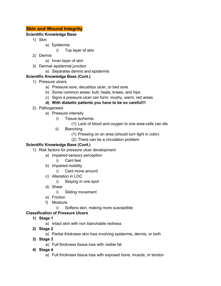
Skin and Wound Integrity Scientific Knowledge Base 1) Skin a) Epidermis i) Top layer of skin 2) Dermis a) Inner layer of skin 3) Dermal–epidermal junction a) Separates dermis and epidermis Scientific Knowledge Base (Cont.) 1) Pressure ulcers a) Pressure sore, decubitus ulcer, or bed sore b) Some common areas: butt, heals, knees, and hips c) Signs a pressure ulcer can form: mushy, warm, red areas d) With diabetic patients you have to be so careful!!! 2) Pathogenesis a) Pressure intensity i) Tissue ischemia (1) Lack of blood and oxygen to one area-cells can die ii) Blanching (1) Pressing on an area (should turn light in color) (2) There can be a circulation problem Scientific Knowledge Base (Cont.) 1) Risk factors for pressure ulcer development a) Impaired sensory perception i) Cant feel b) Impaired mobility i) Cant move around c) Alteration in LOC i) Staying in one spot d) Shear i) Sliding movement e) Friction f) Moisture i) Softens skin, making more susceptible Classification of Pressure Ulcers 1) Stage 1 a) Intact skin with non blanchable redness 2) Stage 2 a) Partial thickness skin loss involving epidermis, dermis, or both 3) Stage 3 a) Full thickness tissue loss with visible fat 4) Stage 4 a) Full thickness tissue loss with exposed bone, muscle, or tendon 5) Process of wound healing a) Primary intention i) Edges are approximated b) Secondary intention i) Heal from the inside out ii) Scar will be created iii) Infection can occur 6) Complications of wound healing a) Hemorrhage i) Hematoma b) Infection c) Dehiscence i) Separation of layers ii) Opening in wound iii) Suture lines opening d) Evisceration i) Organ protruding Nursing Knowledge Base 1) Prediction and prevention of pressure ulcers a) Risk assessment i) ii) iii) Braden scale 6-23 → lower the number higher the risk https://www.in.gov/isdh/files/Braden_Scale.pdf We should anticipate which patient will get a pressure ulcer b) Prevention i) Economic consequences of pressure ulcers (1) Medicare and Medicaid: no additional reimbursement for care related to stage III and stage IV pressure ulcers that occur during the hospitalization Nursing Knowledge Base (Cont.) 1) Factors influencing pressure ulcer formation and wound healing a) Nutrition i) Deficiency can cause impaired healing so vitamins and diet are important b) Tissue perfusion i) Oxygen blood crucial to healing ii) Poor circulation iii) Poor perfusion iv) Healing will get worse c) Infection i) Infection can spread ii) Delays healing d) Age i) May not remember to walk or move around e) Psychosocial impact of wounds i) Body image ii) Self concept Nursing Process: Assessment 1) Skin a) Continually assess skin for signs of breakdown and/or ulcer development b) Especially check bony prominences Assessment (Cont.) 1) Wounds a) Wound appearance (REEDA) i) Redness, Edema, Ecchymosis, discharge, approximation b) Character of wound drainage (COCA) i) Color, Odor, Consistency, Amount c) Drains d) Wound closures e) Palpation of wound i) Looking for scaring, warmth, and bubbles f) Wound cultures i) Gram stains ii) Biopsy iii) To check to see if anything is growing g) Serous i) Clear, watery, yellow h) Purulent i) Infectious, thick, yellow-green, tan i) Serousanguous i) Clear, watery, pale pink (mixture of blood and serous) j) Sanguous i) Deep read color Nursing Diagnosis 1) Nursing diagnoses associated with impaired skin integrity and wounds: a) Risk for infection b) Imbalanced nutrition: less than body requirements c) Acute or chronic pain d) Impaired physical mobility e) Impaired skin integrity f) Risk for impaired skin integrity g) Ineffective peripheral tissue perfusion h) Impaired tissue integrity Planning 1) Goals and outcomes a) Plan interventions according to i) Risk for pressure ulcers ii) Type and severity of the wound iii) Presence of complications iv) PLAN IS TO HEAL 2) Setting priorities a) Preventing pressure ulcers b) Promoting wound healing 3) Teamwork and collaboration (wound care specialist, nutrition, and physician) Implementation (Cont.) 1) Acute Care a) Management of pressure ulcers b) Wound management i) Debridement ii) Education iii) Nutritional status (1) Vitamin and minerals (2) Eat and drink well iv) Protein status v) Hemoglobin (1) Oxygen (2) Should have adequate amount of RBCs Interventions 1) Post and implement a turning schedule a) Repositioning redistributes pressure 2) Obtain and place over the patients mattress a low air loss overlay a) Redistributes the amount of pressure on the bony areas 3) Clean wound and periwound skin; dry periwound skin a) Remove debris and old drainage from site, prevent further wound progression and skin breakdown 4) Apply a hydrocolloid dressing to the wound a) Use of hydrocolloid dressing will support moist wound healing and will protect the wound 5) Determine in collaboration with dietitian an appropriate diet a) Adequate nutrition such as protein intake, increased caloric count, and vitamins aid in wound healing Implementation (Cont.) 1) First Aid for Wounds 2) Hemostasis a) Control bleeding. i) Allow puncture wounds to bleed. ii) Do not remove a penetrating object (1) Can cause more damage b) Bandage 3) Cleaning a) Gentle b) Normal saline 4) Protection 5) Dressings a) Purposes of dressings b) Protects from microorganisms c) Aids in hemostasis d) Promotes healing by absorbing drainage or debriding a wound e) Supports wound site f) Promotes thermal insulation g) Provides a moist environment 6) Types of dressings a) Gauze i) Used the most b) Transparent film i) Keeps moisture in c) Hydrocolloid i) Absorbent dressing d) Hydrogel i) Gets wound slightly moist and absorb e) Foam i) Pull out drainage f) Composite i) Provides barrier, absorption 7) Packing a wound a) Negative-pressure wound therapy 8) Securing a) Tape b) Ties c) Binders Implementation (Cont.) 9) Cleaning skin and drain sites a) Basic Skin Cleaning i) Clean from least contaminated to the surrounding skin ii) Use gentle friction (1) To get bacteria off skin iii) When irrigating, allow the solution to flow from the least to most contaminated area b) Irrigation i) Wound irrigations ii) Normal saline and squirt wound 10) Suture care a) Staple removal i) Faster closure time 11) Drainage Evacuation a) Constant, low-pressure vacuum to remove and collect drainage i) Wound VAC 12) Heat and Cold Therapy a) Heat will open the wound more and vasodilate b) Cold will vasoconstrict c) Make sure there is a barrier between therapy and skin d) Assessment for temperature tolerance i) Not everyone can get cold therapy and not everyone can get hot therapy e) Bodily responses to heat and cold f) Local effects of heat and cold i) Effects of heat application (1) No more than 20 mins on and then 20-30 minutes off ii) Effects of cold application g) Factors influencing heat and cold tolerance i) Exposure time ii) Exposed skin iii) Temperature iv) Age v) Perception of sensory stimuli Evaluation 1) Patient outcomes a) Individualize nursing interventions b) Patients with impaired skin integrity i) Ongoing evaluations ii) Validated risk-assessment tool Safety Guidelines for Nursing Skills 1) Position patient to prevent the patient from rolling over the side of the bed. 2) Keep a plastic bag within reach to discard dressings and prevent cross-contamination. Keep extra gloves within reach to allow a change of gloves if the gloves become soiled. 3) If irrigating a wound, use appropriate PPE. 4) When applying an elastic bandage, check the extremity for temperature or sensation changes. Skin, Hair, and Nails Assessment Structure and Function 1) Skin - three layers a) Epidermis i) Stratum germinativum or basal cell layer ii) Stratum corneum or horny cell layer iii) Derivation of skin color b) Dermis i) Connective tissue or collagen ii) Elastic tissue c) Subcutaneous layer 2) Epidermal appendages a) Hair b) Sebaceous glands c) Sweat glands i) Eccrine glands ii) Apocrine glands iii) Nails (1) Keratin Functions of the Skin 1) Protection from microbial and foreign substance invasion 2) Prevents penetration a) If something is coming towards us 3) Retain fluid and electrolytes a) In skin, gives off how healthy 4) Perception/ Sensation a) Ex: holding an ice cube in your hand. Body is telling you its cold 5) Temperature regulation 6) Identification a) What it looks like b) Different types of cultures 7) Communication/ express emotion a) Smiling 8) Wound repair 9) Absorption and excretion (sweat) 10) Production of vitamin D a) Essential for calcium absorption b) Get vitamin D from the sun Subjective Data—Health History Questions 1) Previous history of skin disease (allergies, hives, psoriasis, or eczema) 2) Change in mole a) Can be cancerous 3) Change in pigmentation (size or color) 4) Excessive dryness or moisture a) Turgor b) Edema c) Sweating 5) Pruritus a) Dry skin, itching 6) Excessive bruising a) May fall a lot b) Balance issue c) Abusive home d) Hemophilia causes easy bruising 7) Rash or lesion 8) Medications a) Blood thinners 9) Hair loss a) Alopecia b) cancer 10) Change in nails a) clubbing 11) Environmental or occupational hazards 12) Self-care behaviors a) How much you care for yourself Objective Data— The Physical Exam 1) Preparation a) External variables that influence skin color i) Emotions- smile, frown, blush ii) Environment- might have dark skin in southern areas and pale in northern areas iii) Physical 2) Equipment needed a) Strong direct lighting b) Small centimeter ruler c) Penlight; adequate lighting d) Gloves Skin—Inspect and Palpate 1) Color a) General pigmentation b) Vascularity i) Circulation problems c) Bruising d) Widespread color change i) Vitiligo ABNORMAL SKIN FINDINGS * 1) PALLOR – pale; indicates anemia, shock a) Hemorrhage due to loss of RBC and skin will turn pale b) When blood is not circulating good 2) CYANOSIS - blue tinge to skin or gray color in lips; indicates lack of oxygen, exposure to cold a) Check lips, fingers, toes 3) JAUNDICE - yellow from increased serum bilirubin; indicates liver disease a) Should be passed out the body so when it is not passed, the skin will turn yellow b) Check eyes or mouth 4) ERYTHEMA - red from increased cutaneous blood flow; indicates inflammation, fever, blushing Skin Color Changes 1) Light skin a) Pallor i) White or pale b) Erythema i) Red or bright pink c) Cyanosis i) Dusky blue d) Jaundice i) Yellow ii) Yellow sclera iii) Hard palate of the mouth 2) Dark skin a) Pallor i) Yellowish-brown or ashen grey b) Erythema i) Purplish tint c) Cyanosis i) Dark, dull and lifeless d) Jaundice i) Yellow sclera ii) Hard and soft palate juncture iii) Palms of the hands Examination: Skin (Cont’d) 1) Inspect for general color & uniformity of color a) Consistent over body surface except vascular areas b) Whitish pink to olive tones to deep brown c) Sun-exposed skin is darker. 2) Inspect skin for localized variations in color. a) Intentional: tattoos, coining patterns b) Normal localized variations: pigmented nevi (moles), freckles, patches, striae (stretch marks secondary to weight gain or pregnancy) NORMAL SKIN VARIATIONS - Abnormal descriptions is what is documented 1) STRIAE : silver or pink “stretch marks” 2) VITILIGO : unpigmented skin 3) MOLES (NEVI) : tan to dark brown, raised or flat; watch for changes 4) FRECKLES : small, flat macules 5) BIRTHMARKS : tan, reddish, or brown & flat Objective Data— The Physical Exam (continued) 1) Skin— Systematically inspect and palpate skin from head to neck to trunk, arms, legs, and back a) Temperature i) Hypothermia ii) Hyperthermia 2) Moisture – warm and dry a) Diaphoresis - sweating b) Dehydration Objective Data— The Physical Exam, cont. 1) Skin—Inspect and Palpate, cont. a) Texture i) Smooth, soft, intact, even surface ii) Calluses on hands, feet, elbows, knees 2) Thickness a) Varies with age and area i) Eyelids thinnest ii) Palms and soles thickest iii) Callus – thick from friction and pressure 3) Vascularity or bruising a) Circulation in body b) Document this Skin Mobility & Turgor 1) Skin mobility & turgor is assessed by a) Pinching up a large fold of skin on the anterior chest under the clavicle (or on the hand). b) Mobility is the skin’s ease of rising. c) Turgor is its ability to return to place promptly when released. i) Poor skin turgor is a sign of dehydration. ii) The skin recedes slowly or “tents” when released and stands by itself. iii) Skin should come down in 3 seconds Examination: Hair 1) Inspect and palpate scalp and hair for surface characteristics, hair distribution, texture, quantity, and color. a) Surface characteristics: smooth without flaking, scaling, redness, or lesions b) Should be shiny and soft c) Quantity and distribution: balding patterns and hair loss; male patterned d) Texture and color 2) Inspect facial and body hair for distribution, quantity, and texture. a) Less and less as you get older 3) Inspect for nails for shape, contour, color, consistency, thickness, and cleanliness. a) Edges: smooth and rounded b) Shape and contour: flat and slightly rounded c) Consistency: note grooves, depressions, pitting, and ridges d) Color: pink, blanched in light-skinned clients; yellow/brown with vertical lines in dark-skinned clients e) Thickness: smooth, uniform f) Inspect for cleanliness g) Capillary refill i) Depress the nail edge to blanch, then release. ii) Watch the color return. iii) Normal is <2 seconds. (1) More than 2 seconds means circulation problem Profile sign 1) Profile sign - viewing the finger from the side in order to detect early clubbing a) Normal angle of nail base – 160 b) Curved nail – normal when angle is 160° c) Early clubbing – angle is 180° (straight line) d) Late clubbing – angle is > 180° Edema 1) Fluid accumulating in the intercellular spaces 2) Not normally present Check for edema: 1) Imprint the thumb firmly against ankle malleolus and tibia 2) Normally the skin surface stays smooth. 3) If the pressure leaves a dent in the skin, pitting edema is present. Pitting Edema * 1) 4-point grading scale a) 1+ Mild pitting - slight indentation, no perceptible swelling of the leg b) 2+ Moderate pitting - indentation subsides rapidly c) 3+ Deep pitting - indentation remains for a short time, leg looks swollen d) 4+ Very deep pitting - indentation lasts a long time, leg is very swollen ABCDE Skin assessment 1) Asymmetry a) If mole was divided in half, they wouldn't be the same on both side 2) Border a) Jagged edges 3) Colour a) Gaining or losing colour 4) Diameter a) More than ½ cm in diameter 5) Evolution a) Moles that have changed size, shape, colour, or risen Activity and Exercise Musculoskeletal System 1) General inspection: a) Gait i) How you walk b) Posture i) Standing ii) Sitting c) Balance i) Without good balance you are at risk for falls d) Body alignment e) Friction Nursing Process: Assessment (Cont.) 1) Mobility a) Gait (a particular manner or style of walking) b) Exercise (physical activity for conditioning the body, improving health, and maintaining fitness) i) 30 min/5x a week c) Activity tolerance i) Physiological (1) Helps us mentally ii) Emotional iii) Developmental 2) Body alignment a) Standing b) Sitting c) Lying d) Prone- stomach down e) Supine- lying on back f) No excessive strain on joints, tendons, or ligaments g) Body should be distributed evenly h) Center of gravity helps with balance-belly button 3) Assess for lordosis, kyphosis, or scoliosis. a) Lordosis i) Lower back b) Kyphosis i) Higher upper back c) Scoliosis i) Most degree ii) Side way curve Nature of Movement 1) Body mechanics a) Coordinated efforts of the musculoskeletal and nervous systems 2) Alignment and balance a) Also refers to posture 3) Gravity a) Weight force exerted on the body 4) Friction a) Force that occurs in a direction opposite to movement Scientific Knowledge Base (Cont.) 1) Overview of exercise and activity: a) Exercise and activity i) Activity tolerance (1) Isotonic exercises: muscle contraction that changes the muscle length (a) Tightens without moving (2) Isometric exercises: tightening muscles without moving body parts (a) Planking (3) Resistive isometric exercises: contraction of the muscle while pushing against a stationary object Principles of Transfer and Positioning Techniques 1) When moving a patient, knowledge of safe transfer and positioning is crucial. 2) Pathological influences on body alignment mobility, and activity: a) Congenital defects b) Disorders of bones, joints, and muscles c) Central nervous system damage d) Musculoskeletal trauma e) Try to move these patients with some help so you dont hurt them 3) Safe patient handling a) Transfer techniques (algorithm pg. 793) Factors Influencing Activity and Exercise 1) Developmental changes 2) Behavioral aspects a) Patients are more likely to incorporate an exercise program if those around them are supportive b) Have a support system 3) Environmental issues a) Work site (incentives given) i) Give a reward or pass to the employee b) Schools (to offset obesity issue) c) Community (public health agencies, parks, etc.) i) Walking trails d) Have to see what motivates them and see what is appropriate 4) Cultural and ethnic influences 5) Family and social support (buddy system) Assessment 1) Thoroughly assess: a) Body alignment and posture with the patient standing, sitting, or lying down b) Normal physiological changes c) Deviations related to poor posture, trauma, muscle damage, or nerve dysfunction d) Patients’ learning needs 2) Through the patient’s eyes a) Assess patient expectations concerning activity and exercise b) People get anxious of when they can start exercising again Lifting Techniques 1) Tighten stomach muscles and tuck pelvis 2) Bend at knees 3) Keep weight lifted close to body 4) Maintain trunk erect and knees bent 5) Avoid twisting 6) Maintain center of gravity HOW TO LIFT PROPERLY 1) When lifting, come close to object 2) Enlarge the base of support 3) Lower center of gravity Implementation 1) Health promotion a) Teach patients to calculate maximum heart rate. i) When they are exercising and if they can see their own HR, they can damage their heart or pass out b) (220 minus their current age) c) Body mechanics (knowledge of equipment) i) If not used properly you can hurt yourself 2) Acute care a) Musculoskeletal system b) Joint mobility (ROM) Range of Motion exercises c) Walking 3) Helping a patient to walk a) Assess patient’s ability to walk safely b) Evaluate environment for safety c) Assist patient to sitting position, dangle patient’s legs over the side of the bed 1 to 2 minutes before standing i) Orthostatic hypotension d) Provide support at the waist so the patient’s center of gravity remains midline (gait belt) 4) Restorative and continuing care a) Implement strategies to assist patient with ADLs b) Restore and promote optimal functioning in patients with heart disease, COPD, diabetes c) With people with cognitive disorders, may be they don't understand d) MAKE SURE TO DO A SAFETY CHECK Range of Motion (ROM) Goals 1) The goal of ROM is to keep patients in the best physical shape possible. 2) Another goal is to increase joint mobility and to increase circulation to the affected part 3) Push patient to resistance but not push to pain Passive ROM 1) The patient is unable to move independently and someone else manipulates body parts. 2) Dependent, helping patient with it Active – Assistive ROM 1) The nurse provides minimal support as the patient moves through ROM. 2) Same thing as passive Active ROM 1) The patient moves independently through a full ROM for each joint 2) Show them what to do and they do independently Assistive Devices for Walking 1) Walkers a) The patient holds the hand grips on the upper bars, takes a step, moves the walker forward, and takes another step. 2) Canes a) Keep cane on stronger side of the body b) Place cane forward 6 to 10 inches, keeping body weight on both legs c) Weaker leg is moved forward, divide weight between cane and stronger leg d) Stronger leg is advanced past cane; divide weight between cane and weaker leg 3) Crutches a) Measuring for crutches i) 2 to 3 fingers widths from axilla and position the tips approximately 2 inches lateral and 4-6 inches anterior to the front of the patient's shoes, elbows slightly flexed at 20-25 degrees ii) MAKE SURE TO TELL THEM TO BEND THEIR ARM b) Crutch Walking on Stairs: Ascending Stairs i) Up with the good c) Crutch Walking on Stairs: Descending Stairs i) Down with the bad Evaluation 1) Patient outcomes a) Reassess the patient for signs of improved activity and exercise tolerance. b) Make comparisons with baseline measures c) Compare actual outcomes with expected outcomes. Immobility Pathological Influences on Mobility 1) Postural abnormalities 2) Muscle abnormalities 3) Damage to CNS 4) Musculoskeletal trauma Nursing Knowledge Base: Factors Influencing Mobility-Immobility 1) Mobility refers to a person’s ability to move about freely, and immobility refers to the inability to do so 2) Bed rest 3) Effects of muscular deconditioning a) Disuse atrophy (loss of muscle tone) b) Physiological c) Psychological d) Social i) Might have a fear of going out e) Complications i) Constipation ii) UTI DVT Systemic Effects 1) Metabolic a) Endocrine, calcium absorption, and GI function 2) Cardiovascular a) Orthostatic hypotension b) Thrombus 3) Muscle effects a) Loss of muscle mass b) Muscle atrophy 4) Urinary elimination a) Urinary stasis b) Renal calculi c) Constipation 5) Respiratory a) Atelectasis (total or partial collapse of lung/lobe) and hypostatic pneumonia 6) Musculoskeletal changes a) Loss of endurance and muscle mass and decreased stability and balance 7) Skeletal effects a) Impaired calcium absorption b) Joint abnormalities 8) Integumentary a) Pressure ulcer b) Ischemia Psychosocial Effects 1) Emotional and behavioral responses a) Hostility, giddiness, fear, anxiety 2) Sensory alterations a) Altered sleep patterns 3) Changes in coping a) Depression, sadness, dejection Nursing Process: Assessment 1) Mobility a) Range of motion 2) Range of motion a) Contractures: develop in joints not moved periodically through their full ROM b) Neck, shoulder, elbow, forearm, wrist, fingers and thumb, hip, knee, ankle and foot, and toes 3) Mobility a) Body alignment is used for: i) Determining normal physical changes ii) Identifying deviations in body alignment iii) Patient awareness of posture iv) Identifying postural learning needs of patients v) Identifying trauma, muscle damage, or nerve dysfunction vi) Obtaining information on incorrect alignment (i.e., fatigue, malnutrition, psychological problems) Nursing Diagnosis 1) Impaired physical mobility 2) Ineffective airway clearance 3) Impaired urinary elimination 4) Risk for disuse syndrome 5) Ineffective coping 6) Risk for impaired skin integrity 7) Social isolation Implementation: Acute Care 1) Metabolic a) Provide high-protein, high-calorie diet with vitamin B and C supplements 2) Respiratory a) Cough and deep breathe every 1 to 2 hours. b) Provide chest physiotherapy. 3) Cardiovascular a) Reducing orthostatic hypotension b) Reducing cardiac workload c) Preventing thrombus formation d) SCDs (Sequential compression devices), thromboembolic disease (TED), hose, and leg exercises i) Try to get out of bed as soon as possible but usually given aspirin Implementation (Cont.) 1) Musculoskeletal system a) Prevent muscle atrophy and joint contractures Implementation 1) Integumentary system a) Reposition every 1 to 2 hours. b) Provide skin care. 2) Elimination system a) Provide adequate hydration. b) Serve a diet rich in fluids, fruits, vegetables, and fiber. 3) Psychosocial a) Talking to pt to make sure pt knows you are there for them Positioning Techniques 1) Trochanter roll a) Prevent rotation of hips b) Roll towel next to side so the leg does not externally rotate 2) Hand roll a) Contract of hand 3) Trapeze bar a) Helps patient move up 4) Supported Fowler’s 5) Supine a) Lying on back 6) Prone a) Lying on stomach 7) Side-lying 8) Sims’ 9) Moving patients a) Safety is first priority b) Ask patient to help as much as possible c) Determine if patient comprehends what is expected d) Determine patient’s comfort level e) Determine if you need assistance in moving the patient 10) Restorative and continuing care a) IADLs b) ROM exercise c) Walking Positioning/Moving a Patient Up in Bed 1) Allow patient to move himself if able to do so. 2) HOB down---don’t move up hill. 3) Position height of bed for nurses’ comfort. 4) Have patient flex knees, chin to chest, arms folded across chest 5) Nurses tightens abdominal girdles, flex knees. 6) Nurses shift weight, moving patient. 7) Reposition HOB, bed in low position. Turning a Patient 1) Determine what patient can do, find assistance if it is needed. 2) Position height of bed for nurses’ comfort. 3) Position patient supine on far side of bed. 4) Patient arms across chest, far leg over near one. 5) Tighten girdle, flex knees. 6) Place one hand on patient shoulder, other on hip. 7) Roll patient toward you. 8) Position patient for comfort, support with pillows if needed. 9) Raise side rails, lower bed. Implementation (Cont.) 1) Wide base of support with one foot in front of other, supporting body weight, then gently lower patient to ground while supporting their head Use Mechanical Devices 1) Lifts will save backs – yours included!!! Evaluation 1) Patient outcomes a) Evaluate effectiveness of specific interventions b) Evaluate patient’s and family’s understanding of all teaching provided Safety Guidelines for Nursing Skills 1) Communicate clearly with members of the health care team 2) Assess and incorporate the patient’s priorities of care and preferences 3) Use the best evidence when making decisions about your patient’s care Sleep Physiology of Sleep: Sleep Regulation 1) Regulated by a sequence of physiological states integrated by central nervous system (CNS) activit 2) Hypothalamus Stages of the Adult Sleep Cycle 1) Four stages of NREM 2) Sleep cycle lasts 90 to 100 minutes 3) Sleep goes through stages 1 to 4, then reversal from 4 to 3 to 2, followed by REM 4) There is REM and NREM Sleep Disorders 1) Insomnia a) Difficulty falling asleep b) Most common c) Can be caused by medical condition 2) Sleep apnea a) Lack of airflow through nose and mouth for periods of 10 second or longer during sleep b) 3 types: central, obstructive, and mixed c) Obstructive i) When muscles or structures of the oral cavity or throat relax during sleep ii) Airway becomes blocked d) Central i) Dysfunction in control center of brain ii) Impulse to breath fails 3) Narcolepsy a) Person suddenly feels an overwhelming wave of sleepiness and falls asleep b) Cataplexy is sudden muscle weakness during intense emotions i) Falls into paralysis and cant move 4) Sleep deprivation a) Stress, medications, environmental disturbances, hospitalization 5) Parasomnias a) Sleep problems that are more common in children b) Sleep walking, night terrors, nightmares, bed wetting, body rocking, and tooth grinding Factors Influencing Sleep 1) Drugs and substances a) Hypnotics, diuretics, narcotics, antidepressants and so on 2) Lifestyle a) Work, social activities, routines 3) Emotional stress a) Worries, physical health, death, losses 4) Exercise and fatigue a) Causes restful sleep 5) Usual sleep patterns a) May be disrupted by social activity or work 6) Environment a) Nosie, routines 7) Food and calorie intake a) Time of day, caffeine, nicotine, alcohol Assessment 1) Through the patient’s eye 2) Sleep assessment a) Sources for sleep assessment = Patient, family b) Tools for sleep assessment 3) Sleep history a) Description of sleeping problems, usual sleep pattern, current life events, physical and psychological illness, emotional and mental status, bedtime routines, bedtime environment, behaviors of sleep deprivation 4) Description of sleeping problems a) Conduct a more detailed history when a patient has a sleep problem. This ensures that you provide appropriate therapeutic care. b) Open-ended questions help a patient describe a problem more fully. c) Ask specific questions related to the sleep problem. 5) Usual sleep pattern a) Have patients describe their normal sleep patterns. Assessment (Cont.) 1) Physical and psychological illness 2) Current life events 3) Emotion and mental status 4) Bedtime routine 5) Bedtime environment 6) Behaviors of sleep deprivation 7) Implementation 8) Health promotion a) Environmental controls b) Promoting bedtime routines c) Promoting safety d) Promoting comfort e) Establishing periods of rest and sleep f) Stress reduction g) Bedtime snacks h) Pharmacological approaches 9) Environment controls 10) Promoting bedtime routines 11) Promoting safety 12) Promoting comfort 13) Establishing periods of rest and sleep 14) Stress reduction 15) Bedtime snacks 16) Pharmacological approaches Implementation (Cont.) 1) Acute care a) Environmental controls b) Promoting comfort c) Establishing periods of rest and sleep d) Promoting safety e) Stress reduction 2) Restorative or continuing care a) Promoting comfort b) Controlling physiological disturbances c) Pharmacological approaches Basic Assessment Techniques Objectives 1) Discuss the basic techniques used in health assessment, including inspection, auscultation, percussion, and palpation. Physical Assessment 1) Perform a physical examination only after proper preparation of the environment and equipment and the patient has been prepared physically and psychologically. 2) Throughout the examination, keep the patient warm, comfortable, and informed of each step of the process. 3) A competent examiner is systematic while combining simultaneous assessment of different body systems. 4) Information from history helps to focus on body systems likely to be affected. Positioning 1) Position depends on type of exam and condition of client. a) Sitting and supine positions are most common. 2) Appropriate draping in positions with adequate exposure is needed for exam. 3) Inability of client to assume position may be a significant finding about physical condition and requires accommodation. Inspection 1) Look, listen, and smell to distinguish normal from abnormal findings 2) Physical exams begin with inspection. a) Visual exam of body, including movement and posture i) Data obtained by smell are also a part of inspection. 3) Examination of every body system includes technique of inspection. 4) Patient is draped appropriately to maintain modesty while allowing sufficient exposure for exam; adequate lighting is essential. 5) Use adequate lighting. 6) Use direct lighting to inspect body cavities. 7) Inspect each area for size, shape, color, symmetry, position, and abnormality. 8) Position and expose body parts as needed so all surfaces can be viewed but privacy can be maintained. 9) When possible, check for symmetry. 10) Patient should be thoroughly observed with a critical eye. 11) Concentration without distraction avoids overlooking potentially important data. 12) Use of equipment may facilitate inspection of certain body systems. a) Penlight, otoscope, ophthalmoscope, and vaginal speculum Auscultation - Bell- low pitches - Diaphragm- high pitch 1) Hear movements of air, blood, or fluid in the body over lungs and abdomen. 2) Learn normal sounds first before identifying abnormal sounds or variations. a) Breath sounds-bronchial, bronchioles, vesicular, tracheal b) Hear wheezing up top c) Crackles-fluid,as the fluid builds up, it gets worse 3) Requires a good stethoscope 4) Requires concentration and practice 5) Stethoscope is used for auscultation to block out extraneous sounds when evaluating condition of heart, blood vessels, lungs, and intestines a) Will get outside noise, if patient is talking you wont be able to hear well 6) Listen for sound and characteristics:intensity, pitch, duration, quality 7) Concentrate; sounds may be transitory or subtle. a) Closing eyes may improve listening. b) Selective listening is isolating specific sounds, such as air during inspiration, or a single heart sound. 8) Optimize quality of auscultation findings. a) Best to auscultate in quiet room where noise cannot interfere. b) Stethoscope must be placed directly on skin because clothes obscure or alter sounds. c) If client is cold and shivers, involuntary muscle contractions may interfere with normal sounds. d) Friction of body hair rubbing against diaphragm could be mistaken for abnormal lung sounds (crackles) Percussion 1) Percussion performed to: a) Evaluate size, borders, and consistency of internal organs. b) Detect tenderness. c) Determine extent of fluid in a body cavity. 2) Tap body with fingertips to produce a vibration. 3) Sound determines location, size, and density of structures. 4) Indirect percussion: tapping over middle finger a) example: percuss over lung tissue 5) Direct percussion: striking finger or hand directly against client’s body. a) Evaluate adult sinus by tapping a finger over sinus. b) Elicit tenderness over kidney by striking costovertebral angle (CVA) directly with fist. 6) Five percussion tones a) Tympany is loud, high-pitched sound heard over abdomen. i) Gasy areas b) Resonance is heard over normal lung tissue. i) Make sure to get in between ribs c) Hyperresonance is heard in overinflated lungs, as in emphysema. d) Dullness is heard over liver. e) Flatness is heard over bones and muscle. Palpation 1) Use of hands to feel texture, size, shape, consistency, location of certain parts, and identify painful or tender areas 2) Requires nurse to move into personal space 3) Gentle touch, warm hands, and short nails to prevent discomfort or injury to client a) Touch has cultural symbolism and significance. b) State purpose, manner, and location of touching. c) Wear gloves when palpating mucous membranes or other areas where contact with body fluids is possible. 4) Using the sense of touch to gather information 5) Skin: Temp, moisture, texture, turgor, tenderness, thickness a) 2-3 seconds for turgor 6) Abdomen: tenderness, distension or masses 7) Palmar surface of fingers and finger pads are more sensitive than fingertips. a) Better to determine position, texture, size, consistency, masses, fluid, crepitus 8) Ulnar surface of hand to fifth finger is most sensitive to vibration. 9) Dorsal surface is better for assessing temperature. 10) Using palmar surfaces of fingers may be light or deep and controlled by amount of pressure. 11) Light palpation accomplished by pressing to a depth of approximately 1 cm, used to assess skin, pulsations, and tenderness. 12) Deep palpation accomplished by pressing to a depth of 4 cm with one or two hands used to determine organ size and contour. 13) Bimanual technique of palpation uses both hands, one anterior, one posterior, to entrap an organ or mass between fingertips to assess size and shape. 14) Light palpation should always precede deep palpation because deep palpation may cause tenderness or disrupt fluids, which may interfere with collecting data by light palpation. Head, Neck, Neuro General Health History: Present Health Status 1) Present health status a) Ask about changes in patients’ overall health or changes to eyes, ears, nose, or mouth. b) Chronic conditions that affect eyes, ears, nose, mouth, head, or neck regions include: i) Cataracts, glaucoma, migraine headaches, hearing loss, oral cancer, hypothyroidism c) Other chronic conditions include: i) Hypertension, human immunodeficiency virus (HIV) infection, diabetes mellitus, autoimmune disorders d) Chronic diseases often impact clinical findings. 2) Medications: what and how often? a) Side effects of medications are common and may explain symptoms or clinical findings associated with head and neck regions. b) Headaches, dizziness, changes in vision, ringing in ears, and dry mouth are all examples of medication side effects. 3) Last routine examinations? Dental, vision, and hearing? a) What corrective devices do you use (e.g., contact lenses, glasses, hearing aids, dentures)? 4) Describe daily practices to maintain health of eyes, ears, and mouth. a) Understand health promotion practices and risks. 5) Any occupational or recreational risks for injury to your eyes, ears, or mouth? a) Assessment of environmental risk factors that can contribute to vision or hearing loss is an important component of a health history. b) patients should be encouraged to take protective action to minimize injury in contact sports. c) Noisy environment d) Living near factory or refinery e) Not wearing goggles f) 6) Do you use nicotine products or drink alcohol? If so, how much? a) Understand potential risks involving head, eyes, ears, and mouth b) Chronic alcohol intake and smoking are risk factors for many problems including cataracts, glaucoma, and cancers of the oropharynx. 7) Have you ever had an injury to your eyes, ears, mouth, or neck? a) Describe when and what. Do you continue to have any problems related to injury? b) Recent or past injuries may provide relevant data to clinical findings. 8) Have you had surgery involving eyes, nose, ears, mouth or neck? To what purpose? a) Knowledge of past surgeries may provide information that may be applied to symptoms or clinical findings. 9) Have you had chronic infections affecting eyes, ears, sinuses, or throat? a) Did this occur during childhood? Adulthood? How was the problem treated? 10) Establish baseline information for persons with history of chronic infections—even if they do not currently have these problems. a) These data may shed light on other findings. Neurological system 1) Responsible for many functions 2) Full assessment requires time and attention to detail 3) Many variables must be considered during evaluation: level of consciousness, physical status, chief complaint 4) Collect all equipment before beginning Neurological System 1) AAOx3: Awake, Alert and Oriented to person, place and time a) It is now x4 b) Awake,alert, oriented to person, place, time, and situation 2) Mental and emotional status a) Mini-Mental State Examination (MMSE) i) Orientation ii) Tells patient to repeat 3 words iii) Identify object iv) Read a sentence b) Cultural considerations c) Delirium d) Can they understand and signs of illness 3) Level of consciousness a) Glasgow Coma Scale 4) Behavior and appearance a) Nonverbal and verbal i) Have to match 5) Language a) Aphasia i) Sensory (receptive) (1) Comprehends language ii) Motor (expressive) (1) Express what is going on 6) Motor function a) Coordination b) Strength i) Pushing up and down on doctors arms or hands c) Balance i) Romberg’s test ii) Positive- they do lose balance iii) Negative- there is no swaying 7) Intellectual function a) b) c) d) Memory Knowledge Abstract thinking Association i) Associating words e) Judgment 8) Cranial nerve function (12) a) Nerves control different parts of the body b) They also impact organs 9) Sensory function a) Dermatomes i) Sense pain in different parts of the body ii) Pain throughout the body 10) Reflexes:** a) 0: no response b) 1+: sluggish/diminished c) 2+: active/expected response-normal d) 3+: more brisk than expected, slightly hyperactive e) 4+: brisk and hyperactive with intermittent or transient clonus f) Position. i) Tap tendon briskly. ii) Compare corresponding sides. Head and Neck 1) Includes assessment of the head, eyes, ears, nose, mouth, pharynx, neck, lymph nodes, carotid arteries, thyroid gland, and trachea. 2) Use inspection, palpation, and auscultation. 3) Assessment of the head and neck a) First step- inspection b) Assess for symmetry of the head c) Look for any gross abnormalities d) Have patient turn head to right and left and observe movement Neck 1) Neck muscles a) Anterior triangle b) Posterior triangle 2) Lymph nodes a) Malignancy 3) Carotid artery a) Should be able to feel a pulse b) Don't feel both carotids at once 4) Jugular vein a) Distended vein means there is fluid backup b) JVD 5) Have patient bend chin to chest and observe movement of neck 6) Palpate the back of the neck as patient leans head forward 7) Palpable Lymph Nodes a) Move to the front of the neck and assess paths of lymph nodes b) Palpate lightly with tips of fingers 8) Thyroid gland 9) Trachea a) Part of the upper respiratory system Nose and Sinuses 1) Nose a) Excoriation b) Polyps 2) Sinuses Otoscope used by the advanced practitioner to examine external ear canal - Check for an inflammation or redness Eyes 1) External eye structure a) Position and alignment b) Eyebrows c) Eyelids d) Lacrimal apparatus e) Conjunctivae and sclerae f) Corneas g) Pupils and irises i) Pinpoint pupils result to opioid use h) PERRLA i) Visual acuity: Snellen Test i) Eye test with the letters as you would see in doctors offices ii) Larger bottom number, the worse your vision is iii) 20/40 vision example iv) What you see at 20 feet others can see at 40 feet v) Read without glasses first and then try again with glasses j) Extraocular movements (EOM) for 6 Cardinal Fields i) Nystagmus- involuntary eye movement, eyes flutter k) Convergence test l) Accommodation eye test i) Person looks at wall behind you ii) Eyes should dilate iii) Then once they look back at you and their eyes converge and pupils constrict m) Snellen test n) EOM test o) Peripheral vision test Ears 1) Auricles a) Texture b) Tenderness c) Lesions d) Color e) Pain f) Cerumen i) Ear wax g) Ear should be normal, smooth, no lesions h) Down syndrome ears are lower 2) Hearing acuity a) Three types of hearing loss i) Conduction (1) Swelling (2) Sound waves from outer and inner ii) Sensorineural (1) Inner and nerves iii) Mixed (1) Total blockage b) Ototoxicity i) Could be from medications Tuning Fork Tests 1) Weber’s test a) Hold fork at base and tap it lightly against heel of palm. b) Place base of vibrating fork on midline vertex of patient’s head or middle of forehead. c) Ask patient if he or she hears the sound equally in both ears or better in one ear (lateralization). d) Impaired ear and hear best at top of head e) Sensory- normal ear can hear better 2) Rinne test a) Place stem of vibrating tuning fork against patient’s mastoid process (see illustration B). b) Begin counting the interval with your watch. c) Ask patient to tell you when she no longer hears the sound; note number of seconds. d) Quickly place still-vibrating tines 1 to 2 cm (1/2 to 1 inch) from ear canal, and ask patient to tell you when she no longer hears the sound. e) Continue counting time the sound is heard by air conduction. f) Compare number of seconds the sound is heard by bone conduction versus air conduction. g) Count: air- 20 seconds, bone- 10 seconds h) Normal is 2 to 1 ratio i) Supposed to hear longer than feeling Mouth and Pharynx 1) Lips a) Color b) Texture c) Hydration d) Contour e) Lesions f) Buccal mucosa g) Gums h) Teeth i) Tongue j) Floor of mouth k) Palate i) Hard ii) Soft l) Pharynx Respiratory Introduction 1) Lungs major function a) Provide continuous gas exchange between inspired air and blood in the pulmonary circulation b) Left side has two lobes c) Right side has three lobes d) Alveolus are damaged the most Anatomy 1) Respiratory tract extends from mouth/nose to alveoli 2) Upper airway filters airborne particles, humidifies and warms inspired gases a) Through nose 3) Lower airway serves for gas exchange Contributors of Respiration 1) Controlled in the brainstem a) Controls respiratory 2) Mediated by muscles of respiration a) Diaphragm primary muscle of inspiration b) Accessory muscles of inspiration i) SCM (Sternocleidomastoid) ii) Scalenes iii) Intercostals 3) Expiration is a passive process from elastic recoil of lung and chest wall, with passive diaphragm relaxation Technique for Respiratory Exam 1) NEED ORDERLY PROCESS 2) Before beginning, if possible: a) Quiet environment b) Proper positioning (patient sitting for posterior thorax exam, supine for anterior thorax exam) c) Bare skin for auscultation d) Patient comfort, warm hands and diaphragm of stethoscope, be considerate of women (drape sheet to cover chest) 3) Inspect 4) Palpate 5) Percuss 6) Auscultate Initial Respiratory Survey 1) Observe the patient’s breathing pattern a) Rate (normal vs. increased/decreased) i) Bradypnea ii) Tachypnea b) Depth (shallow vs. deep) i) Could mean problem c) Effort (any sign of accessory muscle use (retractions, inspect neck) 2) Assess A-P diameter a) Front to back b) Anterior to posterior 3) Assess the patient’s color a) cyanosis Normal Respiratory Rates 1) Newborn- 30-60 2) Infant (6 months)- 30-50 3) Toddler (2 years)- 25-32 4) Child- 20-30 5) Adolescent- 16-20 6) Adult- 12-20 7) Just know babies breath faster and know the adult range . Pertinent History 1) Any chronic conditions a) Asthma, COPD, Heart failure, Diabetes i) Heart failure is when the heart is not pumping well so there can be a backup 2) Exposure to new medication 3) Recent change in diet 4) Substance abuse/Overdose a) Can stop breathing 5) Prior DVT, PE (Deep vein thrombosis or venous thrombotic embolism VTE, pulmonary embolism. ) a) Quick shallow breaths 6) Recent trauma to chest a) Recent damage to lung Inspection 1) Note the shape of the chest and the way it moves a) Deformities or asymmetry i) Increased AP diameter in COPD 2) Abnormal retractions of interspaces during respiration a) Lower interspaces, supraclavicular in acute asthma exacerbation b) May see cyanosis of the lips 3) Impaired respiratory movement a) Flail Chest and paradoxical movement with rib fractures Palpation 1) Identify tender areas a) Bruising with rib fracture 2) Observe for appropriate chest wall expansion (chest excursion) a) Put your hands on both side of ribs to feel the ribs and chest expanding 3) Feel for vocal or tactile fremitus symmetrically a) palpable vibrations transmitted to chest wall b) use ulnar surface of hand, say “ninety-nine” c) decreased with COPD, pleural effusions 4) Palpating for Fremitus 5) Chest Excursion Percussion-tapping on a finger on the chest 1) 2) 3) 4) Helps to identify if underlying tissues are air-filled, fluid-filled, or solid Always percuss symmetrically on chest wall Percuss side to side, avoiding bone Dullness replaces resonance (over lung tissue) when fluid or solid tissue replaces air containing lung a) Pleural Effusions b) Hemothorax i) Blood filling c) Tumor 5) Unilateral Hyperresonance (over inflated tissue) a) Pneumothorax 6) Generalized Hyperresonance a) COPD b) Emphysema Auscultation 1) 8 anterior and 8 lateral locations 2) 10 posterior locations 3) Auscultate symmetrically 4) Should listen to at least 6 locations anteriorly and posteriorly Breath Sounds 1) Normal a) Bronchial b) Bronchovesicular c) Vesicular 2) Abnormal a) Absent b) Decreased c) Bronchial (if heard in other locations of the lung) 3) Adventitious (Abnormal) a) Crackles i) “Pop” like cereal b) Wheeze i) Musical note c) Rhonchi d) Stridor e) Pleural Friction Rub i) Grating sound made when inflamed pleural spaces move during respiration Normal Breath Sounds 1) Created by turbulent air flow 2) Inspiration a) Air moves to smaller airways hitting walls b) More turbulence, Increased sound 3) Expiration a) Air moves toward larger airways b) Less turbulence, Decreased sound 4) Normal breath sounds a) Loudest during inspiration, softest during expiration Physical Examination: Auscultation 1) Normal breath sounds a) Bronchial i) heard over trachea, high-pitched, expiration > inspiration ii) Quick inspiration, long expiration 2) Broncho vesicular a) heard over major bronchi, between the scapulae, around the sternum, mediumpitched, inspiration = expiration 3) Vesicular a) heard over peripheral lung fields, soft-pitched, inspiration > expiration Adventitious Breath Sounds 1) Crackles a) Snap crackle pop b) Heard more commonly with inspiration c) Classified as fine or coarse d) Normal at anterior lung bases i) Maximal expiration ii) Prolonged recumbency e) Crackles caused by air moving through secretions and collapsed alveoli f) Associated conditions i) pulmonary edema and early heart failure ii) Pulmonary edema means fluid in lungs g) Crackles is not good period, but you rather have it at the bottom of the lungs rather than mid way or at the top 2) Wheeze a) Continuous, high pitched, musical sound, longer than crackles b) Heard > with expiration i) Heard more clearly c) Produced when air flows through narrowed airways d) Associated conditions i) Asthma, COPD e) Hear wheezes when vessels are constricting such as smoking 3) Rhonchi a) Loud, low pitched, snoring quality b) Implies obstruction of larger airways by secretions c) Associated condition i) acute bronchitis 4) Stridor a) Inspiratory musical wheeze b) Loudest over trachea c) Suggests obstructed trachea or larynx d) Medical emergency requiring immediate attention e) Associated condition i) inhaled foreign body Causes of Decreased or Absent Breath Sounds 1) Asthma (narrowing of airway) a) Airway can eventually close which would be an emergency 2) Chronic Obstructive Pulmonary Disease (COPD) a) Constricts and closes off airway 3) Pleural Effusion (fluid in the pleural space) 4) Pneumothorax (air leak into pleural space) 5) Adult Respiratory Distress Syndrome (ARDS): fluid in alveoli sacs 6) Atelectasis (collapse of alveoli) 7) Symptoms of Hypoxia a) Early signs RAT- restlessness, anxiety, tachycardia, tachypnea b) Late signs BED-bradycardia, extreme restlessness, dyspnea Nursing Diagnosis 1) Oxygenation-Nursing Process 2) Activity Intolerance 3) Airway clearance, ineffective 4) Breathing pattern, ineffective 5) Impaired Gas Exchange 6) Risk for Infection 7) Fear or Pain Interventions: 1) Incentive Spirometry a) Inhale in order for the ball to move 2) Acapella Device a) Expel, blowing out to strengthen the lungs 3) Pursed lip breathing a) Inhale slow, exhale even slower Coughing & Deep Breathing: 1) Chest physiotherapy (manual) 2) (with vest) Pulmonary Function Test (PFT) 1) Pulmonary function tests are a group of tests that measure how well the lungs take in and release air and how well they move gases such as oxygen from the atmosphere into the body's circulation a) Measures amount of O2 in and CO2 out b) Lung volume and functioning Why Do PFT? 1) Diagnose certain types of lung disease 2) Find the cause of shortness of breath (SOB) 3) Assess the effect of respiratory medication a) Before and after medication to see if the med worked 4) Measure progress in disease treatment Assessment Methods 1) Peak Flow Meter a) Green is good b) Yellow is not so good c) Red is bad d) Tells pt who has asthma if they should do something or not 2) Pulse Oximetry a) 95%-100% 3) Sputum collection a) Best in AM b) Sterile specimen container c) Expectoration i) Cough deeply and expectorate into container Positioning 1) Allows for free movement of the diaphragm and expansion of the chest wall 2) High Fowler Position a) Allows for free movement of the diaphragm and expansion of the chest wall Administering Oxygen 1) Nasal cannula a) 1-6L/min b) Good for low O2 c) Skin breakdown can happen d) Only goes to 6L e) COPD-keep them on less than 2L of O2 2) Face mask (never less than 5L/min – can retain CO2) a) Simple mask 6-12L/min b) Partial rebreather and non-rebreather 10-15L/min c) Venturi mask – 4-10L/ min; most precise flow concentration d) High flow of O2 Suctioning 1) Suction catheter 2) Suctioning via nose or mouth a) Nose is cleaner b) When suctioning what else are you suctioning? i) Air ii) Secretions iii) Mucous c) When suctioning, it is intermittent and not continuous d) Always suction when coming out and not going in

