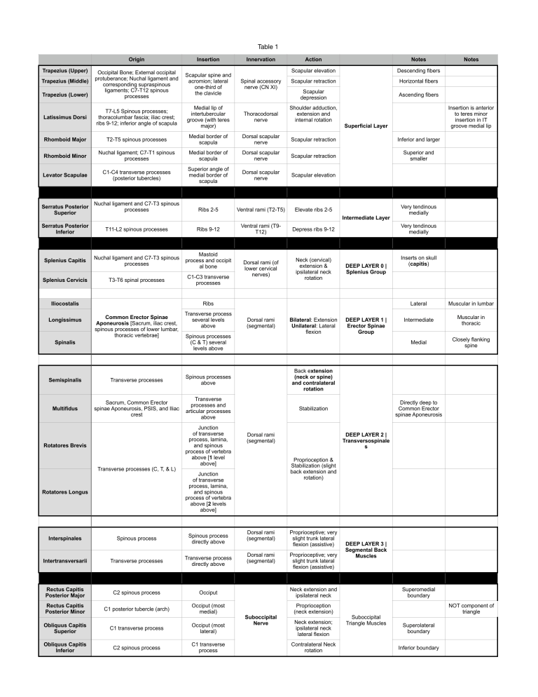
Table 1 Origin Insertion Occipital Bone; External occipital protuberance; Nuchal ligament and corresponding supraspinous ligaments; C7-T12 spinous processes Scapular spine and acromion; lateral one-third of the clavicle Spinal accessory nerve (CN XI) T7-L5 Spinous processes; thoracolumbar fascia; iliac crest; ribs 9-12; inferior angle of scapula Medial lip of intertubercular groove (with teres major) Thoracodorsal nerve Shoulder adduction, extension and internal rotation Rhomboid Major T2-T5 spinous processes Medial border of scapula Dorsal scapular nerve Scapular retraction Inferior and larger Rhomboid Minor Nuchal ligament; C7-T1 spinous processes Medial border of scapula Dorsal scapular nerve Scapular retraction Superior and smaller Levator Scapulae C1-C4 transverse processes (posterior tubercles) Superior angle of medial border of scapula Dorsal scapular nerve Scapular elevation Nuchal ligament and C7-T3 spinous processes Ribs 2-5 Ventral rami (T2-T5) Elevate ribs 2-5 Trapezius (Upper) Trapezius (Middle) Trapezius (Lower) Latissimus Dorsi Serratus Posterior Superior Action Notes Scapular elevation Descending fibers Scapular retraction Horizontal fibers Scapular depression Ascending fibers T11-L2 spinous processes Ribs 9-12 Splenius Capitis Nuchal ligament and C7-T3 spinous processes Mastoid process and occipit al bone Splenius Cervicis T3-T6 spinal processes C1-C3 transverse processes Iliocostalis Ventral rami (T9T12) Depress ribs 9-12 Dorsal rami (of lower cervical nerves) Neck (cervical) extension & ipsilateral neck rotation Transverse process several levels above Dorsal rami (segmental) Bilateral: Extension Unilateral: Lateral flexion Spinalis Spinous processes (C & T) several levels above Semispinalis Transverse processes Spinous processes above Back extension (neck or spine) and contralateral rotation Multifidus Sacrum, Common Erector spinae Aponeurosis, PSIS, and Iliac crest Transverse processes and articular processes above Stabilization Rotatores Brevis Transverse processes (C, T, & L) Rotatores Longus Junction of transverse process, lamina, and spinous process of vertebra above [1 level above] Dorsal rami (segmental) DEEP LAYER 0 | Splenius Group Dorsal rami (segmental) Proprioceptive; very slight trunk lateral flexion (assistive) Dorsal rami (segmental) Proprioceptive; very slight trunk lateral flexion (assistive) Spinous process Spinous process directly above Intertransversarii Transverse processes Transverse process directly above Rectus Capitis Posterior Major C2 spinous process Occiput Neck extension and ipsilateral neck Rectus Capitis Posterior Minor C1 posterior tubercle (arch) Occiput (most medial) Proprioception (neck extension) Occiput (most lateral) Obliquus Capitis Inferior C2 spinous process C1 transverse process Lateral Muscular in lumbar Intermediate Muscular in thoracic Medial Closely flanking spine Directly deep to Common Erector spinae Aponeurosis DEEP LAYER 2 | Transversospinale s Interspinales C1 transverse process DEEP LAYER 1 | Erector Spinae Group Inserts on skull (capitis) Proprioception & Stabilization (slight back extension and rotation) Junction of transverse process, lamina, and spinous process of vertebra above [2 levels above] Obliquus Capitis Superior Very tendinous medially Very tendinous medially Ribs Common Erector Spinae Aponeurosis [Sacrum, iliac crest, spinous processes of lower lumbar, thoracic vertebrae] Notes Insertion is anterior to teres minor insertion in IT groove medial lip Superficial Layer Intermediate Layer Serratus Posterior Inferior Longissimus Innervation Suboccipital Nerve Neck extension; ipsilateral neck lateral flexion Contralateral Neck rotation DEEP LAYER 3 | Segmental Back Muscles Superomedial boundary Suboccipital Triangle Muscles NOT component of triangle Superolateral boundary Inferior boundary Table 1 Rectus Abdominis Origin Insertion Pubic Crest Ribs 5-7 costal cartilage and xiphoid process External Abdominal Oblique Ribs 5-12 Xiphoid process, iliac crest, pubic crest, linea alba, inguinal ligament, and ASIS Internal Abdominal Oblique Inguinal ligament, Iliac crest, and the thoracolumbar fascia. Linea alba, Pubis (conjoint tendon), and ribs 10-12 Transversus Abdominis Pyramidalis Iliac crest, inguinal ligament, thoracolumbar fascia, and costal cartilages 7-12 Xiphoid process, linea alba, pubic crest, and pubis (conjoint tendon) Pubic symphysis and pubic crest Linea alba Innervation Thoracoabdominal nerves (T7T11) and subcostal nerve (T12) Action Trunk flexion; protect abdominal contents Trunk flexion and contralateral trunk rotation; protect abdominal contents Bilateral: Compresses abdomen Unilateral: Ipsilateral trunk rotation Thoracoabdominal nerves (T7T11), subcostal nerve (T12), iliohypogastric (L1) abd ilioinguinal (L2) nerves Subcostal nerve (T12) Compresses abdominal contents Tenses the linea alba Notes Superficial ↓ Deep Notes Surrounded by Separated by anterior and t e n d i n o u s posterior rectus intersections sheaths Fibers run inferiorly and medially Part of complex I S T Fibers run inferiorly and laterally Fibers horizontally run Part of complex Sometimes absent and highly genetically variable IST Table 1 KNEE EXTENSORS & FLEXORS Origin Insertion Innervation Action Tibial tuberosity via the patellar ligament Femoral Nerve Knee extension and hip flexion Anterior femur Tibial tuberosity via the patellar ligament Femoral Nerve Knee extension Lateral femur Tibial tuberosity via the patellar ligament Femoral Nerve Knee extension [1] Medial femur [2] tendon of the adductor magnus Tibial tuberosity via the patellar ligament Femoral Nerve Knee extension and control patella Anterior superior iliac spine (ASIS) Superiomedial surface of the tibia (Pes Anserine) Femoral Nerve [1] Iliac fossa (iliacus) [2] lumbar spine (psoas major) Lesser trochanter L1 to L3 Hip flexion Origin Insertion Innervation Action Pectineal line of superior pubic ramus Pectineal line of the femur [1] Femoral nerve [2] Obturator nerve Hip flexion and adduction Pubic body Distal 2/3 of linea aspera Obturator nerve Hip flexion and adduction Inferior pubic ramus Medial tibia, inferior to Pes Anserine Obturator nerve Hip flexion and adduction [1] Ischial tuberosity [2] ischial ramus [1] Linea aspera [2] adductor tubercle [1] Obturator nerve [2] Sciatic nerve (tibial portion) Hip adduction; flexes or extends hip (depending on fibers) Inferior pubic ramus Femur Obturator nerve Hip flexion and adduction Origin Insertion Innervation Action [1] Ilium posterior to posterior gluteal line [2] sacrum [3] sacrotuberous ligament [4] coccyx [1] Gluteal tuberosity [2] iliotibial (IT) band Inferior gluteal nerve Hip extension and lateral (external) rotation Pelvic surface of the sacrum Greater trochanter Nerve to piriformis Hip extension and lateral (external) rotation Ilium between iliac crest and superior gluteal line Greater trochanter Superior gluteal nerve Hip abduction and medial (internal) rotation Obturator membrane Greater trochanter Nerve to the obturator internus Hip extension and lateral (external) rotation Ischial spine Greater trochanter Nerve to obturator internus Hip extension and lateral (external) rotation Ischial tuberosity Greater trochanter Nerve to quadratus femoris Hip extension and lateral (external) rotation Ischial tuberosity Intertrochanteric crest Nerve to quadratus femoris Hip extension and lateral (external) rotation Ilium between superior and inferior gluteal lines Greater trochanter Superior gluteal nerve Hip abduction and medial (internal) rotation Obturator membrane Trochanteric fossa Obturator nerve Hip adduction and lateral (external) rotation Iliac crest Iliotibial (IT) band Superior gluteal nerve Hip abduction and flexion Origin Insertion Innervation Action Rectus femoris [1] Anterior inferior iliac spine (AIIS) [2] acetabular rim Vastus intermedius Vastus lateralis Vastus medialis Sartorius Iliopsoas ADDUCTOR GROUP Pectineus Adductor Longus Gracilis GLUTEAL GROUP Gluteus Maximus Piriformis Gluteus Medius Obturator Internus Superior Gemellus Inferior Gemellus Quadratus Femoris Gluteus Minimus Obturator Externus Tensor Fascia Latae HAMSTRING GROUP Biceps Femoris (Long Head) Biceps Femoris (Short Head) tibial portion of the sciatic ischial tuberosity head of the fibula common peroneal portion of the sciatic linea aspera Notes Hip flexion and lateral rotation; weak hip abductor and knee flexor Adductor Magnus Adductor Brevis Notes Hip extension and knee flexion Semitendinosis ischial tuberosity Medial surface of tibia inferior to the condyle tibial portion of the sciatic [1] hip extension [2] flex, medially rotate knee Semimembranosis ischial tuberosity Medial condyle of tibia tibial portion of the sciatic [1] hip extension [2] flex, medially rotate knee Pubofemoral Ischiocondylar (adductor) portion (hamstring) portion inserts on femur, inserts on adductor and fibers run more tubercle, and fibers diagonally run vertically Only one to NOT originate on the pelvis Table 1 MUSCLES OF LEG Origin Grouping Notes Insertion Innervation Action medial cuneiform and base of first metatarsal Deep peroneal dorsiflexion and inversion of foot superior 2/3 of the fibula middle and distal phalanges of the lateral four toes Deep peroneal dorsiflexion of the foot and extension of the toes middle 1/3 fibula base of distal phalanx of hallux deep peroneal dorsiflexion of the foot distal end of the fibula base of the fifth metatarsal deep peroneal dorsiflexion of the foot and eversion of the foot calcaneus long extensor tendons of digits 2-4 deep peroneal extension of digits 2-4 calcaneus base of proximal phalanx of hallux deep peroneal extension of the big toe superior 2/3 of the fibula base of the 1st metatarsal and medial cuneiform superficial peroneal eversion and plantarflexion of the foot distal end of the fibula base of the 5th metatarsal superficial peroneal eversion and plantarflexion of the foot calcaneus tibial nerve plantarflexion of foot, flexion of leg (knee) posterior surface of fibula and tibia calcaneus tibial nerve plantarflexion of the foot lateral condyle of femur calcaneus tibial nerve plantarflexion of the foot, flexion of leg Flexor Digitorum Longus tibia distal phalanx of lateral four digits tibial nerve flexion of digits 2-5 and plantarflexion of the foot DICK Flexor Hallucis Longus fibula distal phalanx of big toe tibial nerve flexion of hallux and plantarflexion of the foot HARRY tibia, fibula and interosseous membrane navicular, cuneiform, cuboid and base of the 2nd, 3rd, and 4th metatarsal tibial nerve inversion and plantarflexion of the four Upper end of tibia Lateral condyle of femur Tibial nerve rotation of knee joint, knee flexion Tibialis Anterior superior 2/3 lateral surface of tibia Extensor Digitorum Longus Extensor Hallucis Longus Peroneus Tertius Extensor Digitorum Brevis Extensor Hallucis Brevis Peroneus (Fibularis) Longus Peroneus (Fibularis) Brevis lateral head from lateral condyle Gastrocnemius of femur, medial head from medial condyle of femur Soleus Plantaris Tibialis Posterior Popliteus Anterior Leg Compartment Dorsum of Foot Lateral Leg Compartment Inferior to Peroneal Trochlea Superior to Peroneal Trochlea Medial head slightly more superior SUPERFICIAL Posterior Leg Compartment Tendon between gastrocnemius and soleus DEEP Posterior Leg Compartment TOM Table 1 MUSCLES OF FEET Origin Insertion Innervation Action Medial calcaneus and Abductor hallucis plantar aponeurosis Proximal phalanx of hallux Medial plantar nerve Abduct and flex hallux at MTP joint Medial calcaneus and Flexor digitorum brevis plantar aponeurosis middle phalanges of lateral four toes Medial plantar nerve flexes toes 2 – 5 at MTP & PIP joints Abductor digiti (minimi) Lateral calcaneus and quinti plantar aponeurosis proximal phalanx of 5th digit Lateral plantar nerve abduct and flex little toe at MTP Flexor digitorum longus tendon tibia distal phalanx of lateral four tibial nerve (posterior leg) digits flexion of digits 2-5 at MTP, PIP and DIP joints distal phalanx of big toe tibial nerve (posterior leg) flexion of hallux at MTP & IP joints tendon of the flexor digitorum longus lateral plantar nerve redirect the line of pull of the tendons of the flexor digitorum longus Flexor hallucis longus tendon fibula plantar surface of Quadratus plantae calcaneus [1] medial one by the medial medial borders of tendons bases of prox phalanx 2 – 5 plantar nerve [2] lateral flexes the MTP and extends the PIP & DIP of Lumbricals (4) of FDL and dorsal digital three by the lateral plantar the lateral four toes expansions nerve medial and lateral proximal phalanx of the big toe (sesamoids in tendons) Medial plantar nerve flexes the great toe proximal phalanx of the 5th Flexor digiti quinti base of the 5th metatarsal toe lateral plantar nerve flex the little toe lateral base proximal phalanx of hallux lateral plantar nerve adducts the great toe, assists in maintaining the transverse arch of the foot lateral base proximal phalanx of hallux lateral plantar nerve adducts the great toe, assists in maintaining the transverse arch of the foot medial sides of the 3rd, Plantar interossei (3) 4th, and 5th metatarsals medial sides of the proximal phalanges of the 3rd, 4th, and 5th digits and extensor expansions lateral plantar nerve adduct to the line of the second toe adjacent sides of the two Dorsal interossei (4) metatarsals #1 to medial side of the 2nd lateral plantar nerve digit Flexor hallucis brevis plantar surface of cuboid and lateral cuneiform Adductor hallucis (oblique base of the 2nd, 3rd, and head) 4th metatarsals Adductor hallucis plantar ligaments of the (transverse head) lateral four MPJ #2 to lateral side of the 2nd digit #3 to lateral side of the 3rd digit #4 to lateral side of the 4th digit abducts from line of the 2nd toe Table 1 Origin Insertion Innervation Action NOTES - 1 NOTES - 2 External Intercostals Rib Above (Inferior surface) Rib Below (Superior surface) Internal Intercostals Rib Below (Superior surface) Rib Above (Inferior surface) Intercostal Nerve (Segmental) Elevate Ribs 2-12 External intercostal membrane (ANTERIOR) Fibers run inferiorly and medially Intercostal vessels (Segmental) Intercostal Nerve (Segmental) Depress Ribs 1-11 Internal intercostal Fibers run inferiorly membrane and laterally (POSTERIOR) Intercostal vessels (Segmental) Innermost intercostal membrane (ANTERIOR & POSTERIOR) Innermost Intercostals Rib Above (Inferior surface) Rib Below (Superior surface) Intercostal Nerve (Segmental) Transversus Thoracis Posterior-inferior sternum Costal cartilages 2-6 (internal surfaces) Intercostal Nerve (Segmental; T2-T6) Depress ribs (assist) Subcostales Inferior surface of the lower ribs near rib angle Superior border of rib 2 levels below Intercostal Nerve (Segmental) Depress ribs (assist) Serratus Posterior Inferior T11-L2 spinous processes Ribs 9-12 T9-T12 Ventral Rami Depress ribs (assist) Levatores Costarum C7-C11 transverse processes Superior surface of rib below Intercostal Nerve (Segmental; C8T11) Elevate ribs (assist) Serratus Posterior Superior Nuchal ligament and C7T3 spinous processes Ribs 2-5 T2-T5 Ventral Rami Elevate ribs (assist) Pectoralis Major (Clavicular) Anterior half medial clavicle Lateral lip of inter tubercular groove (more distal) Pectoralis Major (Sternocostal) Sternum, upper 6 costal cartilages, external oblique aponeurosis Lateral lip of inter tubercular groove (more proximal) Pectoralis Minor Ribs 3-5 and costal cartilage Coracoid process Serratus Anterior Upper 9 ribs Medial border of scapula (anterior surface) Long thoracic nerve Scapular protraction and upward rotation Subclavius Rib 1 and costal cartilage Middle third of clavicle (inferior surface) Nerve to subclavius Lateral and medial pectoral nerves Medial pectoral nerve Shoulder flexion, lateral flexion, medial rotation Shoulder extension, lateral extension, lateral rotation Stabilize scapula against thoracic wall Fibers are similar to internal intercostals Blood Supply Intercostal vessels (Segmental) Peel back intercostals to see this muscle Rib Depressors Assist in FORCED Expiration Rib Elevators Assist in FORCED Inspiration Also an inferior abdominal head; superior part covered by platysma Scapular PROTRACTORS Anchors and depresses clavicle Shoulder flexion, medial rotation Deltoid (Anterior) Lateral third of clavicle, acromion, and spine Deltoid tuberosity Teres Major Inferior angle of the scapula (dorsal surface) Medial lip of the intertubercular groove Lower subscapular nerve Shoulder extension, adduction, medial rotation Supraspinatus Supraspinous fossa Greater tubercle (superior surface) Suprascapular nerve Initiate shoulder abduction; stabilize shoulder joint Superior scapula; most common rotator cuff injury Suprascapular artery Infraspinatus Infraspinous fossa Greater tubercle (middle surface) Suprascapular nerve Shoulder lateral rotation; stabilize shoulder joint Posterior scapula Suprascapular and circumflex scapular arteries Teres Minor Lateral border of the scapula (superior part) Greater tubercle (inferior surface) Axillary nerve (C5C6) Shoulder lateral rotation; stabilize shoulder joint Posterior scapula Posterior circumflex humeral artery and the circumflex scapular artery Lesser tubercle Upper subscapular nerve, lower subscapular nerve (C5, C6) Shoulder medial rotation; stabilize shoulder joint Anterior scapula Subscapular artery Deltoid (Middle) Axillary nerve Shoulder extension, lateral rotation Deltoid (Posterior) Subscapularis Subscapular fossa Shoulder abduction NOT a rotator cuff muscle ROTATOR CUFF SITS Table 1 Origin Insertion Innervation Action Mylohyoid Mandible (mylohyoid line) Hyoid bone Mylohyoid nerve Hyoid, tongue, oral cavity floor elevation (swallowing); depress mandible (swallowing) Geniohyoid Mandible (inferior mental spine) Hyoid bone Hypoglossal nerve (CN XII) Hyoid and tongue elevation (swallowing) Stylohyoid Temporal bone (styloid process) Hyoid bone (greater cornu) Facial nerve (CN VII) Hyoid elevation (swallowing) Digastric (Anterior Belly) Mandible (digastric fossa) Digastric (Posterior Belly) Temporal bone (mastoid notch) Sternohyoid Manubrium Hyoid bone Omohyoid (Superior Belly) Intermediate tendon Hyoid bone Omohyoid (Inferior Belly) Upper border of scapula Sternothyroid Manubrium Thyroid cartilage Ansa cervicalis (C1C3) Thyroid cartilage depression Thyrohyoid Thyroid cartilage Hyoid bone Hypoglossal nerve (CN XII) Thyroid elevation; hyoid depression Origin Insertion Innervation Action NOTES Atlas Occiput (basilar part) C1, C2 ventral rami Occiput (jugular process) C1, C2 ventral rami (C3-C5) Anterior tubercle (C1) C2-C4 ventral rami None Given in Chapter Review; Likely PROPRIOCEPTIVE DEEP Anterior Neck Intersegmental Stability Bodies of C5-T3 Bodies of C2-C4 C2-C6 ventral rami Bodies of T1-T3 C5, C6 ventral rami Occiput (basilar part) C1-C3 ventral rami Rib 1 C4-C6 ventral rami Rectus Capitis Anterior Rectus Capitis Lateralis Longus Colli [Superior Oblique (SO), Vertical Intermediate (VI), Inferior Oblique (IO)] Atlas (transverse process) Anterior tubercles Anterior tubercles (C5, C6) Longus Capitis Anterior Scalenes Middle Scalenes Posterior Scalenes Anterior tubercles (C3-C6) Anterior tubercles (C3-C6) Transverse processes (C1-C2); Posterior tubercles (C3-C7) Posterior tubercles (C4-C6) Trigeminal nerve (CN V ) (mandibular Intermediate tendon division via (in muscular sling of mylohyoid nerve) hyoid bone) Facial nerve (CN VII) Hyoid depression Hyoid and larynx depression; hyoid retraction C3-C8 ventral rami Rib 2 C5-C7 ventral rami HYOID Layer - Movements of Hyoid (and tongue) Motor | spinal accessory nerve Sensory | Cervical plexus Proprioceptive | C2, C3 ventral rami Unilaterally | contralateral cervical rotation, ipsilateral cervical flexion; bilaterally | cervical flexion clavicle Base of mandible; skin of cheek and lower lip; angle of muth Cervical branch of facial nerve (CN VII) Draws corners of mouth inferiorly and widens mouth Also draws the skin of the neck superiorly when teeth are clenched Origin Insertion Innervation Action Mandible (inferior mental spine) Hyoid bone Hypoglossal nerve (CN XII) Hyoid and tongue elevation (swallowing) Manubrium Mastoid process Sternocleidomastoid (Sternal) Medial clavicle Subcutaneous tissue above and below Platysma Geniohyoid INTERMEDIATE Anterior Neck - Functional Movement Elevate Rib 2 HYOID LAYER [SEE OTHER TABLE ABOVE] Sternocleidomastoid (Clavicular) INFRAHYOID Group Cervical Flexion @ Atlanto-occipital joint Elevate Rib 1 Rib 1 SUPRAHYOID Group Opens the jaw (during masseter and temporalis relaxation) Ansa cervicalis (C1C3) Intermediate tendon Ansa cervicalis (C1C3) (in muscular sling of clavicle) NOTES SUPERFICIAL Anterior Neck - Functional Movement NOTES Inferior fibers | protrude tongue Genioglossus Hyoglossus Styloglossus Palatoglossus Mandible (superior Underside of tongue; mental spine) hyoid bone Hypoglossal nerve (CN XII) Middle fibers | depress tongue Superior fibers | draw the tip back INTRINSIC Muscles that and down move tongue Hyoid bone Side of the tongue Hypoglossal nerve (CN XII) Tongue depression and retraction Temporal bone (styloid process) Tip and sides of tongue Hypoglossal nerve (CN XII) Tongue elevation and retraction Palatine aponeurosis Tongue Vagus nerve (CN X) Elevation of posterior tongue EXTRINSIC Muscles that move tongue Table 1 Muscle Origin Biceps Brachii (Short Head) Coracoid process Biceps Brachii (Long Head) Supraglenoid tubercle (down inter tubercular groove) Insertion Innervation Action Radial tuberosity AND Bicipital aponeurosis Musculocutaneous nerve Elbow flexion AND Radioulnar supination Brachialis Distal half of anterior humerus Coronoid process AND ulnar tuberosity Coracobrachialis Coracoid process Middle-third of medial humerus Musculocutaneous nerve Elbow flexion Musculocutaneous nerve Shoulder flexion and adduction Grouping Notes Notes Most active in elbow flexion in complete supination Anterior Brachium Compartment Always active in elbow flexion (WORK HORSE) Not an elbow flexor (Does NOT span elbow joint) Co Triceps Brachii (Long Head) Infraglenoid tubercle Triceps Brachii (Lateral Head) Posterior humerus above spiral groove Triceps Brachii (Medial Head) Posterior humerus below spiral groove Olecranon process Lateral olecranon AND posterior ulna Radial nerve Elbow extension Radial nerve runs between these 2 heads Elbow extension AND pulls joint capsule to prevent impingement NOT part of posterior brachium May be partially fused with triceps brachii Radial nerve Elbow flexion Most active in elbow flexion during mid-pronation/ mid-supination Most Lateral (Thumb Side) Anconeus Lateral epicondyle Brachioradialis Proximal 2/3 of lateral supracondylar ridge Lateral distal radius Pronator Teres Medial epicondyle AND coronoid process Middle, lateral radius Median nerve Radioulnar pronation AND elbow flexion (assists) Flexor Carpi Radialis Medial epicondyle 2nd metacarpal base Median nerve Wrist flexion AND radial devotion Medial epicondyle Flexor retinaculum AND palmar aponeurosis Flexor Digitorum Superficialis Palmaris Longus Posterior Brachium Compartment Part that can facilitate shoulder extension and adduction Radial nerve Middle Superficial Anterior Forearm - EXTRINSIC Median nerve Wrist flexion Medial epicondyle Middle phalanges 2-5 Median nerve Wrist flexion; Flexes middle and proximal phalanges of digits 2-5 Flexor Carpi Ulnaris Medial epicondyle Pisiform, hook of hamate, and 5th metacarpal Ulnar nerve Wrist flexion AND ulnar deviation Most Medial (Digiti Minimi Side) Flexor Digitorum Profundus Proximal 3/4 of medial and anterior ulna and interosseous membrane Base of distal phalanges (digits 2-5) Medial half - Ulnar nerve; Lateral Half - Median nerve Flexes distal phalanges of digits 2-5 Medial to FPL Flexor Pollicis Longus Anterior radius and interosseous membrane Base of distal phalanx of thumb (digit 1) Anterior interosseus Flexes phalanges of nerve thumb (pollux) Pronator Quadratus Distal, anterior ulna Distal anterior radius Anterior interosseus nerve Radioulnar pronation Deep Anterior Forearm EXTRINSIC Lateral FDP Fibers run perpendicular to radius and ulna Table 1 Muscle Origin Insertion Innervation Action Extensor Carpi Radialis Longus Lateral supracondyar ridge Base of metacarpal 2 Radial nerve Wrist extension AND radial deviation Extensor Carpi Radialis Brevis Lateral epicondyle Base of metacarpal 3 Deep radial nerve Wrist extension AND radial deviation Extensor Digitorum Communis Lateral epicondyle Extensor expansions of digits 2-5 Posterior interosseous nerve Extends digits 2-5 Extensor Digiti Minimi Ulnar side of extensor digitorum communis Extensor expansion of digit 5 Posterior interosseous nerve Extends digit 5 Extensor Carpi Ulnaris Lateral epicondyle Base of metacarpal 5 Posterior interosseous nerve Wrist extension AND ulnar deviation Anconeus Lateral epicondyle Lateral olecranon AND posterior ulna Radial nerve Elbow extension AND pulls joint capsule to prevent impingement Abductor Pollicis Longus Posterior middle ulna, radius and interosseous membrane Base of metacarpal 1 Posterior interosseous nerve Abducts thumb Extensor Indicis Posterior ulna and interosseous membrane extensor digitorum tendon to index finger Posterior interosseous nerve Extends index finger (digit 2) Extensor Pollicis Longus Middle 1/3 of posterior ulna and interosseous membrane Base of distal phalanx of thumb Posterior interosseous nerve Extends thumb Extensor Pollicis Brevis Middle 1/3 of posterior radius and interosseous membrane Base of proximal phalanx of thumb Posterior interosseous nerve Extends thumb Supinator Lateral epicondyle Lateral, proximal, anterior surface of proximal radius Radial nerve (DEEP branch) Radioulnar supination Grouping Notes Most Lateral (Thumb Side) Superficial Posterior Forearm EXTRINSIC Middle Most Medial (Digiti Minimi Side) Deep Posterior Forearm - EXTRINSIC MUSCLES WITHIN THE HAND - INTRINSIC MUSCLES Abductor Pollicis Brevis Scaphoid and trapezium Lateral base of proximal phalanx Median nerve (recurrent branch) Abducts thumb @ CMC joint Flexor Pollicis Brevis Flexor retinaculum and trapezium Lateral base of proximal phalanx Median nerve (recurrent branch) Flexes thumb Opponens Pollicis Flexor retinaculum and trapezium Lateral first metacarpal Median nerve (recurrent branch) Oppose thumb Deep to APB & FPB Abductor Digiti Minimi Pisiform Base of proximal phalanx of digit 5 Ulnar nerve Abduct digit 5 Furthest from thumb Flexor Digiti Minimi Brevis Hamate Base of proximal phalanx of digit 5 Ulnar nerve Flex digit 5 Opponens Digiti Minimi Hamate Medial fifth metacarpal Ulnar nerve Oppose digit 5 Adductor Pollicis (Oblique Head) Carpal bones and bases of metacarpals 2 and 3 Base of proximal phalanx of thumb Ulnar nerve Adduct thumb Adductor Pollicis (Transverse Head) Shaft if metacarpal 3 Lumbricals 1 & 2 Median nerve; Lumbricals 3 & 4 Ulnar nerve Flexion of MP joints; Extension of proximal IP joints Lumbricals Tendons of FDP Lateral side of extensor expansions Palmar Interossei Sides of metacarpal bones closest to digit 3 Base of proximal phalanges and extensor expansions Ulnar nerve ADDuct fingers Dorsal Interossei Two adjacent flanking metacarpals Proximal phalanges and extensor expansions Ulnar nerve ABDuct fingers Furthest toward thumb Thenar Muscles - INTRINSIC HYPOthenar Muscles - INTRINSIC Medial to APB Lateral to ADMin Deep to ADMin & FDMinB Other Intrinsic Hand Muscles Notes Lateral to ECRB Medial to ECRL Table 1 Temporalis Masseter Lateral Pterygoids Medial Pteryogoids Origin Insertion Temporal fossa Innervation Action Notes Notes Coronoid process of the mandible Mandibular elevation and retraction Anterior fibers: elevation Posterior fibers: retraction Zygomatic arch Mandibular ramus and angle Mandibular elevation Greater wing (Sphenoid) and lateral pterygoid plate Neck of mandible and TMJ disc Bilaterally: mandibular protrusion; Unilaterally: mandibular lateral translation Superior and Inferior Heads Only muscle here UNABLE to elevate mandible Lateral pterygoid plate and maxilla Mandibular angle (medial surface) Bilaterally: mandibular elevation and protrusion; Unilaterally: mandibular lateral translation Superficial and Deep Heads Mandibular nerve, CN V3 Table 1 TABLE 2 Neck (Trapezius Reflected) Suboccipital Region Muscle Nerve Supply Lower Cervical (Dorsal Rami) Lower does NOT Inferior to Splenius Capitis insert on occiput Splenius Capitis C3, C4 (Dorsal Rami) Inserts on occiput Rectus Capitis Suboccipital Posterior Minor Vertebral Rectus Capitis Suboccipital Posterior Major Vertebral Obliquus Capitis Suboccipital Superior Vertebral Obliquus Capitis Suboccipital Inferior Vertebral Trapezius FALSE back muscles Spinal Accessory (CN XI) Rhomboid Minor Dorsal Scapular Triangle contains Suboccipital suboccipital nerve Triangle and vertebral artery B r o a d thoracolumbar fascia Under it = serratus anterior Larger, lowest Insert on medial-to- Smaller, above superior scapula major Levator Scapulae Dorsal Scapular Comes from above Serratus Posterior T 2 - T 5 ( V e n t r a l Superior Rami) Very thin, mostly tendon Serratus Posterior T 9 - T 1 2 ( Ve n t r a l Inferior Rami) Very thin, mostly tendon Iliocostalis Segmental Longissimus Segmental Spinalis Segmental Under Trapezius (Dr. P likes this Q) Inserts on RIBS In Middle Not much to it, along spine Semispinalis Segmental Transversospinali s Segmental Back Muscles Posterior Abdominal Wall Superior NOT part of Suboccipital Triangle Rhomboid Major Dorsal Scapular Serratus Anterior Long thoracic Erector Spinae Notes Splenius Cervicis Latissimus Dorsi Thoracodorsal Back - Superficial and Intermediate Layers - Blood Supply Multifidus Segmental Under Common ES Tendon Rotatores Brevis Segmental NOT ON CADAVER Rotatores Longus Segmental NOT ON CADAVER Interspinalis Segmental NOT ON CADAVER Intertransversarii Segmental NOT ON CADAVER TRUE Back Muscles Psoas Major L 2 - L 4 ( Ve n t r a l Rami) F e m o r a l n e r v e Genitofemoral e m e r g e s f r o m nerve pierces behind through Iliacus Femoral (Nerve to Iliacus) On iliac fossa Psoas Minor L1 (Ventral Ramus) Tendon shiny white on psoas major muscle Subcostal (T12), Quadratus Upper lumbar Lumborum ventral rami Anterior Abdominal Wall T 7 - T 1 1 ( Ve n t r a l Rectus Abdominis Rami) & Subcostal (T12) Medial T 7 - T 1 1 ( Ve n t r a l External Abdominal Rami) & Subcostal Oblique (T12) Lateral Most superficial; fibers = medial and down T 7 - T 1 1 ( Ve n t r a l Internal Abdominal Rami) & Subcostal Oblique (T12) Lateral Intermediate; fibers = medial and up T 7 - T 1 1 ( Ve n t r a l Transversus Rami) & Subcostal Abdominis (T12) Lateral Deepest; horizontal fibers Pyramidalis Subcostal (T12) Medial (at base of NOT ON CADAVER rectus abdominis) Puborectalis Nerve to Levator Ani (S3,S4) Most of pelvic floor Pubococcygeus Nerve to Levator Ani (S3,S4) Levator Ani Group (Name as Levator Most of pelvic floor Ani) Iliococcygeus Nerve to Levator Ani (S3,S4) Most of pelvic floor Ischiococcygeus Nerve to Levator Ani (S3,S4) Pelvic Floor Posterior pelvic floor Table 1 TABLE 1 Gluteals TFL Hip Lateral Rotators Muscle Nerve Supply Blood Supply Gluteus Maximus Inferior Gluteal Inferior Gluteal Gluteus Minimus Superior Gluteal Superior Gluteal Gluteus Medius Superior Gluteal Superior Gluteal Profunda femoris, Superior Gluteal Embedded in IT Inserts on Gerdy’s band Tubercle Piriformis Nerve to Piriformis Inferior Gluteal Sciatic Nerve goes Most Superior [1] under muscle Superior Gemellus Nevre to Obturator Internus Inferior Gluteal Obturator Internus Nevre to Obturator Internus Inferior Gluteal Inferior Gemellus Nerve to Quadratus Femoris Inferior Gluteal Quadratus Femoris Nerve to Quadratus Femoris Profunda femoris Tensor Fascia Lata Superior Gluteal Obturator Externus Obturator Anterior Thigh Compartment Quadriceps Femoris Posterior Thigh Compartment Hamstring Medial Thigh Compartment Adductor Group Deep Posterior Leg Compartment - Toe flexors & ankle inverters Femoral Vastus Intermedius Femoral Femoral Vastus Lateralis Femoral Femoral Vastus Medialis Femoral Femoral Lateral Leg Compartment Everters S e e n a s w h i t e [3] as tendon tendon [4] Below Ob. Int. B e l o w G e m e l l i Most Inferior [5] (LARGE) DEEP Very WHITE Articularis Genus Femoral Femoral M o v e s / b r a c e s Cannot see suprapatellar bursa during knee extension Sartorius Femoral Femoral S G T ; T = m o s t Pes Anserine posterior Semitendinosis Sciatic - Tibial Profunda femoris T h i n t e n d o n a t Pes Anserine knee, SGT Semimembranosis Sciatic - Tibial Profunda femoris Wide tendon at knee Biceps Femoris Sciatic - Tibial (Long Head) Profunda femoris Biceps Femoris Sciatic - Common (Short Head) Peroneal Profunda femoris Superficial Deeper SGT Pes Anserine Gracilis Obturator Obturator Adductor Magnus Obturator (Adductor) Obturator Adductor Magnus Sciatic - Tibial (Hamstring) Obturator, Profunda femoris Adductor Longus Obturator Obturator Adductor Brevis Obturator Obturator Deep to Longus Obturator Most superior adductor Medial head higher Gastrocnemius Tibial Posterior Tibial Soleus Tibial Posterior Tibial Plantaris Tibial Posterior Tibial M o r proprioceptive Flexor Digitorum Tibial Longus Posterior Tibial Te n d o n s a r e insertion of Quadratus plantae; origin of lumbricals Flexor Hallucis Tibial Longus Posterior Tibial Tibialis Posterior Tibial Posterior Tibial Popliteus Tibial Anterior Leg Compartment Dorsiflexors & Toe Extensors [2] Above Ob. Int. Obturator Rectus Femoris Femoral Pectineus Obturator, Femoral Superficial Posterior Leg Compartment Plantarflexors Notes Popliteal Tibialis Anterior Deep Peroneal Anterior Tibial Extensor Digitorum Deep Peroneal Longus Anterior Tibial Extensor Hallucis Deep Peroneal Longus Anterior Tibial Peroneus Tertius Deep Peroneal Anterior Tibial e Unlocks knee during knee flexion Peroneus Longus Superficial Peroneal Peroneal Te n d o n b e l o w peroneal trochlea Peroneus Brevis Superficial Peroneal Peroneal Te n d o n a b o v e peroneal trochlea Abductor Hallucis Medial Plantar Foot - Layer 1 Flexor Digitorum Medial Plantar Brevis Abductor Digiti Lateral Plantar Quinti FDL Tendons FHL Tendon Foot - Layer 2 Foot - Layer 3 Quadratus Plantae Lateral Plantar Look for FDL tendons Lumbricals (Medial Medial Plantar 1) Look for FDL tendons Lumbricals (Lateral Lateral Plantar 3) Look for FDL tendons Adductor Hallucis Lateral Plantar Oblique head more visible Flexor Digiti Quinti Lateral Plantar Flexor Hallucis Medial Plantar Brevis Foot - Layer 4 Dorsal Interossei Lateral Plantar Plantar Interossei Lateral Plantar Lateral head has fabella Table 1 Initial Contact Loading Response Midstance Terminal Stance Preswing Initial Swing Midswing Terminal Swing 0% (Instantaneous) 0-12% 12-31% 31-50% 50-62% 62-75% 75-87% 87-100% Torque Rapid Flexion τ Flexion/Adduction τ Extension/Adduction τ Extension τ ↓ Extension τ ↓ Extension τ → Neutral ↑ Flexion τ ↓ Flexion τ Muscles Hip Extensors Hip Extensors/Abductors ECC Abductors ↓ Posterior TFL; ↑ Anterior TFL Adductor longus CON; Rectus femoris* Iliacus, Gracilis, Sartorius, Adductor Longus Hamstrings Adductor magnus, Glut. Max/Med, TFL; Hamstrings peak ROM 20° Hip Flexion 20° Hip Flexion 0° 20° Hip Extension 10° Hip Extension 15° Hip Flexion 25° Hip Flexion 20° Hip Flexion Torque Brief Extension τ Flexion τ Extension τ Extension τ Flexion τ Flexion τ Flexion τ → Extension τ Extension τ Muscles Hamstrings Counter Extension τ (prevent hypertextension) Quadriceps ECC Triceps Surae Restrain Tibia Triceps Surae Restrain Tibia Gracilis; Rectus femoris* Biceps femoris, Sartorius, Gracilis Biceps femoris controls extension Quadriceps CON; Hamstrings peak ROM 5° Knee Flexion 15° Knee Flexion 5° Knee Flexion 5° Knee Flexion 40° Knee Flexion 60° Knee Flexion 25° Knee Flexion 5° Knee Flexion Torque Plantarflexion τ Plantarflexion τ Dorsiflexion τ MAX Dorsiflexion τ ↓ Dorsiflexion τ Low Plantarflexion τ Negligible Plantarflexion τ Negligible Plantarflexion τ → Neutral Muscles Pretibials ISO Pretibials ECC Triceps Surae ECC MAX Triceps Surae ↓ Triceps Surae; ↑ Pretibials ↑ Pretibials (for DF; clear foot) Pretibials CON Pretibials ISO ROM 0° 5° Plantarflexion 5° Dorsiflexion 10° Dorsiflexion 15° Plantarflexion 5° Plantarflexion 0° 0° Subtalar Pronation OR Supination? Pronation ↓ Pronation Supination Torque Eversion τ Inversion τ; negligible (2°) Eversion ↓ Inversion τ Negligible Inversion τ Negligible Inversion τ Negligible Inversion τ Muscles Anterior & Posterior Tibialis ECC Posterior Tibialis, Soleus, Peroneals ECC [1] Posterior Tibialis & Soleus CON; [2] Peroneals ISO Tibialis Anterior Tibialis Anterior Tibialis Anterior Tibialis Anterior 5° Eversion 5° Eversion 2° Eversion 0° 0° 0° 0° Torque Extension τ ↓ Extension τ Flexion τ & then ↓ Muscles Triceps Surae & Posterior Tibialis CON Triceps Surae 🛑 (STOP) EHL & EDL PERCENT OF GAIT CYCLE HIP KNEE ANKLE SUBTALAR JOINT ROM Metatarsophalangeal MTP Joint PELVIS 0° ↓ Eversion τ ROM 0° 0° 0° 30° Extension 60° Extension 0° 0° 0° Rotation in Horizontal Plane 5° Forward Rotation 5° Forward Rotation To Neutral 0° 5° Backward Rotation & Anterior Tilt 5° Backward Rotation 5° Backward Rotation To Neutral 0° 5° Forward Rotation & Anterior Tilt Heel Rocker Ankle Rocker Forefoot Rocker WEIGHT ACCEPTANCE SINGLE LIMB SUPPORT STANCE PHASE SWING LIMB ADVANCEMENT SWING PHASE


