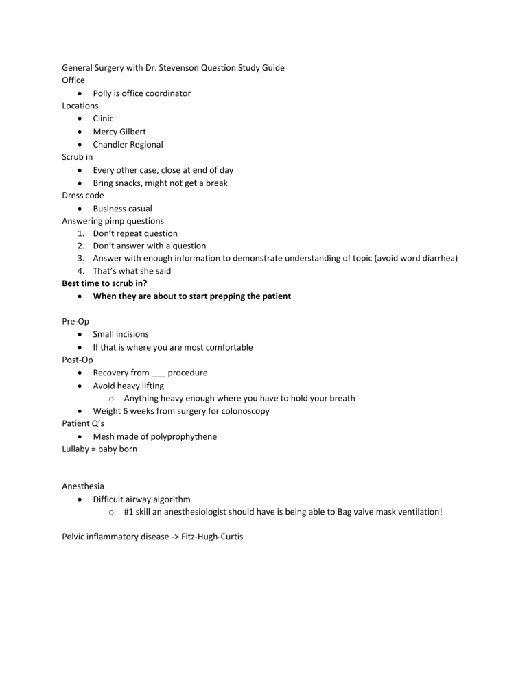
General Surgery with Dr. Stevenson Question Study Guide Office Polly is office coordinator Locations Clinic Mercy Gilbert Chandler Regional Scrub in Every other case, close at end of day Bring snacks, might not get a break Dress code Business casual Answering pimp questions 1. Don’t repeat question 2. Don’t answer with a question 3. Answer with enough information to demonstrate understanding of topic (avoid word diarrhea) 4. That’s what she said Best time to scrub in? When they are about to start prepping the patient Pre-Op Small incisions If that is where you are most comfortable Post-Op Recovery from ___ procedure Avoid heavy lifting o Anything heavy enough where you have to hold your breath Weight 6 weeks from surgery for colonoscopy Patient Q’s Mesh made of polyprophythene Lullaby = baby born Anesthesia Difficult airway algorithm o #1 skill an anesthesiologist should have is being able to Bag valve mask ventilation! Pelvic inflammatory disease -> Fitz-Hugh-Curtis Colon cancer -> mets to Liver (1st), lung (2nd) -Misc: Hypothetical situation -> milky fluid from pancreas -> check amylase/lipase The French scale or French gauge system is commonly used to measure the size of a catheter. It is most often abbreviated as Fr, but can often be seen abbreviated as Fg, FR or F. It may also be abbreviated as CH or Ch (for Charrière, its inventor). However, simply gauge, G or GA generally refers to Birmingham gauge. o The French size is three times the diameter in millimeters. Thus, the French size is roughly equivalent to the circumference of a circular catheter; the true circumference being slightly larger (circumference = diameter * π where π is approximately 3.14). o A round catheter of 1 French has an external diameter of 1⁄3 mm,[2] and therefore the diameter of a round catheter in millimetres can be determined by dividing the French size by 3: o 1 mm = Pi Fr o 1 mm = 3 Fr o 1 Fr = 1/3 mm o Co2 is used for insufflation because it is pretty absorbable and less likely to cause air embolism if vein is nicked. Treatment for air embolism includes Trendelenburg and high flow oxygen Falciform ligament attaches to liver and is a remnant of fetal umbilical vein For port placement: Subclavian central line borders o o o o o https://www.youtube.com/watch?v=_VYHj4sRlkc https://anwresidency.com/simulation/guide/subclavian.html In the normal variant of human anatomy, the subclavian vein occurs bilaterally and is a continuation of the axillary vein (a continuation of the brachial vein) from either upper extremity. At the lateral border of the first rib, the axillary vein becomes the subclavian vein where it passes over the rib in the groove of the subclavian vein. Just posterior to the subclavian vein in this area is the axillary artery which becomes the subclavian artery at the lateral border of the first rib and lies in the groove of the subclavian artery. The subclavian vein continues beneath the clavicle heading towards the sternal notch until at the medial border of the anterior scalene muscle it joins the internal jugular vein and becomes the brachiocephalic vein, also called the innominate vein. Also important to note is the pleural apex of the lung which lies inferior to the medial aspect of the subclavian vein. The left side pleural apex often projects more superiorly than the right leading to an increased risk of pneumothorax with left-sided access. The thoracic duct also terminates at the junction of the left subclavian vein and internal jugular vein. This is importantly related to subclavian venous access because it represents another area of o potential injury. A potential advantage of left-sided access is the easier sweeping curve of the left innominate vein that leads to the superior vena cava located in the right mediastinum https://www.ncbi.nlm.nih.gov/books/NBK482224/ o IJV borders o o Seldinger technique o Technique 1. desired vessel or cavity is punctured using a trocar (hollow needle) 2. soft curved tip guide wire is then inserted through the trocar and advanced into the lumen 3. guidewire is held secured in place whilst the introducer trocar is removed 4. large-bore sheath/cannula/catheter is passed over the guidewire into the lumen/cavity 5. guidewire is withdrawn leaving the introducer sheath in situ through which catheters and other medical devices can be introduced https://radiopaedia.org/articles/seldinger-technique?lang=us Pneumothorax Signs and symptoms 1. Tracheal deviation contralaterally Treatment 1. Needle decompression 2. Chest tube placement For hernia procedures you should know: -the anatomical structures and boundaries of the triangle of pain o The Boundaries are The Gonadal Vessels (Testicular artery and Vein) - Medially The Iliopubic Tract- Superiorly and the Peritoneal Reflection below. Contents of this triangle include Femoral branch of Genito femoral nerve, and Lateral cutaneous nerve of thigh. -the anatomical structures and boundaries of the triangle of doom o Triangle of Doom is an anatomical triangle defined by the vas deferens (male) or round ligament of the uterus (female) medially, spermatic vessels laterally and peritoneal fold inferiorly.[1] This triangle contains external iliac artery and vein, the deep circumflex iliac vein, the genital branch of genitofemoral nerve and hidden by fascia, the femoral nerve. o It bears significance in laparoscopic repair of groin hernia. Surgical staples are avoided here. Circle of death – corona mortis o The circle of death (corona mortis) refers to the anastomotic branches of vasculature in this region formed by the common iliac, internal iliac, obturator, inferior epigastric, and external iliac vessels o o -the anatomical structures and boundaries of hesselbach’s triangle o Medial border: Lateral margin of the rectus sheath.[1][2] o Superolateral border: Inferior epigastric vessels.[1][2] o Inferior border: Inguinal ligament.[1][2] o This can be remembered by the mnemonic RIP (Rectus sheath (medial), Inferior epigastric artery (lateral), Poupart's ligament (inguinal ligament, inferior). o The inguinal triangle contains a depression referred to as the medial inguinal fossa, through which direct inguinal hernias protrude through the abdominal wall o o o o o o o o o An appendix hernia is called Amyand hernia o The term used when someone has ipsilateral direct and indirect hernias at the same time o Pantaloon hernia o o o -know how to differentiate between a direct and indirect hernia o -medial vs lateral to the inferior epigastric vessels Direct Indirect Femoral Umbilical Direct through Hesselbach triangle Internal deep ring, along canal, out superficial ring. (external oblique muscle aponeurosis) Into the scrotum Below inguinal ligament Failure of the umbilical ring to close during fetal development causes congenital umbilical hernia. The midgut develops outside the abdominal cavity , inferior lateral to PT. Medial to the inferior epigastri c artery Medial to epigastric artery Lateral to epigastric artery. (INFERIOR EPIGASTRIC VESSELS) Requires surgical intervent ion (more incarcer ation/str angulati on) Older men: weak wall (transversalis fascia) Commoner in the young. M>F More common in women MDs don't LIe" (MedialDirect and LateralIndirect.) congenital defect of the processus vaginalis when it fails to close (can also form a hydrocele) herniated bowel is covered in external spermatic fascia alone Covered by all 3 layers of spermatic fascia The processus vaginalis descends anterior to the testis via the gubernaculum during embryonic development. Failure of this conduit to obliterate after testicular descent into the scrotum causes outpouching of the parietal peritoneum (and bowel) through the deep inguinal ring, the inguinal canal, and the superficial inguinal ring, leading to an indirect inguinal hernia. These hernias are located until the second trimester, when it physiologically herniates back into the abdomen. If the umbilical ring fails to close or the fascia in this region is underdeveloped, abdominal content may bulge through the umbilicus. Umbilical hernias are more common in children with chromosomal abnormalities (e.g., Down syndrome, Edwards syndrome) or congenital hypothyroidism, as seen here. Most congenital umbilical hernias resolve spontaneously by 5 years of age. outside the Hesselbach triangle, lateral to the inferior epigastric vessels, and can manifest with a communicating hydrocele, as seen in this patient. -the layers of the abdominal wall below the arcuate line o Components of the anterior abdominal wall o Layers (from superficial to deep) 1. Skin and subcutaneous tissue 2. Superficial fascia 3. Superficial fatty layer (Camper fascia) 4. Deep membranous layer (Scarpa fascia) 5. External oblique muscle 6. Internal oblique muscle 7. Transversus abdominis muscle 8. Deep fascia (transversalis fascia): fuses with the deep fascia of the thigh (fascia lata) 9. Preperitoneal adipose tissue 10. Parietal peritoneum o For lap choles you should know: -how to define the critical view o Critical view of safety: method for secure identification of the cyst duct and cystic artery during laparoscopic cholecystectomy o https://www.youtube.com/watch?v=K62kqwDjY_Q o https://www.sciencedirect.com/science/article/pii/S221026121400368X o https://ales.amegroups.com/article/view/3940/4771 o https://www.sages.org/safe-cholecystectomy-program/ -the structures of the portal triad o The portal triad contains the extrahepatic segments of the portal vein, hepatic artery, and bile ducts. o The portal triad is contained within the hepatoduodenal ligament and contains the portal vein (posterolateral), hepatic artery (medial), and bile ducts (lateral) o -that you need to be careful not to damage the hepatic duct o -the ligaments of the liver o -also note the pars flaccida of the hepatogastric ligament o o o o o o o The hepatogastric ligament or gastrohepatic ligament connects the liver to the lesser curvature of the stomach. It contains the right and the left gastric arteries. In the abdominal cavity it separates the greater and lesser sacs on the right. It is sometimes cut during surgery in order to access the lesser sac. The hepatogastric ligament consists of a dense cranial portion and the caudal portion termed the pars flaccida. -blood supply to the gallbladder o -important lymph nodes of the gallbladder o o Cystohepatic triangle of calot For bariatric surgeries you should know: -all of the blood supply to the stomach o -identify the most likely locations for H. pylori infection Antrum H. pylori (Helicobacter pylori) are bacteria that can cause an infection in the stomach or duodenum (first part of the small intestine). It's the most common cause of peptic ulcer disease. o For any cancers you will be asked: -the size of the margins to be excised o -6-8 cm for gastric o -5 cm for colorectal o -5 cm proximal and 1 cm distal for rectal -know how many lymph nodes must be excised o -15-16 minimum for gastric o -12 for colorectal The National Quality Forum specifies that the presence of at least 12 lymph nodes in a surgical resection is one of the key quality measures for the evaluation of colorectal cancer. Therefore, the harvesting of a minimum of twelve lymph nodes is the most widely accepted standard for evaluating colorectal cancer. https://www.hindawi.com/journals/grp/2018/1985031/ o Types of GI cancers o Gastrointestinal stromal tumor (GIST) most common in the stomach but can be found throughout GIT Size of margins: **** Treatment: Resection and Imatinib (Gleevec) [Tyrosine kinase inhibitor of the cKIT receptor) Presentation Overt or occult GI bleeding – 28 percent (small intestine) and 50 percent (gastric) Incidental finding (asymptomatic) – 13 to 25 percent Abdominal pain/discomfort – 8 to 17 percent Acute abdomen – 2 to 14 percent Asymptomatic abdominal mass – 5 percent -know the management of rectal cancer o -rectal cancer is primarily done by neoadjuvant therapy first Neoadjuvant chemotherapy is chemotherapy that a person with cancer receives before their primary course of treatment. The aim is to shrink a cancerous tumor using drugs before moving onto other treatments, such as surgery. Neoadjuvant chemotherapy helps doctors target cancerous growths more easily at a later stage o -major rectal cancer procedure types and the different indications for each -TES Transanal endoscopic surgery (TES) is an emerging technique that offers transanal access to resecting benign, premalignant, or early malignant lesions in the mid - to proximal rectum -TME Total mesorectal excision (TME) is a common procedure used in the treatment of colorectal cancer in which a significant length of the bowel around the tumor is removed. TME addresses earlier treatment concerns regarding adequate local control of rectal cancer when an anterior resection is performed -LAR Low anterior resection (LAR) is a surgery that's done to treat rectal cancer. During LAR surgery, the part of your rectum with the cancer will be removed. The remaining part of your rectum will be reconnected to your colon. You'll be able to have bowel movements (poop) as usual once you recover from your surgery. -APR An abdominoperineal resection (APR) is a surgery in which the anus, rectum and sigmoid colon are removed. This procedure is most often used to treat rectal cancers located very low in the rectum. Often this surgery occurs after you have completed radiation and/or chemotherapy treatments. Bariatric surgeries https://asmbs.org/patients/bariatric-surgery-procedures Sleeve Gastrectomy Roux-en-Y Gastric Bypass Adjustable Gastric Band Biliopancreatic Diversion with Duodenal Switch Single Anastomosis Duodeno-Ileal Bypass with Sleeve Gastrectomy Goodsall’s rule PART 2 ● Know subcutaneous and subcuticular closures ● ● ● Know abdominal compartment syndrome ○ Abdominal compartment syndrome (ACS) occurs when the abdomen becomes subject to increased pressure reaching past the point of intra-abdominal hypertension (IAH). ACS is present when intra-abdominal pressure rises and is sustained at > 20 mmHg and there is new organ dysfunction or failure. Liquid diet -> shrinks liver Hiatal hernia -> tighten Ghrelin decreased, restriction with sleeve, issues with malabsorption H. pylori - Incisura angularis (angular notch), lesser curvature of stomach before the pylorus - Typical site for H. pylori - Can also be in the pyloric antrum or body of the stomach Laparoscopic Cholecystectomy - laparoscopic is preferred due to less postoperative pain, better cosmetic, short hospital stay Indications - symptomatic cholelithiasis w/ or w/o complications - asymptomatic cholelithiasis in patients’ risk for gallbladder carcinoma/gallstone complication - acalculous cholecystitis - gallbladder polyps >0.5cm - porcelain gallbladder Contraindications - absolute: inability to tolerate anesthesia, peritonitis w/ hemodynamic compromise, refractory coagulopathy - relative: previous abdominal surgery, pregnancy, morbid obesity and severe comorbidities Preoperative - elevated total bilirubin, ALK PHOS intraoperative - critical view of safety - Lap Chole’s ○ ○ Safe method for gaining sufficient view of Calot’s triangle identification of cystic duct, cystic artery and common hepatic duct, it’s the critical view so that you won’t accidentally cut the CHD ○ you need to see this view before taking out the gallbladder= if cant: cholangiography/open procedure ○ gallbladder extraction via umbilical insicion ● calot's triangle ○ Cystic duct (CD) ○ Common hepatic duct ○ Cystic artery (CA) ● Calot’s node (adjacent and anterior to the cystic artery) Complications - Vascular injury, gallbladder perforation, mesenteric injury, bile duct injury Post complications - Bile duct injury, bile leaks, bleeding, bowel injury , retained CBD stones US findings for cholecystitis: stones/sludge, GB wall thickening >3mm, pericholecystic fluid, dilation of the CBD - Nuclear cholescintigraphy/magnetic resonance cholangiopancreatography (MRCP), endoscopic retrograde cholangiopancreatography (ERCP) Triangles of doom/pain for inguinal hernias ● angle of His ○ Esophagogastric angle ○ Can be obtuse in hiatal hernia ○ Acid reflux barrier?? Laparoscopic Inguinal Hernia Repair - Lap. surgery is approached form the posterior aspect and repair involves placing the mesh in the preperitoneal space - Two approaches transabdominal preperitoneal hernia repair (TAPP) and totally extraperitoneal hernia repair (TEP) TEP TAPP - Performed in the preperitoneal space (trocar never enter the intraabdominal cavity). Space is made b/w the peritoneum and the anterior abdominal wall so that the perineum is not damaged during the repair Mesh is placed in front of the inguinal canal You enter into the abdominal cavity and make an incision into the preperitoneum Repair involves the placement of mesh in a preperitoneal (inside the perineum) which the periteneum keeps the mesh away from bowel Damage intra-abdominal organs, adhesions resulting in intestinal obstruction or bowel herniation Complications -intraabdominal adhesions ● TAPP vs TEP procedures for inguinal hernias ○ TEP = totally extraperitoenal ■ Peritoneal cavity not entered; mesh used to seal hernia from outside ○ TAPP ■ Mesh through the peritoneal cavity, mesh used to seal hernia from outside Indication - Complicated hernia (develops bowel strangulation/bowel obstruction/incarcerated hernia that is not reduceable) - Inguinal hernia w/ moderate/severe symptoms - Acute incarcerated inguinal hernia w/ failed reduction - Femoral hernias - Hernia that causes groin pain w/ exertion, inability to perform daily acitivities due to pain/discomfort from the hernia, inability to reduce the hernia (chronic incarcerated) - Uncomplicated hernias (not causing pain/issues)= watchful waiting Contraindicated - Patients that can’t tolerate general anethesia, patient’s that may be active groin infections/systemic sepsis, ascites, pregnant patients, prior pelvic surgeries in the preperitoneum space Mesh -polypropylene w/ oven mesh (Marlex, Prolene, SurgiPro) is tacked on the abdominal wall Complications - Persistent groin pain, post-surgical hernia neuralgia, testicular complications (interference with the blood supply to the testicles/damage to the vas deferens), sexual dysfunction, mesh infection/migration/erosion into adjacent structures Laparoscopic ventral hernia repair -abdominal hernia is an organ that project through the body wall, usually through incisional sites but can include epigastric, umbilical Indication -incarcerated/strangulated hernia, symptomatic ventral hernia Contraindications -inability to tolerate pneumoperitoneum or unable to access the perineum -active skin infection Complications - Postoperative pain due nerve entrapment - Wound complications - Iatrogenic enterotomy - Hernia recurrence Open LAP for inguinal hernia repair - You approach from the outside-in - Can damage the ilioinguinal nerve (runs with the spermatic cord) - Mesh is attached to the abdominal wall (internal oblique muscle) and the external oblique muscle is closed over the hernia - Tacked in the medial pubicle ligament, laterally to the cojoined tendon o Indications - Same as the other hernias indications Contraindications - Inability to tolerate the anesthesia, coagulopathy, BMI>35 , tobacco use Complications - Recurrence of hernia, pain due to nerve damage ● ● ● ● Know complications/indications for each procedure. Engage the anesthesiologists and they will let you intubate. blood supply to all abd organs different fundoplication procedures - An inguinal/direct hernia at the same time is called pantaloon hernia A hernia from the appendix is called Amyand hernia Appendectomy o Keep appendix in vs taking it out o Must be <65 o Must be in hospital The 5 W's of post-op fever - Wind - atelectasis or pna - 24-48 hrs - Wound - infection >day 3, staph infection MC Water- UTI’s, 2-3 days (or IV line infection) Walking – DVT/ thrombophlebitis Wonder drugs/Whopper - check all the pt.’s meds (anesthetic, antibiotics, salfa) Parkland formula - The Parkland formula for the total fluid requirement in 24 hours is as follows: 4ml x TBSA (%) [think rule of 9s] x body weight (kg); 50% given in first eight hours; 50% given in next 16 hours https://www.youtube.com/watch?v=iHsilm0Ybxw ● colorectal cancer facts; → need 5-7cm minimum for colectomy, minimum 12 lymph nodes Colorectal carcinoma - painless bleeding and change in bowel habits in a patient 50-80Y - you will see apple core lesion on barium enema, adenoma is the MC “premalignant” and evolve to adenocarcinoma of the colon - Tumor marker: CEA - If malignant: sessile, >1cm and villous - Less malignant: pedunculated, <1cm and tubular - 25% change of an asymptomatic person that goes for colon screening may have an adenoma, increases with age. Tx - Resection and chemo Colonoscopy guidelines ● ● IBD facts sources and % blood to the liver ○ Blood Supply to the Liver Portal Triad - R/L hepatic duct “biliary duct”, R/L hepatic artery, portal vein - Drains at the hepatic vein then into the inferior vena cava - Blood supply starts at celiac trunk-common hepatic artery-proper hepatic artery-R/L hepatic artery - Cystic artery “blood supply to the gallbladder” branches off the R. hepatic artery - Located in the hepatoduodenal ligament Laparoscopic Colectomy: Indications: Benign & malignant conditions Contraindications: Emergent (obstruction, perforation, massive bleeding) Complications Vascular injury (MC during abdominal access) Laceration of inferior epigastric artery Abdominal wall hematoma Hemorrhage Mechanical compression and application of hemostatic agents are appropriate initial strategies Clips, suture ligation, electrosurgical methods Failure to convert to open procedure with hemorrhaging Damage to organs like stomach can be avoided with decompression (NG or OG tube) Nerve injury, surgical site infection, upper urinary tract injury, port site metastasis Anesthesia Port-a-cath - Central venous access, can allow other device access: pulm. art. Cath, plasmapheresis cath, hemodialysis cath, extracorporeal life support cannula, inferior vena cava filter Can get access internal jugular vein, subclavian vein, illiac vein, common femoral vein, brachicephalic vein, superior/inferior vena cava Lateral clavicle Finger in substernal notch, thumb out along clavicle Indicated Sick people Inadequate peripheral venous access (infusion regime- long term admin. Of continuous meds, chemotherapy, parenteral nutrition Hemodynamic monitoring (central venous pressure, venous oxyhemoglobin sat., cardiac parameters Extracorporeal therapies- large bore venous access (hemodialysis/plasmapheresis) Procedures: vena cava filters, venous thrombolytic therapy, pul art cath, pacemakers/defibrillators Contraindicated Coagulopathy/thrombocytopenia, INR >3, if emergency do it but get platelets/fresh frozen plasma Site complications: skin infection, proximity to wound/burn/tracheostomy Complications Bleeding, arterial puncture, air embolism, thoracic duct injury, cath. Malposition, Pneumothorax/hemothorax S/S Pnuemo -> JVD, normal heart, decreased breath sounds, hyperresonance, tracheal deviation contralterally. Chest tube/Needle decompression, Midclavicular line 2nd intercostal space Arrhythmia - Triggering SA node → A-fib Different thyroid cx Thyroid follicular epithelial derived cancers (4 types) Papillary (85%), follicular (12%),=differentiated cancers anaplastic (undifferentiated)-3%, medullary thyroid cancer Cancers that metastasize to the thyroid: breast, colon, renal and melanoma Surgery for differentiated thyroid cancers “papillary/follicular” and after endocrinologist handle them Preoperative: U/s evaluation of the central and later neck lymph nodes Decision based on side of the tumor, <1cm w/ no extrathyroidal extension/no lymph=thyroid lobectomy, tumor >1-4cm= total thyroidectomy w/ possible radioiodine therapy to ablate thyroid tissue Post-operative Thyroid medication “levo” if they are not going to get ablation/radioiodine scanning Obtain serum TSH/non stim serum thyroglubulin (Tg) in 4-6wks after postop to determine status <5ng/mL after thyroidectomy, <30ng/mL after thyroid lobectomy If high, suspect metastasis Complications Hypoparathyroid, recurrent laryngeal nerve injury F/U Ultrasounds, TSH and Tg to monitor thyroid activity Goal in high risk <0.1 TSH, low risk 0.1-0.5 TSH - TSH suppression leads to menopause, tachycardia, osteopenia, osteoporosis, a-fib What is shock? Lack of tissue oxygenation -> Anaerobic metabolism -> make lactic acid (metabolic acidosis) How to determine hemodynamic stability o Urine (non-invasive) o Lactic acidosis o Base deficit o Blood pressure Give LR (crystalloid) 1st as fast as you can Different types of shocks and tx circulatory failure no getting enough blood delivered to the tissues=tissue hypoxia- multiple organ dysfunction= death Distributive shock (systemic peripheral vasodilation) Septic shock: MC, dysregulated response to infection leading to organ failure Gram+ bac MC cause S. Entero Sys. Inflam. Resp. Synd. (SIRS): robust inflammatory response- can be infectious or non-infectious Pancreatitis, burns, hypoperfusion due to trauma, blunt trauma, amniotic fluid embolism, air embolism, fat embolism, idiopathic systemic capillary leak syndrome Neurogenic shock: severe TBI, spinal cord injuries, interrupted autonomic pathways causing decreased vascular resistance and altered vagal tone Anaphylactic shock Drug/toxin induced shock Endocrine shock Cardiogenic shock Cardiomyopathic Arrhythmic Mechanical Hypovolemic Hemorrhagic Non-hemorrhagic Obstructive Pulmonary vascular Mechanical FAST (focused assessment of U/S for Trauma) Helps identify free intraperitoneal, pericardial fluid, hemoperitoneum, pneumothorax, hemothorax, hemopericardium w/ or w/o tamponade, traumatic hypovolemia, rib fractures in blunt trauma injuries. Indications: blunt trauma, penetrating cardiac/chest trauma, trauma in pregnancy, pediatric trauma, undifferentiated hypotension, patients with ascites. 10 structures/spaces are imaged in 4 windows. 1. Cardiac (subxiphoid) Percardium, heart chambers (right ventricle) 2. RUQ Morrison pouch (hepatorenal recess) Liver tip (right paracolic gutter) Lower right thorax 3. LUQ Subphrenic space Splenorenal recess Spleen tip (left paracolic gutter) Lower left thorax 4. Pelvic Rectovesical pouch (male patients) Rectouterine “Pouch of Douglas” (females) Fluid Replacement Normal saline Lactated Ringers PORT Vagus Nerve Stim. for epilepsy Indicated when the patient has resistance/refractory antiseizure drugs= usually refractory to three drugs and having more than 6 seizures/mo Mechanism of effect is not well established but think afferent vagal projects through the pontine parabrachial nucleus and thalamus to the seizure generating regions in the basal forebrain/insular cortex Effects the locus ceruleus (recieves vagal afferent signal from nucleus tractus solitarius) Dysynchronization of hypersynchronized cortical activity which is dependent on the stimulant frequency/strength of the electrical current Cortical inhibition secondary to the release of inhibitory NTs such gylcine/GABA Indicated Children >12Y/adults in well documented medically refractory seizures who do not want intracranial surgery/are not candidates for surgery/refractory to antiseizure drugs Focal/generalized seizures (>6/mo)= receiving high stimulation experienced >50% reduction in seizures Contraindicated Cardiac condition disorders “arrhythmias” due to potential efferent conduction through the vagus nerver, especially on the right side can worsen cardiac conduction “think bradycardia” Sleep apnea Procedure Cricoid cartilage is palpated- halfway b/w sternal notch and mandible Incision is made 3cm medial to the SCM Incision for the battery device in made 4cm (3-4fingers inferior to the L infraclavicular medial to the sternum subQ Device Battery powered device similar to a cardiac pacemaker, stimulating leads are surgically placed around the left vagus nerve in the carotid sheath (behind the SCM (internal carotid artery, internal jugular vein, vagus nerve), and connected to an infraclavicular subcutaneous programmable pacemaker Stimulation consist of a 30Hz stimulus delivered 30 seconds every five minutes It detects high heart rate which is associated when a seizure occurs and the device detects and stimulates the vagus nerve to disrupt the seizure. Pt that have auras, on demand stimulation can be used with a magnet Complications - Voice alterations (hoarseness, throat pain, cough, SOB, tingling, muscle pain) Implant site infection (staph aureus) Bradycardia Vocal cord paralysis “manipulation of the recurrent laryngeal nerve) NIH Criteria for Weight Loss Adults that have a BMI >40, BMI of >35 w/ comorbidities DMT2, heart disease, GERD or sleep apnea, BMI >30 with DMT2 that is difficult to control w/ meds/lifestyles changes Restrictive procedures Limit caloric intake by reducing the stomach’s reservoir capacity via resection, bypass, creation of a proximal gastric outlet Vertical banded gastroplasty/laparoscopic adjustable gastric band= limit solid food intake by restriction of the stomach size Malabsorptive Shortening the small intestine by decreasing the effectiveness of nutrient absorption Combination of restrictive and malabsorptive Roux en Y gastric bypass Small proximal pouch (30mL) that is divided and separated from the stomach and anastomosed to a roux limb of small bowel that is 75-150cm in length- helps to restrict caloric intake. Major digestion and absorption occurs in the common channel (gastric acid, pepsin, intrinsic factor, pancreatic enzymes and bile mix with ingested food The small intestine is divided at a distance of 50-150cm distal to the ligament of treitz Biliopancreatic limb and roux limb are connected 75-150cm distally from the gastrojejunostomy (connection of stomach pouch and jejunum) Side effects Lightheadedness, nausea, diaphoresis, abdominal pain, diarrhea with a high sugar meal in ingested Hormones Ghrelin, a peptide hormone is secreted in the foregut (stomach/duodenum) that stimulate the early phase of meal consumption. Its inhibited in gastric bypass which leads to inhibit appetite GLP-1 and CCK are increases after RYGB Weight loss 70% weight loss after two years Restrictive procedures Sleeve gastrectomy Majority of the greater curvature/fundus of the stomach is removed and a tubular stomach is created The tubular stomach is small in its capacity and is resistant to stretching due to the absence of the fundus, aiding in weight loss Creating this type of procedure causes high pressure= GERD, leaks Hormones Ghrelin decreases (found in the fundus), GLP (secreted in the SI) , PKK increase (peptide YY) Weight loss 60% weight loss after two years Relative Contraindications Anesthesia risk, severe uncontrolled psychiatric illness (eating d/o), and coagulopathy, Barrett’s esophagus, uncontrolled GERD Pre-operative EGD to exclude ulcers, polyps, masses, dysplastic changes Complications -bleeding (w/i the greater curvature of the stomach- short gastric art., spleen injury) -leak from the staples and high pressure in the stomach, usually in the gastroesophageal junction (eso-stomach junction) -strictures -GERD -portal vein thrombosis Malabsorption Biliopancreatic diversion with duodenal switch, used in super obese >50 or reserve for revisional procedures for failure to lose weight Gastric sleeve removal Remove the 2st part duodenum Distal ileum is anastomosed to the stomach and the proximal ileum, with the output from liver, pancreas, and duodenum (biliopancreatic limb) anastomosed to the terminal ileum 50-100cm away from the ileocecal valve Fat/starch malabsorption 70-80% weight loss Single anastomosis doudenoileal bypass with sleeve gastrectomy (SADI-S) -variant of BPD-DS but with the preservation of the pylorus Stomach Blood Supply Foregut Stomach, esophagus, proximal duodenum, liver, gallbladder and pancreas get their supply from CELICAC truck (T12), there is also the superior mesenteric artery (SMA-L1)) and inferior mesenteric artery (IMA-L3) Coming off the celiac truck, the left gastic artery anastamose with common hepatic artery to supply the lesser curvature of the stomach Drain in the gastric/splenic veins Child Pugh Score for Cirrhosis Mortality Progressive hepatic fibrosis that disrupts the function of the liver and is considered irreversible in late stages Complications (decompensated cirrhosis) Variceal hemorrhage, ascites, spontaneous bacterial peritonitis, hepatic encephalopathy, hepatocellular carcinoma, hepatorenal syndrome, hepatopulmonary syndrome Operative High risk complications Classification Serum albumin, bilirubin, ascites, encephalopathy, and prothrombin time. Score is from 5-15. 5-6= Child Pugh Class A cirrhosis (compensated), 7-9 = Child Pugh Class B cirrhosis (significant functional compromise) and 10-15 = Child Pugh Class C cirrhosis (decompensated cirrhosis) Class A mortality 10%, Class B mortality 30% mortality, and Class C 82% mortality Also classifies mortality if the patient does not do surgery 1yr survival with class A/B/C= 100%, 80% and 45% MELD score Predicts the prognosis in patients with cirrhosis and prioritization of patient’s waiting for liver transplant Cervical Lymph Node Excision Primary/secondary lymphedema Indications Localized primary lesions (microcytic/macroscopic lymphatic malformations) Failed nonoperative management Recurrent cellulitis Leakage of lymph in the body cavities, organs or extremities Limitations of function Deformity or disfigurement Pain Diminished QOA Staging of lymphedema “Campisi staging system” Operation approach, physiologic vs. reductive techniques Physio- used in early stage lymphedema prior to deposition of excess fat and tissue fibrosis (campisi stage 1/2/3) Reductive- used in advance lymphedema (stage 4/5) Complications Pain, wound healing complications, infections, lymphatic fistulas, suboptimal cosmetic results
