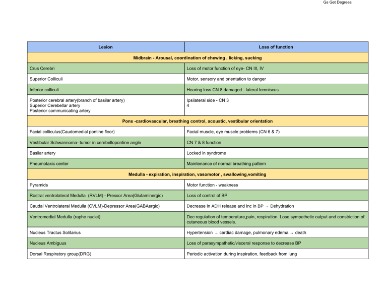
Gs Get Degrees Lesion Loss of function Midbrain - Arousal, coordination of chewing , licking, sucking Crus Cerebri Loss of motor function of eye- CN III, IV Superior Colliculi Motor, sensory and orientation to danger Inferior colliculi Hearing loss CN 8 damaged - lateral lemniscus Posterior cerebral artery(branch of basilar artery) Superior Cerebellar artery Posterior communicating artery Ipsilateral side - CN 3 4 Pons -cardiovascular, breathing control, acoustic, vestibular orientation Facial colliculus(Caudomedial pontine floor) Facial muscle, eye muscle problems (CN 6 & 7) Vestibular Schwannoma- tumor in cerebellopontine angle CN 7 & 8 function Basilar artery Locked in syndrome Pneumotaxic center Maintenance of normal breathing pattern Medulla - expiration, inspiration, vasomotor , swallowing,vomiting Pyramids Motor function - weakness Rostral ventrolateral Medulla (RVLM) - Pressor Area(Glutaminergic) Loss of control of BP Caudal Ventrolateral Medulla (CVLM)-Depressor Area(GABAergic) Decrease in ADH release and inc in BP → Dehydration Ventromedial Medulla (raphe nuclei) Dec regulation of temperature,pain, respiration. Lose sympathetic output and constriction of cutaneous blood vessels. Nucleus Tractus Solitarius Hypertension → cardiac damage, pulmonary edema → death Nucleus Ambiguus Loss of parasympathetic/visceral response to decrease BP Dorsal Respiratory group(DRG) Periodic activation during inspiration, feedback from lung Gs Get Degrees Ventral respiratory group(VRG) Respiratory rhythm generation decreases control of expiration(dorsal VRG) inspiration(rostral VRG) Pre-Botzinger complex in medulla Respiratory rhythm generator Phrenic Nucleus Diaphragmatic contraction Internal capsule (corticospinal tract) - projection tract Opposite side motor function is weak Cerebellum- compares motor output with the sensory feedback. Coordinated and steady movement Cerebellum - Flocculus(CN 8) Uncoordinated, unsteady movement Lateral cerebellum Fine precise control of movements of fingers Medial cerebellum (vermis) Up and down movements - control of trunks and limbs Tonsil herniation Brain herniates through the foramen magnum Vermis herniation Prone to damage medulla Hypothalamus - integrates endocrine, behavioral info Posterior Hypothalamus (normal function = Increases body temperature) “it’s hot in the back” Decreased body temperature Hypothermia(decrease in metabolism and motor activity,peripheral vasodilation due to anterior hypothalamus intact) Anterior Hypothalamus(normal function = decreases body temperature) Increased body temperature Hyperthermia (inc metabolism, shivering, peripheral vasoconstriction -posterior hypothalamus still intact) Ventromedial hypothalamus (normal function = inhibits feeding) obesity Lateral hypothalamus(normal function = increases feeding) Decreased feeding (inhibitory neuronal link between arcuate nucleus and lateral hypothalamus → suppresses food consumption → weight loss) Pterygoid canal 1. 1. vasodilation (b/c sympathetics is lost (deep petrosal nerve)) no secretion from nasal, palatine, and lacrimal gland Gs Get Degrees 2. loss of general sense and taste sensation of palate (damaged Greater petrosalanterior 2/3 tongue gone) Brain swelling Uncus compresses CN III - dilated pupils, sleepiness Lenticulo striate artery(branch of MCA) Paralysis of upper body Intracerebral hemorrhage → obstructive hydrocephalus(Inc ICP) Corticobulbar tract Loss of motor function of CN Suprasacral spinal transection(Above T12) Automatic bladder- bladder is filled until a threshold is reached → reflexing emptying Midsacral afferents (cauda equina) Atonic bladder → loss of afferents - bladder is filled and leads to urinary retention due to loss of reflexive arc(micturition center) → urine dribbling Lateral brain- MCA dominant Language deficit(understanding decreases-Wernicke’s and- Broca’s), weak mouth and face Medial brain- ACA dominant in anteromedial PCA is posteromedial ACA- left Leg, foot weakness, loss of voluntary control of bowel and bladder (incontinence) PCA - CN3 - Central vision is preserved but cannot see periphery, amnesia Anterior spinal artery (ventral 2/3rd of spinal cord) - somatosensation (Lateral column) Cannot tell pain and temperature(ASA) Paralysis(lateral column) Posterior spinal artery Dorsal column of the ipsilateral side Herpes Temporal lobe Prolactinoma -pituitary gland tumor Compresses optic chiasm - Decrease in endocrine cells,Optic nerve dysfunction Fornix (connects hippocampus to mamillary body) Amnesia, Korsakoff Dorsal-column medial lemniscal system Detect position, vibration Spinothalamic tract Pain and temperature Corticospinal(cortical layer V) Voluntary movement -strength decreases Circumventricular organs - Subfornical organs, organum vasculosum lamina terminalis Decreased secretion of ADH, decreased control of regulation of electrolytes Amygdala Decreased emotion and fear response No response to olfactory stimulus Gs Get Degrees Solitary Nucleus Decreased response to pain,temperature and special afferents(taste) from CN 7,9,10 Anterior Paracentral Weakness in contralateral lower extremity Mastoiditis Petrous part of temporal bone Inferior frontal gyrus(pars operacularis and triangularis) Lose Broca’s speech area Lateral funiculus Weakness in arm and leg Middle meningioma Parieto-occipital sulcus Forebrain Autonomic control● Circumventricular organs - Subfornical organ , organum vasculosum of lamina terminalis Lack BBB Detect electrolyte changes - project to hypothalamus e.g. Regulate ADH secretion ● Insular cortex: Receives visceral pain, temperature, taste sensations via thalamus and integrate with emotion ● Anterior cingulate cortex: anterior portion of the limbic lobe. Controls autonomic output via connections with insula, prefrontal cortex, amygdala, hypothalamus & brainstem. ● - Amygdala - regulates stress and fear response. Receives direct input from olfactory system & other solitary nucleus Output fibers to hypothalamus via stria terminalis & to brainstem including periaqueductal grey & reticular formation ● Hypothalamus : integrates autonomic, endocrine, motivated behavior. Basic life processes - BP, body temp, energy metabolism, reproduction, emergency response Hypothalamic control of anterior pituitary Parvocellular neuroendocrine cells within Paraventricular, arcuate nucleus → terminate in primary capillary plexus of superior hypophyseal artery of the infundibulum(pituitary stalk). Pathway is called tubero-infundibular tract Primary capillary plexus → neuroendocrine substances enter blood, neurohormones of post pituitary act on non endocrine cells. ---> portal veins → anterior pituitary → secondary capillary plexus Gs Get Degrees ● Feeding behavior regulated by hypothalamus Ventromedial nucleus of medial hypothalamus - suppresses feeding Lateral hypothalamus - promotes feeding(inhibited by leptin release from adipose tissue) ● - Regulation of body temp Anterior hypothalamus : dec body temp(integrate autonomic control - dilation of skin arterioles, sweating; motivated behavior-seeking cooler environment, remove clothing) Posterior : inc body temp(constriction of skin arterioles; motivated behavior-shivering,seeking warmer environment, add clothing) Autonomics Lecture 20 ● Bladder control : CNS at supratentorial level, posterior fossa and spinal cord Gs Get Degrees Lecture 21 : Neurons and Glia ● ● ● ● Primary sensory afferents : pseudounipolar Central or peripheral special sensory : bipolar Long axons(golgi type 1) e.g. motor neurons, short axons(golgi type II) e.g. local interneurons - multipolar ● - Radial Glia - neuronal, astrocytic, oligodendrocyte progenitors. Scaffold for neuronal migration Become bergmann glia in cerebellum Become muller cells of the retina ● Astrocytes: Regulate Ions Synaptic transmitters Regional cerebral blood flow Neuroprotection: BBB Limit oxidative damage Supply lactate Release gliotransmitter Form gliotic scars ● Microglia: immunocompetent and phagocytic - protect neurons from micro-organisms and toxic effects of cellular debris. Secrete neurotrophic or neuron survival factors upon activation Release cytotoxic molecules : pro-inflammatory cytokines, reactive oxygen intermediates, proteases Contribute to pathological neuronal degeneration Gs Get Degrees - Tanycytes: derived from radial glia. Functional interface between blood and CSF around 3rd ventricle Present near circumventricular organs. Also secrete CSF and transport of hormones Blood Supply: Circle of WIllis - surrounds optic chiasm,optic tract, mammillary body, ventral hypothalamus - anastomosis at diencephalon Circle of Willis is formed by proximal branches of posterior cere- bral artery, posterior communicating arteries, a part of internal carotid artery prior to its bifurcation, proximal part of anterior cerebral artery, and anterior communicating arteries Brachiocephalic trunk→ right common carotid Aortic arch → left common carotid Common carotid → internal and external division at the thyroid Internal carotid artery→ ophthalmic, anterior choroidal, p osterior communicating artery branches → middle cerebral artery (terminal branch of ICA) : runs laterally over insula & supplies temporal & parietal lobes(Wernicke’s, Broca’s) → L enticulo striate artery supplies basal ganglia, internal capsule(rupture=intracerebral hemorrhage) →A nterior cerebral artery (terminal branch of ICA):runs medial to optic chiasm and supplies frontal Anterior communicating artery(common spot for aneurysm) supplies frontal, parietal lobes, septum pellucidum, corpus callosum lobe. Subclavian artery → Vertebral artery → Posterior inferior cerebellar arteries(PICA) & Anterior spinal artery (ASA) supplies pyramids, PSA(supplies dorsal column) → combine to form B asilar artery → A nterior inferior cerebellar artery(AICA)(CN 6,7&8) , Labyrinthine artery, Pontine, superior cerebellar, posterior cerebral(pons and midbrain junction) Midbrain: 1. Posterior cerebral artery 2. Superior cerebellar artery 3. Posterior communicating artery Pons : Long and short circumferential, paramedian branches of basilar artery Medulla : 1. Posterior inferior cerebellar 2. Anterior inferior cerebellar(CN 7&8), 3. ASA (medial area) ● ● Interhemispheric : Corpus callosum Intrahemispheric : association e.g arcuate fasciculus Gs Get Degrees Lecture 24: ● Hyperosmolality weakens BBB but its reversible so it can permit the delivery of lipid-insoluble drugs to the CNS ● ● ● - Sensory receptors express receptor potential Other neurons express postsynaptic potentials Greater length constant → lesser decrement. (increase in diameter) Lower intracellular resistance Greater membrane resistance ● - Initiation zone: populated by voltage gated sodium channels Multipolar: IZ is at the axon hillock and origin of graded potential: dendrites bipolar : IZ and graded receptor potential at the presynaptic terminal ● ● Compared to plasma - CSF has high Cl- , Mg2+ and high to normal Na+ Lumbar Puncture for adults : L3/L4. For children L4/L5. Measure CSF pressure - normal 65-200mm H2O (<15mmHg) ● Glucose and L-DOPA cross BBB through facilitated diffusion but dopamine weakly accesses the brain ● a) b) c) d) e) f) g) Factors that weaken BBB: Hypertension Hyperosmolarity : may permit delivery of lipid-insoluble drugs to the CNS Trauma Ischemia Inflammation Pressure infection Gs Get Degrees ● Sensory neurons- IZ is close to sensory ending Lidocaine: block voltage gated Na+ channels - disrupts AP (not graded potentials) → treat pain Botulinum toxin : blocks Ach release. Binds to cholinergic nerve endings after ingestion → enters the cell via retrograde transport(dynein driven) → suppresses docking by cleaving synaptobrevin → prevents vesicular release of ACH irreversibly → weakness!! Tetanus toxin : enters PNS via wound → retrograde transport → enters gkycinergic interneurons → suppresses docking by cleaving synaptobrevin → irreversible suppression of glycine release → disinhibits LMN → m uscular spasm “opisthotonus” Aminoglycoside antibiotics: Neomycin inhibits exocytosis Ach at motor nerve terminals → block presynaptic calcium channels. Effects of both antibiotics are reversed by elevated extracellular Ca2+ Gs Get Degrees 4-Aminopyridine : K+ channel blocking drug prolongs duration of the impulse → enhances Ca2+ entry into motor nerve endings → Inc AcH release → Inc quantum content GABAa binding agonist Benzodiazepines Barbiturates Neurotransmitter Achetylcholine Nuclei ( → projects into) 1) 2) Glutamate Receptor Basal forebrain: Basal nucleus of Meynert → cortex, limbic system - involved with Alzheimer's Dorsolateral pontine tegmental → brainstem, thalamus, hypothalamus, basal ganglia, cerebellum : motor control of movement Ubiquitous (everywhere) GABA 1) 2) - Small GABAergic neurons are ubiquitous modulators Longer GABAergic pathways arise from varied nuclei Striatum → substantia nigra Substantia nigra → superior colliculus and thalamus Medial vestibular nuclei → spinal cord Cerebellar cortex → deep cerebellar nuclei Glycine - Neurons are small local regulators Found near spinal and bulbar motor nuclei a) AMPA/Quisqualate kainate : ionotropic cationic : Na+ influx, K+ efflux b) NMDA : glutamate binds, glycine has to occupy the strychnine-insensitive binding sites Non-NMDA receptor mediated depolarization - Mg2+ removed → Na+, Ca2+ influx and K+ efflux Aspartate uses same receptor GABAa - ionotropic - Cl- passing receptor > benzodiazepines works with GABA(a) to allow Cl> barbiturates can work independently GABAb - metabotropic: inc K+ efflux and Ca2+ dec influx → axoaxonic → reduce NT release - Dopamine 1. 2. Substantia nigra pars compacta → via nigrostriatal pathway → caudate and putamen (motor function) - Parkinson's affects midbrain Ventral tegmental area situated medial to substantia nigra Structurally and functionally similar to GABAa receptors - Cl- conducting (ionotropic) Blocked by strychnine Metabotropic : D1 like (D1 & D5): excitatory coupled to cAMP D2 like (D2, D3, D4) - inhibitory coupling to cAMP Gs Get Degrees - Prefrontal cortex (mesocortical pathway) - increase schizophrenia Nucleus accumbens and limbic structures(mesolimbic pathway) happy center 3. Hypothalamic arcuate nucleus → hypothalamic median eminence for dumping of DA into hypophyseal portal system → inhibits prolactin Norepinephrine 1. 2. Serotonin (Nuclei- only one found in brainstem) Locus coeruleus → diencephalon, limbic system, cerebral lobes and cerebellum ( indicated in depression) Other clusters of pontomedullary noradrenergic nuclei project → NTS and spinal targets Array of midline brainstem(raphe) Mesencephalic and pontine nuclei → thalamus , limbic areas, cortex (Depression) Medullary serotonergic cells → within the medulla and to spinal cord Metabotropic : a1 and B1 excitatory a2 & b2 inhibitory Metabotropic 5-Ht1, 5-Ht5 : inhibitory 5-Ht2: excitatory 5-HT3: excitatory ionotropic (cation -permeable) 5-HT4, 5-HT6, 5-HT7 : excitatory Rheumatic fever case: Age: young children ~ 6 year olds No milestone delays, development is normal. Everything else is NORMAL. Sensory: & neural examination : normal Deep tendon reflexes : normal’ MRI : normal Symptom: unusual hyperactivity, involuntary movement (hyperkinesia) → getting worse, tongue protrusion, recurrent sore throat, enlarged tonsils Acute Rheumatic fever - Carditis, arthritis major things found. Lesion? - BASAL GANGLIA What constitutes it? - Caudate Nuceus, Putamen, Globus pallidus Gs Get Degrees Sydenham Chorea MRI: abnormal Inflammation
