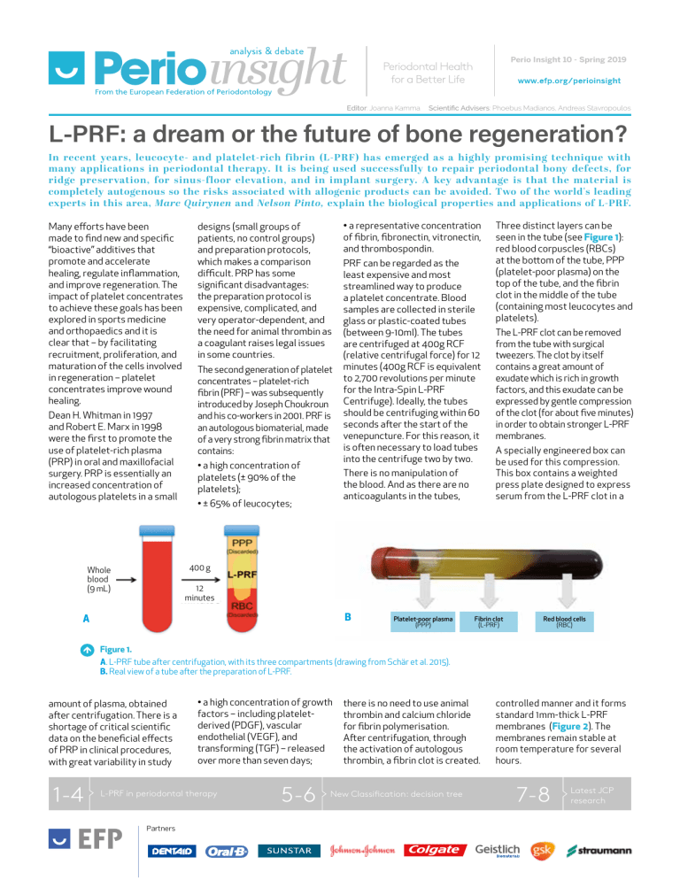
Perio Insight 10 - Spring 2019 Editor: Joanna Kamma Scientific Advisers: Phoebus Madianos, Andreas Stavropoulos L-PRF: a dream or the future of bone regeneration? In recent years, leucocyte- and platelet-rich fibrin (L-PRF) has emerged as a highly promising technique with many applications in periodontal therapy. It is being used successfully to repair periodontal bony defects, for ridge preservation, for sinus-floor elevation, and in implant surgery. A key advantage is that the material is completely autogenous so the risks associated with allogenic products can be avoided. Two of the world's leading experts in this area, Marc Quirynen and Nelson Pinto, explain the biological properties and applications of L-PRF. Many efforts have been made to find new and specific “bioactive” additives that promote and accelerate healing, regulate inflammation, and improve regeneration. The impact of platelet concentrates to achieve these goals has been explored in sports medicine and orthopaedics and it is clear that – by facilitating recruitment, proliferation, and maturation of the cells involved in regeneration – platelet concentrates improve wound healing. Dean H. Whitman in 1997 and Robert E. Marx in 1998 were the first to promote the use of platelet-rich plasma (PRP) in oral and maxillofacial surgery. PRP is essentially an increased concentration of autologous platelets in a small designs (small groups of patients, no control groups) and preparation protocols, which makes a comparison difficult. PRP has some significant disadvantages: the preparation protocol is expensive, complicated, and very operator-dependent, and the need for animal thrombin as a coagulant raises legal issues in some countries. The second generation of platelet concentrates – platelet-rich fibrin (PRF) – was subsequently introduced by Joseph Choukroun and his co-workers in 2001. PRF is an autologous biomaterial, made of a very strong fibrin matrix that contains: • a high concentration of platelets (± 90% of the platelets); • a representative concentration of fibrin, fibronectin, vitronectin, and thrombospondin. PRF can be regarded as the least expensive and most streamlined way to produce a platelet concentrate. Blood samples are collected in sterile glass or plastic-coated tubes (between 9-10ml). The tubes are centrifuged at 400g RCF (relative centrifugal force) for 12 minutes (400g RCF is equivalent to 2,700 revolutions per minute for the Intra-Spin L-PRF Centrifuge). Ideally, the tubes should be centrifuging within 60 seconds after the start of the venepuncture. For this reason, it is often necessary to load tubes into the centrifuge two by two. There is no manipulation of the blood. And as there are no anticoagulants in the tubes, • ± 65% of leucocytes; Three distinct layers can be seen in the tube (see Figure 1): red blood corpuscles (RBCs) at the bottom of the tube, PPP (platelet-poor plasma) on the top of the tube, and the fibrin clot in the middle of the tube (containing most leucocytes and platelets). The L-PRF clot can be removed from the tube with surgical tweezers. The clot by itself contains a great amount of exudate which is rich in growth factors, and this exudate can be expressed by gentle compression of the clot (for about five minutes) in order to obtain stronger L-PRF membranes. A specially engineered box can be used for this compression. This box contains a weighted press plate designed to express serum from the L-PRF clot in a 400 g Whole blood (9 mL) 12 minutes B A Platelet-poor plasma (PPP) Fibrin clot (L-PRF) Red blood cells (RBC) Figure 1. A. L-PRF tube after centrifugation, with its three compartments (drawing from Schär et al. 2015). B. Real view of a tube after the preparation of L-PRF. amount of plasma, obtained after centrifugation. There is a shortage of critical scientific data on the beneficial effects of PRP in clinical procedures, with great variability in study 1-4 • a high concentration of growth factors – including plateletderived (PDGF), vascular endothelial (VEGF), and transforming (TGF) – released over more than seven days; L-PRF in periodontal therapy Partners 5-6 there is no need to use animal thrombin and calcium chloride for fibrin polymerisation. After centrifugation, through the activation of autologous thrombin, a fibrin clot is created. New Classification: decision tree controlled manner and it forms standard 1mm-thick L-PRF membranes (Figure 2). The membranes remain stable at room temperature for several hours. 7-8 Latest JCP research Spring 2019 2 Figure 2: Process of preparing L-PRF clots and membranes. A: A specially designed box (Xpression tray from Intra-Lock) is used to compress L-PRF clots into L-PRF membranes with a consistent thickness of 1mm. A piston and cylinder assembly (left side) can be used to create L-PRF plugs, suitable for filling extraction sockets. B: L-PRF clots before compression. C: L-PRF membranes after gentle compression: the red area of the membrane represents the face side, where most leucocytes and platelets are concentrated. These autologous membranes (with a dense fibrin network) are strong – a single membrane can withstand a load of ± 500g before rupture. They also have excellent biological properties – they are rich in platelets, growth factors, and cytokines – which opens many new clinical avenues. L-PRF membranes remain solid and intact in vitro and continuously release large quantities of growth factors for between seven and 14 days. Applications of L-PRF In various countries, L-PRF is used for the treatment of non-responding skin ulcers A B including diabetic foot ulcers (DFU), pressure ulcers (PU), acute surgical wounds, and venous leg ulcers (VLUs). In a pilot study carried out by Nelson Pinto and colleagues and published in 2018, L-PRF application gave 100% wound closure for VLU ≤ 10 cm2, and at least 10 of the 15 larger wounds could be closed. For DFU, again L-PRF scored highly, with 100% wound closure in sites where standard wound care failed. The beneficial effect of L-PRF membranes in the healing of chronic leg ulcers can be explained by the high concentration of platelets and leucocytes, together with the long-term release of C growth factors. The progressive release of growth factors (e.g. TGFß-1, TGFß-2, PDGF-AB, VEGF, and IGF), matrix glycoproteins (trombospondin-1 [TSP-1], fibronectine and vitronectine), pro-inflammatory cytokines (IL-1ß, IL-6, TNF-α), and an antiinflammatory cytokine (IL-4) for up to seven days seems to be crucial. Within dentistry, there are various applications of L-PRF, including the treatment of periodontal bony defects, ridge preservation, sinus-floor elevation, in implant surgery, and to create L-PRF bone blocks. Periodontal bony defects A series of clinical studies has evaluated the benefits of applying L-PRF on its own during open-flap debridement. All these studies reported an adjunctive improvement, when L-PRF was used, on parameters such as probing pocket depth (PPD) reduction (1.1 ± 0.5mm extra reduction), clinical attachment gain (CAL, 1.2 ± 0.6mm extra gain), and bonedefect fill (1.5 ± 0.3mm or 46% ± 12.8% extra bone fill). For more information on this, see Part A of the 2017 systematic review and meta-analysis by Ana B. Castro and colleagues. Ridge preservation A B C D E F Figure 3: The use of L-PRF as a filling material of a tooth socket, aiming to maintain the alveolar bone dimensions. A: Preparation of envelope (circa 2mm in width) between bony borders of socket and surrounding soft tissues (this is needed to slide in the L-PRF membranes at the end, in order to prevent the fast ingrowth of connective tissue and to force the epithelium to grow over the membranes). B&C: Placement, one by one, of the L-PRF plugs (± 3-5 plugs or membranes) in the socket, followed by vigorous compression. D&E: Final coverage of socket with at least a double layer of L-PRF membranes (slide borders of membrane into prepared envelope). F: Tension-free suturing with, for example, a modified internal mattress or external mattress technique; primary closure is not necessary at all. The application of L-PRF in an extraction socket significantly reduces the horizontal/vertical ridge resorption (Figure 3), even at sites with bone dehiscence. The observed reduction in bone resorption is comparable to the current “best performing” clinical procedures (using a bone substitute in combination with connective-tissue graft or membrane). L-PRF as a filling material after third-molar extraction has a beneficial effect on both postoperative pain and soft-tissue healing. The L-PRF enables fast neo-angiogenesis and promotes bone regeneration via the release of growth factors and a good cloth stability. Marc Quirynen graduated in 1980 as a dentist and in 1984 completed his training in periodontology at the department of periodontology (both Catholic University of Leuven, Belgium). In 1986 he presented his PhD and in 1990 he was appointed professor at the Faculty of Medicine of the Catholic University of Leuven to teach periodontology and anatomy. His research deals mainly with oral microbiology, oral malodour, simplification/optimisation of periodontal therapy including implant surgery, and the benefits of L-PRF. He has published more than 400 full papers in international peer-reviewed journals and is is member of the editorial board of the Journal of Clinical Periodontology (associate editor), Clinical Oral implants Research, and Journal of Dental Research. Spring 2019 3 Sinus-floor elevation L-PRF can be used successfully as a “sole” filling material during sinus augmentation – but only if simultaneous with the placement of implants (the latter act as posts of a tent to keep the space for bone regeneration). L-PRF can be applied either via a lateral window technique (Figure 4) or through a trans-alveolar approach. Several studies have confirmed a natural bone regeneration around the implants (± 10mm vertical bone gain with the window technique, ± 4mm for transalveolar approach). It is important to note that all gain in radio-opaque area in the sinus, observed on the CBCT (cone-beam computed tomography) scan, is “vital” bone, with 0% of substitute remnants. More information about this can be found in Part B of the 2017 Castro study. A C Implant surgery A recent (2015) study by Elif Öncü concluded that implants coated with L-PRF (the membrane or its exudate) and placed in osteotomies treated with L-PRF showed a statistically higher implant stability quotient (ISQ) than the control group – in particular up to four weeks of healing (a difference of 15 ISQ units). Furthermore, when L-PRF is used during one-stage implant placement, the amount of initial bone remodelling (first three months after implant placement) can be reduced significantly. L-PRF bone block Relatively strong bonesubstitute blocks, that can resist some pressure, can be created via a special procedure. A fibrin network (Figure 5) can be created around the bone-substitute particles by combining three elements: B D A B Figure 4. Sinus-floor augmentation via a lateral-window, with simultaneous implant placement using L-PRF as “sole” substitute (also to cover the window). A: CBCT immediately after surgery. B: X-ray after one year (dotted line represents initial position of floor of the sinus). (I) chopped L-PRF membrane parts (with activated platelets); (II) fibrinogen-rich liquid obtained via three minutes of centrifugation of blood at same speed as for L-PRF membranes, but now using plastic tubes without coating on the inside; (III) a bone substitute. A kind of “bone block” (with the strength of “gummy bears” and an intrinsic memory shape) will be formed within a minute. Initial observations (by Simone Cortellini and co-workers in a 2018 study) of the use of these so-called “L-PRF bone blocks” – either in horizontal bone regeneration or in sinus augmentation – are extremely promising, but they need to be confirmed through well-designed clinical trials. One important advantage is the ease of handling and this approach might reduce the need of reinforced membranes or more expensive tools to maintain the space under the flaps for bone regeneration. Figure 5. Protocol for L-PRF bone block. A: Mix two chopped membranes and 0.5g bone substitute in a Ti dish. B: Spray liquid fibrinogen over the homogeneous mix and stir gently while shaping it to the desired form. C: After 30 seconds, a stable block is formed and this L-PRF bone block is now ready for use. D: Placement of L-PRF bone block over an implant with buccal dehiscence. Nelson Pinto, who graduated in 1985, is the founder and chairman of the Research Center for Tissue Engineering and Regenerative Medicine in Chile. For the past 30 years he has maintained an active private practice, specialising in advanced oral implantology, and he is professor in the department of periodontics and implantology at the University of the Andes (Chile). Prof Pinto is a world-leading expert in L-PRF, soft- and hard-tissue regeneration, and wound healing. Among his many accomplishments are the development of Natural Guided Regeneration Therapy, for which he has received awards including: Best Oral Research Presentation (World Union of WoundHealing Societies,2012), Best Contribution to Clinical Research, 2012 to 2016 (Journal of Wound Care & World Union of Wound Healing Societies, 2016). In 2018, he received the Punyaarjan Humanitarian Service Award. Spring 2019 4 Conclusions The benefit of using the first generation of platelet concentrates was very much debatable and the subject of controversy, but the second generation – L-PRF – produces more consistent and predictable results. The advantages of using L-PRF are its autologous nature, simple collection, ease of chair-side preparation, and simple clinical application without the risks associated with allogeneic or animalderived products. The biological properties of L-PRF clearly show an interesting surgical versatility and all the characteristics that can support faster tissue regeneration and high-quality clinical outcomes. L-PRF can stimulate osteogenesis as well as angiogenesis and it provides a scaffold that allows cellular migration. These are certainly the fundamental aspects for bone regeneration. L-PRF has already been investigated in many periodontal treatments, including the placement of implants. However, for areas such as implant coating, horizontal/vertical bone augmentation, ridge preservation of extraction sites with large bone dehiscence, more welldesigned clinical studies are needed. Clearly the arrival of L-PRF does not mean that the whole way of thinking and acting within oral surgery should be changed. Nonetheless, in many situations, the 100% autogenous L-PRF can replace bone substitutes and/or membranes. Andy Temmerman, Ana B. Castro, Simon Cortellini, and Wim Teughels (all from the Catholic University of Leuven) contributed to this article. Select bibliography >> Boora, P., Rathee, M., & Bhoria, M. (2015). Effect of Platelet Rich Fibrin (PRF) on Peri-implant Soft Tissue and Crestal Bone in One-Stage Implant Placement: A Randomized Controlled Trial. J Clin Diagn Res, 9, ZC18-21. doi:10.7860/JCDR/2015/12636.5788 >> Boswell, S. G., Cole, B. J., Sundman, E. A., Karas, V., & Fortier, L. A. (2012). Platelet-rich plasma: a milieu of bioactive factors. Arthroscopy, 28, 429-439. doi:10.1016/j.arthro.2011.10.018 >> Castro, A. B., Meschi, N., Temmerman, A., Pinto, N., Lambrechts, P., Teughels, W., & Quirynen, M. (2017a). Regenerative potential of leucocyte- and platelet-rich fibrin. Part A: intra-bony defects, furcation defects and periodontal plastic surgery. A systematic review and meta-analysis. J Clin Periodontol, 44, 67-82. doi:10.1111/jcpe.12643 >> Castro, A. B., Meschi, N., Temmerman, A., Pinto, N., Lambrechts, P., Teughels, W., & Quirynen, M. (2017b). Regenerative potential of leucocyte- and platelet-rich fibrin. Part B: sinus floor elevation, alveolar ridge preservation and implant therapy. A systematic review. J Clin Periodontol, 44, 225-234. doi:10.1111/jcpe.12658 >> Choukroun, J. (2001). Une opportunité en paro-implantologie: le PRF. Implantodontie, 42, 55-62. >> Cortellini, S., Castro, A. B., Temmerman, A., Van Dessel, J., Pinto, N., Jacobs, R., & Quirynen, M. (2018). Leucocyte- and platelet-rich fibrin block for bone augmentation procedure: A proof-of-concept study. J Clin Periodontol, 45, 624-634. doi:10.1111/jcpe.12877. Epub 2018 Apr 10. PubMed PMID: 29421855. >> Dohan Ehrenfest, D. M., Andia, I., Zumstein, M. A., Zhang, C. Q., Pinto, N. R., & Bielecki, T. (2014). Classification of platelet concentrates (Platelet-Rich Plasma-PRP, Platelet-Rich Fibrin-PRF) for topical and infiltrative use in orthopedic and sports medicine: current consensus, clinical implications and perspectives. Muscles Ligaments Tendons J, 4, 3-9. >> Dohan Ehrenfest, D. M., Bielecki, T., Jimbo, R., Barbe, G., Del Corso, M., Inchingolo, F., & Sammartino, G. (2012). Do the fibrin architecture and leukocyte content influence the growth factor release of platelet concentrates? An evidence-based answer comparing a pure platelet-rich plasma (P-PRP) gel and a leukocyte- and platelet-rich fibrin (L-PRF). Curr Pharm Biotechnol, 13, 1145-1152. >> Dohan Ehrenfest, D. M., de Peppo, G. M., Doglioli, P., & Sammartino, G. (2009). Slow release of growth factors and thrombospondin-1 in Choukroun’s platelet-rich fibrin (PRF): a gold standard to achieve for all surgical platelet concentrates technologies. Growth Factors, 27, 63-69. doi:10.1080/08977190802636713 >> Eshghpour, M., Dastmalchi, P., Nekooei, A. H., & Nejat, A. (2014). Effect of platelet-rich fibrin on frequency of alveolar osteitis following mandibular third molar surgery: a double-blinded randomized clinical trial. J Oral Maxillofac Surg, 72, 1463-1467. doi:10.1016/j.joms.2014.03.029 >> Marenzi, G., Riccitiello, F., Tia, M., di Lauro, A., & Sammartino, G. (2015). Influence of Leukocyte- and Platelet-Rich Fibrin (L-PRF) in the Healing of Simple Postextraction Sockets: A Split-Mouth Study. Biomed Res Int, 2015, 369273. doi:10.1155/2015/369273 >> Marx, R. E., Carlson, E. R., Eichstaedt, R. M., Schimmele, S. R., Strauss, J. E., & Georgeff, K. R. (1998). Platelet-rich plasma: Growth factor enhancement for bone grafts. Oral Surg Oral Med Oral Pathol Oral Radiol Endod, 85, 638-646. >> Oncu, E., & Alaaddinoglu, E. E. (2015). The effect of platelet-rich fibrin on implant stability. Int J Oral Maxillofac Implants, 30, 578- 582. doi:10.11607/jomi.3897 >> Pinto, N. R., Ubilla, M., Zamora, Y., Del Rio, V., Dohan Ehrenfest, D. M., & Quirynen, M. (2017). Leucocyte- and platelet-rich fibrin (L-PRF) as a regenerative medicine strategy for the treatment of refractory leg ulcers: a prospective cohort study. Platelets, 29, 468-475. doi:10.1080/09537104.2017.1327654 >> Schar, M. O., Diaz-Romero, J., Kohl, S., Zumstein, M. A., & Nesic, D. (2015). Platelet-rich concentrates differentially release growth factors and induce cell migration in vitro. Clin Orthop Relat Res, 473, 1635-1643. doi:10.1007/s11999-015-4192-2 >> Temmerman, A., Vandessel, J., Castro, A., Jacobs, R., Teughels, W., Pinto, N., & Quirynen, M. (2016). The use of leucocyte and platelet-rich fibrin in socket management and ridge preservation: a split-mouth, randomized, controlled clinical trial. J Clin Periodontol, 43, 990-999. doi:10.1111/jcpe.12612 >> Whitman, D. H., Berry, R. L., & Green, D. M. (1997). Platelet gel: an autologous alternative to fibrin glue with applications in oral and maxillofacial surgery. J Oral Maxillofac Surg, 55, 1294-1299. Spring 2019 5 EFP New Classification toolkit offers clinicians decision tree for staging and grading periodontitis One of the key elements of the EFP toolkit on the new classification of periodontal and peri-implant diseases and conditions is the “decision tree” for assessing periodontitis. Created by Maurizio Tonetti and Mariano Sanz, this tool is designed to guide clinicians through the sequential decisions needed to be able to assign cases to the correct periodontal diagnosis. There are four steps involved: 1. The first step enables the clinician to discriminate between periodontal health, gingivitis, and suspect periodontitis when assessing a new patient. 2. The second step is a confirmation step to provide differential diagnosis between periodontitis and other conditions characterised by attachment loss. 3. The third step assesses the severity and complexity of management of the periodontitis case (staging, involving stages I-IV). 4. The fourth step assesses the risk profile of the case (grading, involving three grades, A-C). Professors Tonetti and Sanz have published an article in the Journal of Clinical Periodontology explaining the process behind the creation of the decision tree. “Implementation of the new classification of periodontal diseases: Decision-making algorithms for clinical practice and education” describes how they developed empiric decisionmaking algorithms based on the new classification to discriminate effectively between the key diagnoses of periodontal health and disease. STEP 1 New patient When seeing a patient for the first time, we should first ask if there is a full-mouth radiograph of adequate quality. If yes, we should assess whether there is detectable marginal bone in any area of the dentition. If bone loss (BL) is detectable, the patient is suspected of having periodontitis. At the same time, irrespective of radiographic records, we must clinically explore the patient and assess interdental clinical attachment loss (CAL). If CAL is detectable, the patient is a possible case of periodontitis. If interdental CAL is not detected, we must evaluate the presence of buccal recessions with probing pocket depths (PPD) greater than 3mm. If such recessions are present, the patient is a possible periodontitis case. If there are no buccal PPD greater than 3mm, we must evaluate full-mouth bleeding on probing (BoP). If this is present in more than 10% of the sites, the patient is diagnosed with gingivitis and if present in less than 10% of sites, the patient is diagnosed with periodontal health. Optimising diagnosis Discussing the scientific rationale for the study, they point out that implementing the new classification “requires modification of the current way of thinking to optimize diagnosis in clinical practice and education.” They say that introducing this new classification in clinical practice and education “requires careful planning and the introduction of a novel, yet simple, way of thinking, which introduces new diagnostic tools and a new decision-making algorithm to support the process leading to diagnosis and case definition.” Their JCP paper notes that World Workshop (Chicago, 2017) – organised by the EFP and the American Academy of Periodontology to create the new classification – indicated bleeding on probing (BoP) as the “most reliable and validated diagnostic tool” for assessing gingival X-Rays available New patient Diagnostic quality & full-mouth Yes No inflammation. The workshop therefore incorporated BoP in the case definitions of periodontal health, gingivitis, reduced but healthy periodontium, and gingival inflammation in a treated periodontitis patient. Assessing BoP thus becomes part of the routine dental examination to identify cases of periodontal health and gingivitis and monitor treated subjects. Tonetti and Sanz point out that the classification requires the identification of 10% BoP sites to distinguish between periodontal health and gingivitis and therefore requires full-mouth assessment and recording. They say that this approach “represents an important opportunity since, besides the need for diagnosis, recording of BoP together with plaque represents the key approach to assess the patient oral hygiene and to plan individualized preventive programmes.” Yes Detectable marginal bone loss No No Yes Assess interdental CAL loss Yes No Yes Buccal or oral rec & PPD ˃3mm Suspect periodontitis Yes Localised gingivitis No BoP 10-30% Periodontal health <10% Measure BoP ≥10% Gingivitis BoP ˃30% Generalised gingivitis Proceed to Step 2 Spring 2019 6 The classification workshop also highlighted the need to establish clinical attachment loss (CAL) as the primary definition of periodontitis, although noting that when marginal alveolar bone loss is apparent on diagnosticquality radiographs, it may be an adequate proxy measure of CAL. clinical conditions associated with CAL, “such as gingival recession, vertical root fractures, endo-periodontal lesions, loss on the distal of the lower second molars associated with impacted wisdom teeth, or attachment loss secondary to cervical decay or restorations.” The authors say that this is important because “the use of probing pocket depths (PPDs) does not allow discrimination of periodontal health, gingivitis, periodontitis, reduced but healthy periodontium, [and] gingival inflammation in a periodontitis patient.” Clinicians are advised to recognise the signs of CAL and discriminate them from other Validating the decision tree Periodontal chart In relation to the four-step clinical-decision algorithm, professors Tonetti and Sanz write: “Differentiating between periodontal health and diseases must be performed during routine dental examination in primary care settings in all subjects undergoing dental care. In this manner, the < 30% Periodontitis case 30% or more Periodontitis case CAL loss, bone loss, periodontal tooth loss Pocket depth, intrabony defects, furcation, tooth hypermobility, secondary occlusal trauma, <10 occluding pairs Stage II periodontitis Stage III periodontitis They suggest that this research should be performed with clinicians with different levels of experience (students, general dental practitioners, specialists), operating in different settings (primary care, dental hospitals, specialist practice) and in different areas of the world. Further reading: Periodontitis: clinical decision tree for staging and grading. Guidance for clinicians. EFP New Classification guidance note, authors: Mariano Sanz and Maurizio Tonetti. Available at: http://www.efp.org/publications/ projects/new-classification/ guidance/report-02b.pdf Extent (% teeth) Severity & complexity Stage I periodontitis specific research that evaluates their diagnostic accuracy and their costeffectiveness. Tonetti MS, Sanz M. Implementation of the new classification of periodontal diseases: Decision-making algorithms for clinical practice and education. J Clin Periodontol. 2019;46: 398-405. https://doi.org/10.1111/jcpe.13104 Periodontal history (PTL) Periodontal & bone appraisal Complexity of management The authors state that the practical implications of these four diagnostic steps are that they allow correct case assignment for periodontal health, gingivitis, and periodontitis. However, they add that the validity of the proposed decision trees needs to be assessed through Periodontitis case Full mouth x-rays Severity of breakdown time that dentists can devote to gathering complex diagnostic data and the associated costs will be limited to those subjects requiring such data for subsequent management.” They say that in this context it makes sense to suggest that “the diagnostic process leading to case definition should be performed in a sequence of steps.” Stage IV periodontitis Proceed to grading STEP 3A Patient is a periodontitis case whose stage needs to be established To establish the stage of an individual case of periodontitis, the following information is needed: full mouth x-rays, a periodontal chart, and a periodontal history of tooth loss (PTL). First, we assess the extent of the disease, by assessing whether the CAL/ BL affects less than 30% of the teeth (local) or 30% or more (generalised). Then, we define the stage of the disease by assessing severity (using CAL, BL, and PTL) and complexity (by assessing PPD, furcation and intrabony lesions, tooth hypermobility, secondary occlusal trauma, bite collapse, drifting, flaring, or having fewer than 10 occluding pairs of teeth). Spring 2019 7 Latest research from the EFP’s Journal of Clinical Periodontology The Journal of Clinical Periodontology (JCP) is the official scientific publication of the European Federation of Periodontology. Edited by Maurizio Tonetti, the JCP aims to convey scientific progress in periodontology to those concerned with applying this knowledge for the benefit of the dental health of the community. The journal is aimed primarily at clinicians, general practitioners, periodontists, as well as teachers, students, and administrators involved in the organisation of prevention and treatment of periodontal disease. The JCP is published monthly and has an impact factor of 4.046. The six articles summarised below were published in the JCP in February, March, and May 2019. PERIODONTAL DISEASES New insights on the link between malocclusion and periodontal disease This study investigated the association between malocclusions and periodontal disease by comparing it to that of smoking in subjects recruited from the “Study of Health in Pomerania” populationbased cross-sectional study. Sagittal intermaxillary relationship, variables of malocclusion, and socio-demographic parameters of 1,202 dentate subjects (20-39 years of age) were selected. Probing depth (PD) and attachment loss (AL) were assessed at four sites by tooth in a half-mouth design. Analyses were performed with multilevel models at subject, jaw, and tooth level. The research found that distal occlusion determined in the canine region, ectopic position of canines, anterior spacing, deep anterior overbite, and increased sagittal overjet were associated with AL. Deep anterior overbite with gingival contact and anterior crossbite were associated with PD. Regarding crowding, only severe anterior crowding was compatible with a moderate to large association with PD. Compared to smoking, the overall effect of malocclusions was about one half for AL and one-third for PD. The research concluded that malocclusions or morphologic parameters were associated with periodontal disease. Authors: Olaf Bernhardt, Karl-Friedrich Krey, Amro Daboul, Henry Völzke, Stefan Kindler, Thomas Kocher, Christian Schwahn Published in Journal of Clinical Periodontology Volume 46, Number 2 (February 2019). Full article: https://doi.org/10.1111/jcpe.13062 PERIODONTAL THERAPY Association of non‐surgical periodontal therapy on patients’ oral health‐related quality of life: a multi‐centre cohort study The aim of this study was to investigate the association of non-surgical periodontal therapy (NST) on oral-health-related quality of life (OHRQoL), both in general and related to the severity of periodontal disease and to treatment modalities. and six to eight weeks after NST using a standardised and validated OHRQoL instrument (Oral Health Impact Profile-G14, OHIP-G14). Another questionnaire was filled out by the dentists to evaluate the influence of treatment modalities and disease severity. The study involved 172 patients with periodontal disease from 18 dental practices, measured before Overall, the mean value of the OHIP baseline improved significantly after NST, while a significant negative association between the severity of periodontitis and OHRQoL could be detected, and only patients with moderate and severe periodontitis showed a significant improvement of OHIP mean values. The results also indicated a significant association of practitioners and treatment modalities (favouring systemic antibiotics) in relation to improvements in the patients’ OHRQoL. Authors: Stefanie A. Peikert, Wilhelm Spurzem, Kirstin Vach, Eberhard Frisch, Petra RatkaKrüger, Johan P. Woelber 240 ± 12 months, both groups – split-mouth and parallel – showed significant attachment gain. The study failed to show significant attachment-gain differences between the two groups after 240 months. Krüger, Erik Neukranz, Peter Raetzke, Peter Eickholz, Katrin Nickles Published in Journal of Clinical Periodontology Volume 46, Number 5 (May 2019). Full article: https://doi.org/10.1111/jcpe.13093 PERIODONTAL THERAPY Infrabony defects 20 years after OFD and GTR This randomised controlled trial evaluated results 20 years after open-flap debridement (OFD) and guided tissue regeneration (GTR) of infrabony defects. Periodontal surgery was performed on 44 infrabony defects in 16 periodontitis patients (baseline examination), and polylactide acetyltributyl citrate barriers were randomly assigned to 23 out of these 44 defects (parallel). Ten of these patients (GTR) exhibited a second, contra-lateral defect (OFD) (split-mouth). At 12, 120, and Perio Insight is published quarterly by the European Federation of Periodontology (EFP) © European Federation of Periodontology Editor: Joanna Kamma (EFP contents editor) Scientific advisers: Phoebus Madianos and Andreas Stavropoulos (EFP scientific affairs committee). Authors: Hari Petsos, Petra Ratka- Published in Journal of Clinical Periodontology Volume 46, Number 5 (May 2019). Full article: https://doi.org/10.1111/jcpe.13110 Associate editor: Paul Davies (EFP editorial co-ordinator) Design and layout: Editorial MIC EFP office: Avenida Doctor Arce 14, Office 38, 28200 Madrid, Spain, www.efp.org Spring 2019 8 EFP full-member societies Latest research Austria Österreichische Gesellschaft für Parodontologie PERIODONTAL THERAPY Incorporating antibiotics into platelet-rich fibrin: a novel antibiotics slow‐release biological device This in vitro study sought to explore the possibility of using platelet-rich fibrin (PRF) as a local sustained-released device for antibiotics. Platelet-rich fibrin was prepared with the addition of antibiotics (5mg/ml metronidazole; 150mg/ml clindamycin; 1mU/ml penicillin) or saline prior to centrifugation, while collagen sponges served as control. PRF’s anti-bacterial properties were examined in an antibiogram assay with Staphylococcus aureus or Fusobacterium nucleatum at different time intervals after PRF preparation. The addition of antibiotic solutions at volumes of 2ml or 1ml led to significant changes in PRF’s physical properties, while the addition of a 0.5ml solution did not. PRF with saline showed minor anti-bacterial activity, while all PRFs with antibiotics showed significant anti-bacterial activity. No differences were observed between raw (clot) and pressed (membrane) forms of PRF. Collagen sponges with and without antibiotics showed similar results to PRF. PRF and collagen sponges with antibiotics preserved their antibacterial properties four days after preparation. Platelet-rich fibrin incorporated with antibiotics showed long-term anti-bacterial effect against F. nucleatum and S. aureus. This modified PRF preparation may be used to reduce the risk of post-operative infection in addition to the beneficial healing properties of PRF. Authors: David Polak, Navit Clemer-Shamai, Lior Shapira Published in Journal of Clinical Periodontology Volume 46, Number 2 (February 2019). Full article: https: //doi.org/10.1111/jcpe.13063 IMPLANT THERAPY Clinical effects of the adjunctive use of a 0.03% chlorhexidine and 0.05% cetylpyridinium chloride mouth rinse in the management of peri‐implant diseases: a randomised clinical trial This randomised, double-blinded, clinical trial evaluated the efficacy of a 0.03% chlorhexidine and 0.05% cetylpyridinium chloride mouth rinse, as an adjunct to professionally and patient-administered mechanical plaque removal, in the treatment of peri-implant mucositis (PiM). Forty-six of the 54 patients included attended the final visit (22 in control and 24 in test group). In the test group, there was a 24.49% greater reduction in BoP at the buccal sites than in controls; 58.3% of test implants and 50% controls showed healthy peri-implant tissues at final visit. Patients displaying PiM in at least one implant were included and subjects received professional prophylaxis (baseline and six months) and were instructed in regular oral-hygiene practices and told to rinse, twice daily for one year, with the test or placebo mouth rinses. Clinical, radiographic, and microbiological outcomes were evaluated at baseline, six, and 12 months. Disease resolution was defined as absence of bleeding on probing (BoP). Data were analysed by repeated measures ANOVA, Student’s t, and chi-square tests. Researchers concluded that the test mouth rinse demonstrated some adjunctive benefits in the treatment of PiM, but complete disease resolution could not be achieved in every case. Authors: Alberto Pulcini, Juan Bollaín, Ignacio SanzSánchez, Elena Figuero, Bettina Alonso, Mariano Sanz, David Herrera Published in Journal of Clinical Periodontology Volume 46, Number 3 (March 2019). Full article: https://doi.org/10.1111/jcpe.13088 IMPLANT THERAPY The amount of keratinized mucosa may not influence peri‐implant health in compliant patients: a retrospective five‐year analysis This study investigated the influence of the keratinized mucosa (KM) on peri-implant health or disease to identify a threshold value for the width of KM for peri-implant health. and parameters for peri-implant diseases was negligible and no threshold value was detected for the width of mid-buccal KM in relation to periimplant health. In 87 patients, data were extracted at baseline (prosthesis insertion) and five years including the width of mid-buccal KM, bleeding on probing, probing depth, plaque index, and marginal bone level. The width of KM around dental implants correlated to a negligible extent with parameters for peri-implant diseases. No threshold value for the width of KM to maintain peri-implant health could be identified. Depending on the definition of peri-implant diseases, the prevalence of peri-implantitis ranged from 9.2% (bleeding on probing threshold: <50% or ≥50%) to 24.1% (threshold: absence or the presence). The prevalence of peri-implant mucositis was similar, irrespective of the definition (54%-55.2%). The width of KM Partners Authors: Hyun-Chang Lim, Daniel B. Wiedemeier, Christoph H. F. Hämmerle, Daniel S. Thoma Published in Journal of Clinical Periodontology Volume 46, Number 3 (March 2019). Full article: https://doi.org/10.1111/jcpe.13078 Belgium Société Belge de Parodontologie / Belgische Vereniging voor Parodontologie Croatia Hrvatsko Parodontološko Društvo Czech Republic Ceská Parodontologická Spolecnost Denmark Dansk Parodontologisk Selskab Finland Suomen Hammaslääkäriseura Apollonia France Société Française de Parodontologie et d’Implantologie Orale Germany Deutsche Gesellschaft für Parodontologie Greece Ελληνική Περιοδοντολογική Εταιρεία Hungary Magyar Parodontológiai Társaság Ireland Irish Society of Periodontology I srael Israeli Society of Periodontology and Osseointegration Italy Società Italiana di Parodontologia e Implantologia Lithuania Lietuvos Periodontolog Draugija Netherlands Nederlandse Vereniging voor Parodontologie Norway Norsk periodontist forening Poland Polskie Towarzystwo Periodontologiczne Portugal Sociedade Portuguesa de Periodontologia e Implantes Romania Societatea de Parodontologie din Romania Serbia Udruzenje Parodontologa Srbije lovenia Združenje za ustne bolezni, parodontologijo S in stomatološko implantologijo Spain Sociedad Española de Periodoncia y Osteointegración Sweden Svensk förening för Parodontologi och Implantologi Switzerland Société Suisse de Parodontologie / Schweizerisch Gesellschaft für Parodontologie / Società Svizzera di Parodontologia Turkey Türk Periodontoloji Dernegi United Kingdom British Society of Periodontology EFP associate-member societies Azerbaijan Azərbaycan Parodontologiya Cəmiyyəti Georgia Georgian Association of Periodontology Morocco Société Marocaine de Parodontologie et d’Implantologie ussia Российской Пародонтологической R Ассоциации Ukraine Асоціація лікарів-пародонтологів України EFP international associate members Argentina Sociedad Argentina de Periodontología Australia Australian Society of Periodontology Brazil Sociedade Brasileira de Periodontologia Lebanon Lebanese Society of Periodontology Mexico Asociación Mexicana de Periodontología Taiwan Taiwan Academy of Periodontology
