International Journal of Trend in Scientific Research and Development, Volume 1(4), ISSN: 2456-6470 www.ijtsrd.com
“Determination of Rate of Shell Accretion in Pesticide Induced Fresh
Water Bivalve, Parreysia Corrugata”
Dr. Pramod Phirke
Department of Zoology, Fergusson College,
Pune, Maharashtra, India
Dr. S.P. Zambare
Department of Zoology,
Dr. Babasaheb Ambedkar Marathwada University,
Aurangabad, Maharashtra, India
ABSTRACT
Rate of shell accretion was studied in the freshwater
bivalve, Parreysia corrugata exposed to chronic
treatment of pesticides; quinalphos (0.108 ppm) and
thiodan (0.0708 ppm). The thickness of the accreted
part was maximum at umbo, moderate in the middle
and was minimum at the margin of the shell in
bivalves of all three groups. Rate of shell accretion
was reduced in pesticide exposed bivalves than in
normal bivalves. Accreted piece of the shell found to
become brittle with dull and poor nacre.
KEYWORD: Fresh Water Bivalve, Quinalphos,
Thiodan, Shell Accretion
INTRODUCTION
Water is the basic natural resource and the habitat of
many living organisms. Various water bodies such as
seas, rivers, and lakes have been used as dumping
grounds for the wastes from domestic activities,
industrial activities and agricultural activities.
Pesticides and other toxic chemicals cause harmful
effects on varieties of aquatic organisms like, frogs,
fishes, crabs, mussels, etc. The nature of toxic effects
of pesticides varies greatly as they are not specific in
their action (Moore, 1969). Toxicants such as
pesticides routinely discharged in the water bodies
create stressful situation, which influences growth and
development of aquatic flora and fauna. Enadu (1994)
reported that 46 types of pesticides were used to
control the pests in India with increasing amount as
432 tonnes in 1954; 58,900 tonnes in 1977 – 78;
70,781 tonnes in 1990 – 91.
The genera of mussels such as Lamellidens, Indonaia,
Parreysia, Corbicula, Unio, etc. are very common
Indian freshwater forms. Living bivalves provide
integrated information about concentration of
elements accumulated from their aquatic environment
in the proportion to those in the water (Lutz and
Rhoads 1980). Amount of incorporation of elements
can be assessed by measuring their relative
concentration in the valves. The shell of the Parreysia
is of nacro – prismatic type (Tevesz and Carter,
1980). Its outermost layer is periostracum and inner
calcified surface includes outer prismatic layer and
inner nacreous layer. Nacreous layer contains organic
material such as protein, scleroproteins, polypeptides
and perhaps mucopolysaccharides (Wilbur and
Younge, 1964). Dodd, (1967); Carriker, (1978);
Carriker, et. al. (1978) reported that, the trace and
minor elements can be incorporated in shell as
substitutes for calcium in the calcitic or aragonitic
crystal lattice as constituents of separate mineral
phases and organic compounds and are absorbed on
exterior surfaces.
The shells of mussels are economically important as
these are used in the production of toys, ornaments,
utility articles, lime, cement, paint, etc. These are also
most successfully used for the pearl production. The
shells of Cypraea (cowrie) were used as money as
well as ornaments. Since economically and
commercially important bivalves have been
continuously exposed to pesticides like Quinalphos,
Thiodan, etc. which interfere with the formation and
the growth of shells, present investigation work was
considered
to
determine
the
effect
of
organophosphosphate pesticide “quinalphos” and
organochlorine pesticide “thiodan” on the rate of shell
accretion in fresh water bivalve, Parreysia corrugata.
MATERIALS AND METHODS:
The fresh water bivalves, Parreysia corrugata
collected from dam were acclimatized in laboratory
conditions. The shell of the bivalves was drilled with
643
IJTSRD | May-Jun 2017
Available Online @www.ijtsrd.com
International Journal of Trend in Scientific Research and Development, Volume 1(4), ISSN: 2456-6470 www.ijtsrd.com
dentist drill of 1 mm bore at three different sites such
as umbo, middle level and the margin. They were
kept in three separate troughs for 5 months (i.e. 153
days). One group was maintained as a control, the
second group was treated by sub lethal concentration
(LC50/10 value of 96 hrs.) of quinalphos (0.108 ppm)
and the third group was treated by sub lethal
concentration of thiodan (0.0708 ppm). Water was
changed after every 24 hours from both the control
and experimental troughs. After the chronic treatment
of 5 months the bivalves were sacrificed and their
shells were removed. They were washed in distilled
water and dried at room temperature. The accreted
part from each site was removed and the thickness of
the part was measured by ocular micrometer. The rate
of shell accretion per month was calculated by the
formula-
OBSERVATIONS:
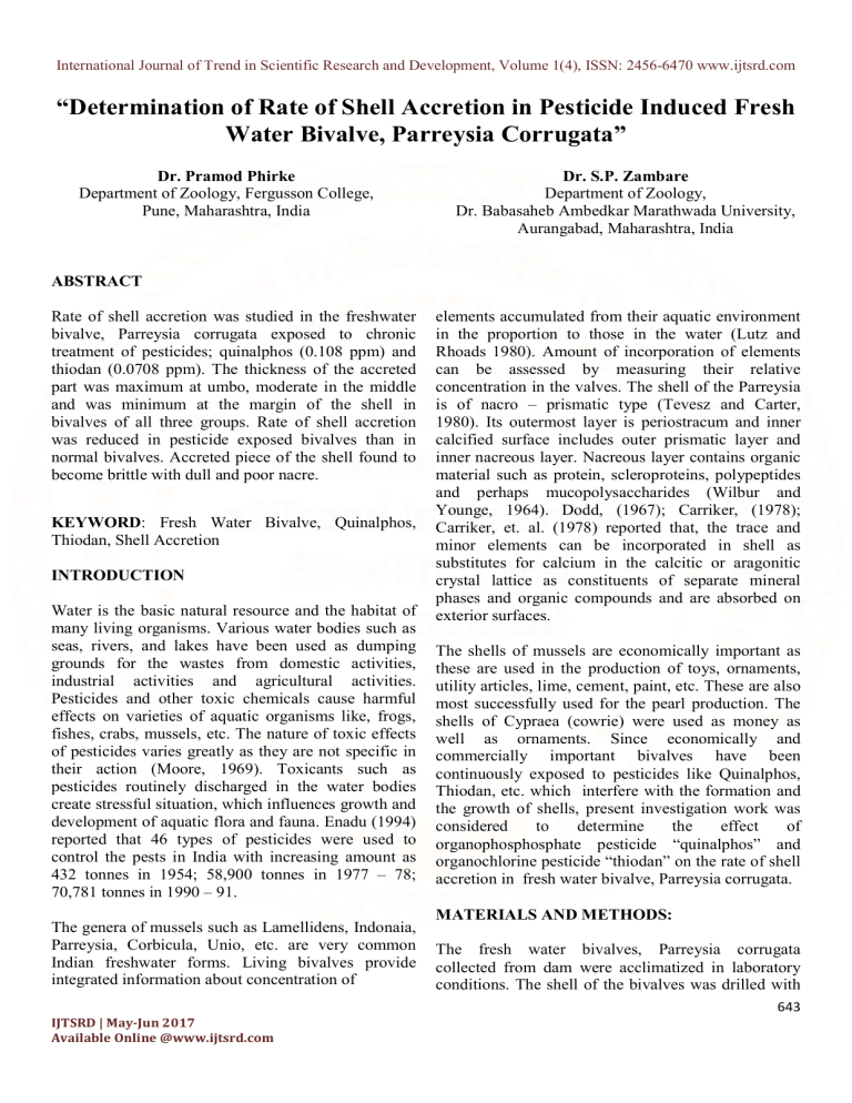
The results regarding the rate of shell accretion per
month in Parreysia corrugata shown in figure 1 and
are cited in table number 1. It is maximum at umbo
region than in middle and at marginal region. The rate
of shell accretion in the bivalves of control group is
32.142 µ / month at umbo region, 31.338 µ / month in
the middle and 24.780 µ / month at the marginal
region. Whereas in bivalves exposed to quinalphos,
the rate of shell accretion is 31.153µ / month at umbo,
24.923 µ / month in the middle and 22.107 µ / month
at the marginal region. The rate of shell accretion in
thiodan exposed bivalves found to be 27.674 µ /
month at umbo, 21.060 µ / month in the middle and
20.300 µ / month at margin. Accreted piece of the
shell became brittle with dull and poor nacre. The
thickness of the accreted part was maximum at umbo,
moderate in the middle and was minimum at the
margin of the shell in bivalves of all three groups.
Table 1: Rate of shell accretion in freshwater bivalve, Parreysia corrugata
after chronic exposure to quinalphos and thiodan
Dose
1.
2.
3.
4.
Rate of Accretion (microns per month)
Control
Umbo
32.142±1.23
Middle
31.338±1.58
Margin
24.780±1.16
Quinalphos
31.153±1.59NS
24.923±1.98**
22.107±1.45*
(0.108 ppm)
(-3.076)
(-20.47)
(-10.78)
Thiodan
27.674±1.58*
21.06±1.74**
20.3±1.68*
(0.0708 ppm)
(-13.90)
(-32.79)
(-18.07)
Thickness of accreted shell expressed as µm/ month.
± indicates standard deviation of three independent replications.
(+) or (-) % variation over control
Significance: * P < 0.05; ** P < 0.01; *** P 0.001; NS = Non-significant
644
IJTSRD | May-Jun 2017
Available Online @www.ijtsrd.com
International Journal of Trend in Scientific Research and Development, Volume 1(4),
1(4), ISSN: 2456-6470 www.ijtsrd.com
Figure 1: Photographs showing shell accretion in Parreysia corrugata
of control, quinalphos and thiodan exposed groups.
(A) Shell accretion in Parreysia corrugata of Control group
(B) Shell accretion in Quinalphos exposed
Parreysia corrugata
(C) Shell accretion in Thiodan exposed
Parreysia corrugata
DISCUSSION:
Shell valves in the bivalves are protective device
which protect internal soft and delicate body from
adverse situation in the environment. Freshwater
bivalves have been continuously exposed to pollutants
and toxicants discharged in the water bodies. It ma
may
influence the formation and growth of the shell.
Annuli on the surface of shell provide information
about age and growth rate in bivalve molluscs. There
are
different views regarding the development of annuli
Many
on the shell surface of the bivalvres. M
investigators reported formation of one growth
annulus in each year (Lutz and Rhoads, 1980; El
Moghraby and Adam, 1984). This view is supported
by few workers (Negus, 1966; Ghent et. al., 1978;
Haukiojo and Hakala, 1978). In median length
ilis radiata, formation of only half of an
mussel, Lampsilis
external annulus was formed per year and no external
645
IJTSRD | May-Jun 2017
Available Online @www.ijtsrd.com
International Journal of Trend in Scientific Research and Development, Volume 1(4), ISSN: 2456-6470 www.ijtsrd.com
annuli was formed even after several years (Downing
et. al., 1992). Jones (1983) stated that internal annuli
may be more reliable than external growth lines in
estimation of age and growth. The distance between
these rings indicates the annual growth performance
of individual bivalves (Mac. Curdy, 1954). Calcium
plays an important role in shell growth and normal
metabolism in marine bivalve molluscs. Vander and
Puymbroeck (1966) found 80% calcium incorporation
in freshwater gastropod, Lymnea stagnalis from food
rich surrounding water. Calcium incorporation in
excised mantle tissue and shell in marine bivalve was
also studied by Jordery (1953).
and Volovirta, 1997). The shell of the Parreysia is of
nacro – prismatic type (Tevesz and Carter, 1980). Its
outermost layer is periostracum and inner calcified
surface includes outer prismatic layer and inner
nacreous layer. Nacreous layer contains organic
material such as protein, scleroproteins, polypeptides
and perhaps mucopolysaccharides (Wilbur and
Younge, 1964). The stressful situation created due to
pesticide exposure in the water body, disturbs the
secretary activities of the mantle and in turn the
formation of different layers including nacreous layer;
the mother of pearl, the rate of shell accretion
decreases.
Spreading of elements in the shell depends on
structural (Carriker et. al., 1980 a) and chemical
(Buchardt and Fritz, 1978; Carriker et. al., 1980 b)
changes in the shell of bivalve caused due to
environmental fluctuations. Fouling, adsorption and
weathering of shell surface also influence the
spreading of elements in the shell (Rosenberg, 1980).
Incorporation of calcium in the shell is however
affected by contamination in the water. Cellular
volume of outer mantle epithelium reduces in
freshwater bivalve, Anodonta cygnea incubated with
pollutants for 8 months. It leads to significant
decrease in secretory activity of mantle due to
exposure to toxic agents and this has implication for
the shell calcification process (Manuel Lopes – Lima
et. al. 2006). Many trace and minor elements in water
body are adsorbed to, or incorporated in, suspended
particulate matter (Bopp and Bigs, 1981). Metals are
concentrated in the shell to a greater extent when they
are combined with suspended materials (Imlay, 1982).
Moura et. al. (2001) concluded that, the increase of
calcium contents with Cu and Cd incubation in
Anodonta cygnea might be due to acidosis which can
create abnormal conditions for shell formation.
The pollutants and toxic substances including
pesticides interfere with the spreading of elements and
deposition of calcium in the shells of bivalves and
also alter the cellular organization of the mantle
epithelium leading to reduced secretion of nacreous
layer. The lustrous quality of the nacre is also poor.
Overall it is resulted into decrease in the rate of shell
accretion with dull and brittle accreted piece in
pesticide exposed bivalves than in normal bivalves.
The chemical constitution of shell of bivalves is
dependent on concentrations of dissolved elements
in the culture water (magnesium, Lorens and Bender,
1977; Calcium, Sick et. al., 1979). Calcium, which is
the major cation in the shell, probably influences the
incorporation of other elements in shell, but the
intensity of its effect and a type of elements affected
are not yet studied (Odum, 1957; Romeril, 1971).
The mussel strongly collects calcium even from soft
water and deposits it mainly in the mantle in the form
of extensive masses of small spherules (Pekkarinen
REFERENCES:
1) Bopp, F. III, Biggs, R. B. 1981. Metals in
estuarine sediments: factor analysis and its
environmental significance. Science, N.Y. 214:
441-443.
2) Carriker, M. R. 1978. Ultrastructural anlysis of bis
solution of shell of the bivalve Mytilus edulis by
the accessory boring organ of the gastropod
Urosalpinx cinerea. Mar. Biol., 48: 105-134.
3) Carriker, M. R., Van Zandt, D. and Grant T. J.
1978.
Penetration of molluscan and nonmolluscan minerals by the boring gastropod
Urosalpinx cinerea, Bio. Bull. Mar. Biol. Lab.,
Woods hole, 155:511-526.
4) Dodd, J. R. 1967. Magnesium and strontium in
calcareous skeletons: a review. J. Paleontol. 41:
1313-1329.
5) Downing, W. L., Shostell, J. and Downing J. A.
1992. Non-annual external annuli in the fresh
water mussels Anodonta grandis grandis and
Lampsilis radiata sililquoidea. Freshwater Biology
28: 309-317.
6) El Moghraby, A. I. and Adam, M. E. 1984. Ring
formation and annual growth in Corbicula
consorbia Caillaud, 1827 (Bivalvia, Corbiculidae).
Hydrobiologia , 110 : 219-225.
646
IJTSRD | May-Jun 2017
Available Online @www.ijtsrd.com
International Journal of Trend in Scientific Research and Development, Volume 1(4), ISSN: 2456-6470 www.ijtsrd.com
7) Ghent, A. W., Singer, R. and Johnson-Singer, L.
1978. Depth distribution determines with SCUBA
and associated studies of the fresh water unionid
clams Elliptio complanata and Anodonta grandis
in Lake Bernard, Ontario. Canadian J. of Zoology
56:1654-1663.
8) Haukioja E. and Hakala T. 1978. Measuring
growth from shell rings in populations of
Anodonta piscinalis. Annali Ziilogica Fennici,
15:60-65.
9) Imlay, M. 1982. Use of shells of freshwater
mussels in monitoring heavy metals and
environmental stresses: a review. Malac. Rev. 15:
1-14.
10) Jodrey, L.H. 1953. Studies on shell formation. III.
Measurement of calcium deposition in shell and
calcium turnover in mantle tissue using the
mantle-shell preparation and Ca45. Biol. Bull.
Mar. boil. Lab., Woods Hole, 103: 269-276.
11) Jones, D. S. 1983. Sclerochronology: reading the
record of the molluscan shell. American Scientist,
71:384-391.
12) Lorens, R. B. and Bender, M. L. 1977.
Physiological exclusion of magnesium from
Mytilus edulis calcite. Nature, Lond., 269:793794.
13) Lutz, R. A. and Rhoads D. C. 1980. Growth
patterns within the molluscan shell. Skeletal
Growth of Aquatic Organismi: Biological Records
of Environmental Change (Eds.D. C. Rhoads and
R. A. Lutz), Plenum Press, New York, 203-254.
14) MacCurdy, E. 1954. The notebooks of Leonardo
da Vinci. Braziller, New York.
15) Manuel Lopes-Lima, Gabriela Moura, Boonyarath
Pratoomchat, Jorge Machado. 2006. Correlation
between the morpho-cytohistochemistry of the
outer mantle epithelium of Anodonta cygnea with
seasonal variations and following pollutant
exposure. Marine and Freshwater Behaviour and
Physiology, Vol. 39, Issue 3, pp 235 – 243.
16) Moore, N. W. 1969. Rural medicine, 1:1
17) Moura G., Almeida, M.J., Machado, M. J. C. and
Machado, J. 2001. Effects of heavy metal
exposure on ionic composition of fluids and nacre
of Anodonta cygnea (Unionidae) Haliotis 30: 3344.
18) Moura G., Almeida, M.J., Machado, M. J. C. and
Machado, J. 2001. Interaction of mineral elements
in sea water and shell of oysters [ Crossostrea
virginica (Gmelin)] cultured in controlled and
natural systems. J. exp. mar.Biol. Ecol. 46: 279296.
19) Negus, C. L. 1966. A quantitative study of growth
and production of unionid mussels in the River
Thames at Reading. J. of Animal Ecology, 35:
513-532.
20) Odum, H. T. 1957. Biogeochemical deposition of
strontium, Inst. mar. Sci., 4: 38-114.
21) Pekkarinen, M. and Valovirta, I. 1997.
Histochemical and X-ray studies on tissue
concretions and shells of Margaritifera
margaritifera (Linnaeus), J. of Shellfish Research
16(1): 169-177.
22) Romeril, M. G. (1971): Some limnological
characteristics of the Nozha hydrome, near
Alexandria, Egypt. Hydrobiologia, 41: 477-500.
23) Rosenberg, G.D. 1980. An ontogenic approach to
the environmental significance of bivalve shell
chemistry. In: Ed. By D.C. Rhoads and R.A. Lutz.
Skeletal growth of aquatic organisms. : Biological
records of environmental change, Plenum Press,
New York, 133-168.
24) Sick, L.V., Johnson, C.C. and Siegfried, C.A.
1979. Fluxes of dissolved and particulate calcium
in selected tissues of Crossostrea virginica.
Marine Biology 54: 293-299.
25) Tevesz, M.J.S. and Carter, J.G. 1980.
In:
D.C.Rhoads, R.A. Lutz (eds.) Skeletal growth of
aquatic organisms: Biological records of
environmental change (Plenum Press, London).
295-322.
26) Van der Borght, O. and Van Puymbroeck S. 1966.
Calcium metabolism in a freshwater mollusc:
quantitative importance of water and food as
supply for calcium during growth. Nature, Lond.
210: 791-793.
27) Wilbur, K.M. and Younge, C.M. 1964. (Eds.)
Physiology of mollusca 1. (Academic Press,
London), 243-282.
647
IJTSRD | May-Jun 2017
Available Online @www.ijtsrd.com


