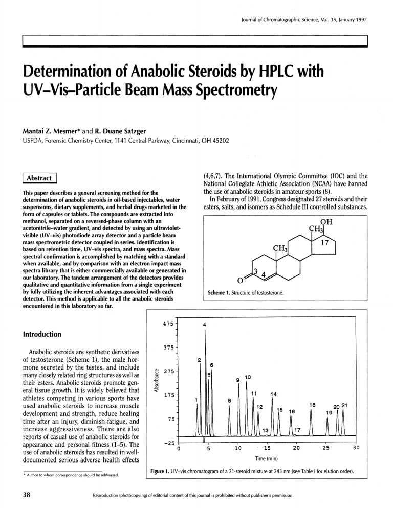
Journal of Chromatographic Science, Vol. 35, January 1997 Determination of Anabolic Steroids by HPLC with UV-Vis-Particle Beam Mass Spectrometry Mantai Z. Mesmer* and R. Duane Satzger U S F D A , Forensic Chemistry Center, 1141 Central Parkway, Cincinnati, O H 45202 Abstract This paper describes a general screening method for the determination of anabolic steroids in oil-based injectables, water suspensions, dietary supplements, and herbal drugs marketed in the (4,6,7). The International Olympic Committee (IOC) and the National Collegiate Athletic Association (NCAA) have banned the use of anabolic steroids in amateur sports (8). In February of 1991, Congress designated 27 steroids and their esters, salts, and isomers as Schedule III controlled substances. form of capsules or tablets. The compounds are extracted into methanol, separated on a reversed-phase column with an acetonitrile-water gradient, and detected by using an ultravioletvisible (UV-vis) photodiode array detector and a particle beam mass spectrometric detector coupled in series. Identification is based on retention time, UV-vis spectra, and mass spectra. Mass spectral confirmation is accomplished by matching with a standard when available, and by comparison with an electron impact mass spectra library that is either commercially available or generated in our laboratory. The tandem arrangement of the detectors provides qualitative and quantitative information from a single experiment by fully utilizing the inherent advantages associated with each Scheme 1 . Structure of testosterone. detector. This method is applicable to all the anabolic steroids encountered in this laboratory so far. Introduction Anabolic steroids are synthetic derivatives of testosterone (Scheme 1), the male hor­ mone secreted by the testes, and include many closely related ring structures as well as their esters. Anabolic steroids promote gen­ eral tissue growth. It is widely believed that athletes competing in various sports have used anabolic steroids to increase muscle development and strength, reduce healing time after an injury, diminish fatigue, and increase aggressiveness. There are also reports of casual use of anabolic steroids for appearance and personal fitness (1-5). The use of anabolic steroids has resulted in welldocumented serious adverse health effects Figure 1. UV-vis chromatogram of a 21-steroid mixture at 243 nm (see Table I for elution order). * Author to whom correspondence should be addressed. 38 Reproduction (photocopying) of editorial content of this journal is prohibited without publisher's permission. Journal of Chromatographic Science, Vol. 35, January 1997 This action expanded the analyses of steroids from the toxicological to the forensic area (9). Also, several states have placed anabolic steroids on their controlled-substances lists. The legislative control of anabolic steroids, together with the increase in demand for these drugs, has led to a growth in the clandestine market for steroid dosage forms (5) and coun­ terfeiting of these products. Some of these anabolic steroids are approved for veterinary use and can easily be diverted for abuse by humans. Products seen in the illegal market are mostly oil- Figure 2. UV-vis chromatogram based injectables, water suspensions, dietary supplements, and herbal drugs marketed as capsules or tablets, which often do not contain the ingredients declared on the label. The analysis of steroids is complicated by the sheer number of known steroids (over 7000), the close structural similarities common to the entire class, their thermal lability, low ultra­ violet extinction coefficients, and their poor volatility (9). There are reports of the analysis of anabolic steroids in forensic samples by high-performance liquid chromatography with ultraviolet-visible detection (HPLC-UV-vis) (9-11), micellar electrokinetic capillary chro­ matography (9), and gas chromatographymass spectrometry (GC-MS) (10,12). Sandra et al. reported the analysis of four anabolic steroids by HPLC with parallel detection, particle beam mass spectrometry, and UV-vis spectrophotometry (13). Particle beam LC-MS provides several advantages when compared to other LC-MS techniques. It is compatible with many LC mobile phases, it can easily be converted from electron ionization (EI) to chemical ionization (CI) mode of operation, and its ability to provide library-researchable EI mass spectra makes it the preferred tech­ nique in the identification of unknowns (14,15). In criminal and forensic cases, matched EI spectra are critical in estab­ lishing the identity of an unknown. We have developed a general screening method for of a 21-steroid mixture at 210 nm (see Table I for elution order). the determination of anabolic steroids in oilbased injectables, water suspensions, dietary supplements, and herbal drugs marketed in the form of capsules or tablets. The steroids were extracted into methanol, separated on a reversed-phase column with an acetonitrile-water gradient, and detected by using a UV-vis photodiode array detector and a par­ ticle beam MS detector coupled in series. Identification was based on retention time, UV-vis spectra, and mass spectra. Mass spec­ tral confirmation was accomplished by matching with a standard when available and by comparison with an EI mass spectra library that was either commercially avail­ able or generated in our laboratory. Experimental Apparatus Figure 3. Total ion chromatogram of a 21-steroid mixture (see Table I for elution order). The mass spectrometer used was a Hewlett-Packard (Palo Alto, CA) 5989A equipped with a particle beam interface. The HPLC was a Hewlett-Packard model 1090L equipped with an autosampler, a Rheodyne injector, and a photodiode array detector. 39 Journal of Chromatographic Science, Vol. 35, January 1997 Chromatographic and MS conditions Table I. Elution Order of Steroids The HPLC conditions were set as follows. A Waters (Milford, MA)μBondapakC 30-cm × 2.0-mm column was used. The injection volume was 5μL.The mobile phase consisted of 60% acetonitrile in water increased to 90% at 20 min and to 95% at 22 min, with an equilibration time of 7 min between runs. The flow rate was 0.35 mL/min. The mobile phase for the flowinjection experiments was 25% methanol in water. MS parameters were set as follows. The ion source tempera­ ture was 250°C, the quadrupole temperature was 100°C, the ionization mode was electron impact, the electron multiplier voltage was 2072 V, the monitored mass range (m/z) was 56-460, the desolvation chamber temperature was 55°C, and the nebulizer helium pressure was 30 psi. 1. Formebolone (2-formyl-17-methyl-1,4-androstadien-11,17-diol-3-one), C H O 2 1 2 8 18 4 2. Fluoxymesterone (9α-fluoro-11 β-hydroxy-1 7α-methyltestosterone), C H FO 20 29 3 3. Trenbolone (l7β-hydroxyestra-4,9,11-trien-3-one), C H O 1 8 4. Nandrolone (17-hydroxyestr-4-en-3-one), C H O 1 8 2 6 2 2 2 2 5. Testosterone (17β-hydroxyandrost-4-en-3-one), C H O 1 9 2 8 2 6. Methyltestosterone (17-hydroxy-1 7-methylandrost-4-en-3-one), C H O 2 0 3 0 2 7. Mesterolone (1α-methylandrostan-17β-ol-3-one), C20H32O2 8. Progesterone, C H O 2 1 3 0 2 9. Testosterone acetate, C H O 2 1 3 0 3 10. Methenolone acetate (primobolan, primonabol), C H O 2 2 11. Testosterone propionate, C H O 2 2 3 2 3 2 4 Reagents 3 Steroids were obtained from the following sources: flu­ oxymesterone, testosterone propionate, mesterolone, trenbolone,β-estradiol-3-benzoate,β-estradiol 17-valerate, testos­ terone 17-phenylpropionate, testosterone isocaproate, β-estradiol 17-cypionate, testosterone decanoate, boldenone undecylenate, nandrolone phenpropionate, and nandrolone were obtained from Sigma (St. Louis, MO). Testosterone, pro­ gesterone, nandrolone decanoate, testosterone 17β-cypionate, and methyltestosterone are United States Pharmacopeia refer­ ence standards. Formebolon was obtained from LPB Instituto Farmaceutico S.P.A. (Milan, Italy), testosterone acetate was obtained from K&K Labs (Hollywood, California), and metheno­ lone acetate was obtained from Schering AB/Berlin Bergkamen (Berlin, Germany). Nandrolone and nandrolone phenpropionate were obtained from Steraloids (Wilton, NH). HPLC-grade methanol and acetonitrile were obtained from Fisher Scientific (Pittsburgh, PA), HPLC-grade water was pre­ pared using a Milli-Q water purification system from Millipore (Bedford, MA) and a Culligan Aqua-Cleer reverse osmosis system from Culligan (Northbrook, IL). The sonicator is a model 08849-00 from ColeParmer (Niles, IL). 12. β-Estradiol-3-benzoate (17β-estra-1,3,5-[10]-triene-3,17-diol-3-benzoate), C H O 2 5 2 8 3 13. β-Estradiol-17-valerate (1,3,5-[10]-estratriene-3,17β-diοl-17-valerate), C H O 2 3 3 2 3 14. Nandrolone phenpropionate (4-estren-17β-ol-3-one-phenylpropionate), CHO 27 34 3 15. Testosterone 17-phenylpropionate (4-androsten-17β-οl-3-οne-17phenylpropionate), C H O 2 8 3 6 3 16. Testosterone isocaproate (4-androsten-17β-οl-3-οne-1 7-[4-methylpentanoate]), C H O 2 5 3 8 3 17. β-estradiol 17-cypionate (1,3,5-[10]-estratriene-3, 17β-diol-17cyclopentylpropionate), C H O 2 6 3 6 3 18. Testosterone 17β-cypionate (17β-[3-cyclopentyl-1-oxopropoxy] androst-4-en-3-one), C H O 2 7 4 0 3 19. Boldenone undecylenate, C H O 3 0 4 4 3 20. Nandrolone decanoate (17β-[(oxodecyl)oxy]-estr-4-en-3-one), C H O 2 8 4 4 3 21. Testosterone decanoate (4-androsten-17β-οl-3-οne-17-caprate), C H O 2 9 4 6 3 Table II. Linear Dynamic Ranges and Detection Limits Detection limits (ng)* Linear dynamic range UV-vis SIM-MS Fluoxymesterone 3 orders 2 25 Methyltestosterone 3 orders 3 20 Testosterone acetate 3 orders 3 25 Methenolone acetate 3 orders 5 20 Testosterone propionate 3 orders 2 10 Nandrolone phenpropionate 3 orders 3 50 Testosterone 17β-cypionate 3 orders 1 40 Boldenone undecylenate 3 orders 1 40 Nandrolone decanoate 3 orders 1 90 Testosterone decanoate 3 orders 1 180 * Detection limits are defined as the amount of analyte that would produce a signal 2.5 times the peak-to-peak noise in the baseline. The response at 243 nm was used for calculation of detection limits. The particle beam-MS mode exhibited poor linear dynamic ranges, typically less than two orders of magnitude. Responses drastically deviated from linearity at lower concentrations, which is consistent with reports in the literature (14). Selected ion monitoring (SIM) provided modest improvement in sensitivity, an increase anywhere from 2- to 5-fold over scanning. 40 Sample preparation Oil-based samples were prepared by soni­ cating the appropriate amount (typically 20 μL) in 4 mL of methanol for 20 min and fil­ tering through 0.5-μm or 0.2-μm nylon fil­ ters. Tablets were prepared by sonicating an appropriate amount (typically between 20 and 100 mg) in 4 mL of methanol and fil­ tering as mentioned above. Results and Discussion Retention times and UV-vis spectra were obtained for 36 commonly encountered anabolic steroids using the gradient shown above. The EI mass spectra of 36 steroid stan­ dards were generated by the flow-injection Journal of Chromatographic Science, Vol. 35, January 1997 Figure 4.Total ion chromatogram of five steroids spiked in sesame oil. Peaks: 1, nandrolone; 2, methyltestosterone; 3, sesamin; 4, mesterolone; 5, sesamin; 6, estradiol-3-benzoate; 7, testosterone decanoate. Figure 5. Total ion chromatogram of a veterinary drug sample. Peaks: 1, testosterone propionate; 2, testosterone 17-phenpropionate; 3, testosterone isocaproate; 4, testosterone decanoate. introduction of 200-1000-ng amounts of each steroid into the mass spectrometer and stored in a unique library. This library is used fre­ quently for confirmation of the presence of anabolic steroids in forensic samples. Acetonitrile appeared to be a better mobile phase than methanol for the particle beam MS detector because, as the percentage of meth­ anol goes above 90, there is a large increase in the baseline of the total ion chromatogram, whereas an increasing percentage of acetonitrile has no noticeable effect on the baseline. In addition, acetonitrile has a lower UV cutoff. There is no significant difference in sensitivity when using either solvent. There were no sig­ nificant differences between EI spectra obtained in methanol-water and methanolacetonitrile. Under the gradient conditions used, 21 of the anabolic steroids used in this study were separated. The tandem nature of the analysis provided quantitative and qualitative infor­ mation from a single experiment. Figures 1 and 2 show the UV-vis chromatograms of a 21-steroid mixture monitored at 243 nm and 210 nm, respectively; Figure 3 shows the cor­ responding total ion chromatogram. Note that the mass spectrometer, which is down­ stream from the UV-vis detector, detected all 21 steroids in the mixture. The UV-vis detector detected 20 of the steroids at either one of the two lines monitored, except for trenbolone, which was only detected by the mass spectrometer. The elution order of the 21-steroid mixture is listed in Table I in increasing order of retention time. The flow rates for the 2-mm-i.d. column (0.3-0.5 mL/min) are compatible with the optimum flow rate of the particle beam inter­ face facilitating the direct in-series coupling of the UV-vis detector with the mass spectrometer. The linear dynamic ranges and detection limits obtained using UV-vis detection for 10 representative anabolic steroids are given in Table II. The correlation coefficient for nine of these steroids was 0.9999 or better and 0.9998 for testosterone acetate. The photodiode array detector was used for quantitative analysis because of its better sensitivity, reproducibility, and wide linear dynamic ranges when compared with the particle beam MS detector. Figure 4 shows the total ion chromatogram of five steroids spiked in sesame oil. Figure 5 shows the total ion chromatogram of a veterinary drug sample that contained four steroids, and Figure 6 shows the mass spectrum of testos­ terone decanoate in the drug sample. The 41 Journal of Chromatographic Science, Vol. 35, January 1997 Acknowledgment rd This study was presented at the 43 ASMS Conference on Mass Spectrometry and Allied Topics in May, 1995; Atlanta, GA. References Figure 6. Mass spectrum of testosterone decanoate in sample (peak 4 in Figure 5). recoveries for three representative steroids (nandrolone, β-estradiol-3-benzoate, and testosterone decanoate) spiked into sesame oil were better than 96%. We applied this method to the analysis of oil-based steroid-containing veterinary drugs, food supple­ ments, and herbal drugs. The library of mass spectra generated in our laboratory proved to be useful because mass spectra of some of the steroids encountered in this laboratory are not avail­ able in commercially available mass spectral libraries. The min­ imum amount of analyte needed to provide positive spectral identification ranged from 20 to 200 ng, except for testosterone decanoate, which required 450 ng. The direct coupling of the photodiode array detector to the MS detector fully utilized the advantages associated with each detector. 8. 9. 10. 11. 12. 13. Conclusion This study provides a simple method for the determination of anabolic steroids in pharmaceutical and clandestine prepara­ tions. The tandem arrangement of the detectors provides qual­ itative and quantitative information from a single experiment by fully utilizing the inherent advantages associated with each detector. This method has been successfully applied to all the anabolic steroids encountered in this laboratory so far. 42 14. 15. 1. W.V. Moore. Anabolic steroid use in adoles­ cence (editorial). JAMA 260: 3404-06 (1988). 2. C. Birdsong. W h y athletes use drugs. Am. Pharm. 26:43 (1986). 3. M.B. Mellion. Anabolic steroids in athletes. Am. Fam. Phy. 30: 113-19 (1984). 4. M.W. Kibble and M.B. Ross. Adverse effects of anabolic steroids in athletes. Clin. Pharm. 6:686-692 (1987). 5. S. Penn. Muscling in as ever more people try anabolic steroids; traffickers take over. Wall Street Journal October 1,1988, p. 1 . 6. T.J. Limbird. Anabolic steroids in the training and treatment of athletes. Compt. Ther. 1 1 : 25-30 (1985). 7. R.H. Strauss, M.T. Liggett, and R.R. Lanese. Anabolic steroid use and perceived effects in ten weight-trained women athletes. JAMA 253: 2871-73 (1985). J.C. Wagner. Substance-abuse policies and guidelines in amateur and professional sports. Am. J. Hosp., Phar. 44: 305-10 (1987). I.S. Lurie, A.R. Sperling, and R.P. Meyers. The determination of an­ abolic steroids by M E C C , gradient HPLC and capillary G C . Forensic Sci., JFSCA 39(1): 74-85 (1994). M.J. Walters, R.J. Ayers, and D.J. Brown. Analysis of illegally dis­ tributed anabolic steroid products by liquid chromatography with identity confirmation by mass spectrometry or infrared spectrom­ etry. JAOAC 73(6): 904-26 (1990). F.T. Noggle, Jr., C.R. Clark, and J . DeRuiter. Liquid chromato­ graphic and spectral analysis of 17-hydroxy anabolic steroids. J. Chromatogr. Sci. 28: 162-66 (1990). F.T. Noggle, Jr., C.R. Clark, and J . DeRuiter. Liquid chromato­ graphic and mass spectral analysis of anabolic 17-hydroxy steroids esters. J. Chromatogr. Sci. 28: 263-68 (1990). P. Sandra, P. Courselle, G . Steenbeke, and M . Schelfaut. Dual channel detection - diode array and mass spectroscopy (EI or CI) - inHPLC.J . of High Res. Chromatogr. 12: 544-46 (1989). A. Apffel and M.L. Perry. Quantitation and linearity for par­ ticle-beam liquid chromatography-mass spectrometry. J. Chro­ matogr. 554: 103-118 (1991). F.R. Brown and W . M . Draper. The matrix effect in particle beam liquid chromatography/mass spectrometry and reliable quantita­ tion by isotope dilution. Biol. Mass Spectrom. 20: 515-21 (1991). Manuscript accepted August 15, 1996.


