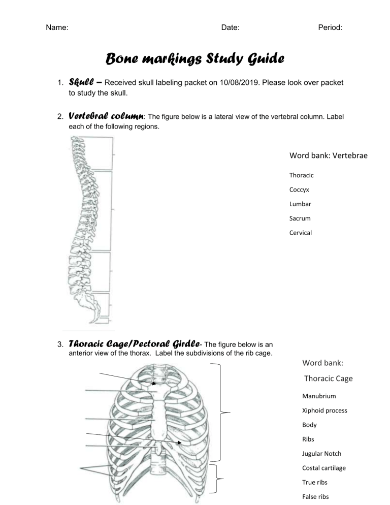
Name: Date: Period: Bone markings Study Guide 1. Skull – Received skull labeling packet on 10/08/2019. Please look over packet to study the skull. 2. Vertebral column: The figure below is a lateral view of the vertebral column. Label each of the following regions. Word bank: Vertebrae Thoracic Coccyx Lumbar Sacrum Cervical 3. Thoracic Cage/Pectoral Girdle- The figure below is an anterior view of the thorax. Label the subdivisions of the rib cage. Word bank: Thoracic Cage Manubrium Xiphoid process Body Ribs Jugular Notch Costal cartilage True ribs False ribs How many pairs of ribs do women have? _______ pairs How many pairs of ribs do men have? _______ pairs 4. Upper limbs I. The figure below is an anterior view (a) and posterior view (b) of the humerus. Word bank: Humerus Greater Tubercle Medial epicondyle Lateral epicondyle Intertubercular sulcus Deltoid tuberosity Head Trochlea Anatomical neck Olecranon fossa Coronoid fossa Lateral epicondyle Surgical neck Capitulum Lesser tubercle II. The figure below is an anterior view of the radius and ulna. Word bank: Radius/Ulna Olecranon process Radius styloid process Ulna Trochlear notch Head of radius Coronoid process Radius Head of ulna Radial tuberosity Ulna styloid process III. The figure below is a diagram of the hand. Identify the following bones Word bank: Hand Phalanges Radius Metacarpals Carpals Ulna Proximal phalanx Middle phalanx Distal phalanx 5. Pelvic Girdle – The figure below is an anterior view (a) and posterior view (b) of the pelvic girdle. Look on page 239 (Figure 7.48) in your textbook to label the figure. 6. Lower limbs I. The figure below is an anterior view (a) and posterior view (b) of the femur Word bank: Femur Lateral condyle Lesser trochanter Head Intercondylar fossa Medial condyle Fovea capitis Greater trochanter Linea aspera Neck Patellar surface Gluteal tuberosity Medial epicondyle Lateral epicondyle II. The figure below is an anterior view of the fibula and tibia Word bank: Fibula/Tibia Tibia Medial malleolus Lateral condyle Anterior crest Fibula Tibial tuberosity Lateral malleolus Head of fibula Intercondylar eminence Medial condyle III. The figure below is an anterior view (a) and posterior view (b) of the foot Word bank: Foot Phalanges Calcaneal tuberosity Navicular Fibula Tibia Talus Calcaneus Metatarsals Medial cuneiform 7. The figure below is an anterior view (a) and posterior view (b) of the major bones of the skeleton


