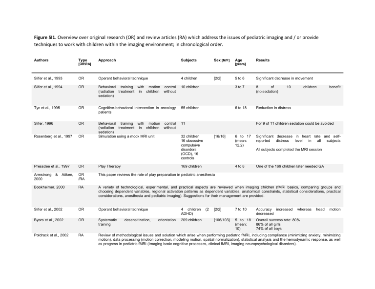
Figure SI1. Overview over original research (OR) and review articles (RA) which address the issues of pediatric imaging and / or provide techniques to work with children within the imaging environment; in chronological order. Authors Type Approach Subjects Sex [M/F] [OR\RA] Age Results [years] Slifer et al., 1993 OR Operant behavioral technique 4 children Slifer et al., 1994 OR Behavioral training with motion (radiation treatment in children sedation) control without Tyc et al., 1995 OR Slifer, 1996 OR 5 to 6 Significant decrease in movement 10 children 3 to 7 8 of (no sedation) Cognitive-behavioral intervention in oncology patients 55 children 6 to 18 Reduction in distress Behavioral training with motion (radiation treatment in children sedation) Simulation using a mock MRI unit 11 Rosenberg et al., 1997 OR control without [2/2] 10 children benefit For 9 of 11 children sedation could be avoided 32 children 16 obsessive compulsive disorders (OCD), 16 controls [16/16] 6 to 17 (mean: 12.2) Significant decrease in heart rate and selfreported distress level in all subjects All subjects completed the MRI session Pressdee et al., 1997 OR Play Therapy Armstrong 2000 OR /RA This paper reviews the role of play preparation in pediatric anesthesia Bookheimer, 2000 RA A variety of technological, experimental, and practical aspects are reviewed when imaging children (fMRI basics, comparing groups and choosing dependent variables, regional activation patterns as dependent variables, anatomical constraints, statistical considerations, practical considerations, anesthesia and pediatric imaging). Suggestions for their management are provided. Slifer et al., 2002 OR Operant behavioral technique Byars et al., 2002 OR Systematic training Poldrack et al., 2002 RA Review of methodological issues and solution which arise when performing pediatric fMRI, including compliance (minimizing anxiety, minimizing motion), data processing (motion correction, modeling motion, spatial normalization), statistical analysis and the hemodynamic response, as well as progress in pediatric fMRI (Imaging basic cognitive processes, clinical fMRI, imaging neuropsychological disorders). & Aitken, 169 children desensitization, 4 children ADHD) orientation 209 children 4 to 8 (2 One of the 169 children later needed GA [2/2] 7 to 10 Accuracy increased decreased whereas [106/103] 5 to 18 (mean: 10) Overall success rate: 80% 86% of all girls 74% of all boys head motion Wilke et al., 2003 RA An overview over current and future applications of fMRI is given, and typical problems, pitfall, and benefits of doing fMRI in pediatric age group are discussed. This is done on the background of fMRI basics, current research applications brain plasticity and current clinical applications. Davidson et al., 2003 RA Important issues relevant to developmental and clinical neuroimaging research are discussed, including anatomical, physical and psychological differences between children and adults, as well as general issues. Additionally, the development of age appropriate and scanner appropriate tasks for children are discussed, also in the context of an empirical study of development and learning in healthy children and adults. Overy et al., 2005 RA Behavioral preparation techniques Sury et al., 2005 RA Review - problems of painless imaging (Ultrasound & echocardiography, computer tomography, nuclear medicine imaging, positron emission tomography, magnetic resonance imaging) and patient management techniques (behavioral techniques, natural sleep, sedation, anaesthesia) DeAmorim e Silva et al., 2006 OR Practice magnetic resonance unit Kotsoni et al., 2006 RA Review of methodological and theoretical issues (developmental differences in anatomy & physiology and developmental and clinical differences in ability) and provides possible approaches (acclimation to, and modification of, the imaging environment for pediatric population) Epstein et al., 2007 OR Mock scanner training Operant feedback using a video system 45 participants (23 youth, 22 parents / ADHD and non-ADHD) [24/21] adults: 47 youth: 17 10% data loss (excessive movement > 2mm) No in between group differences Hallowell et al., 2008 OR (1) behavioral technique (2) use of a practice MRI unit 291 children [142/ 149] 3.6 to 17 (mean 7.9) 74.9% pass at practice 12 % borderline pass 96% diagnostic useful images of children entering MRI machine O'Shaughnessy et al., 2008 RA Review considering principles of fMRI, issues relevant to imaging children (anatomy, development, response variability, task selection, cooperation and movement), research using fMRI to examine cognitive processing in pediatric population (executive functions, visual spatial processing, facial expression and special focus on language studies) and applications to patient care Hunt & Thomas, 2008 RA Review aiming to provide a foundation for investigators aiming to use (f)MRI in research. Special consideration is given towards basic concepts of MRI physics, typical MRI components, scan types, experimental design factors, work with pediatric and special population. 33 children 134 children (retrospective evaluation) 5 to 7 [63/71] 4.1 to 16.1 (mean 7.7) Mean performance similar during practice and MRI session (no adverse effect due to MRI environment after training) Overall success rate: 90% of all children passed the practice session 98% of those had a clinical non-GA MRI 94% of those with success Reliable data in 95% of typically developing children > 8 years 80% of children aged 4 to 5 Raschle et al., 2009 P Video Protocol for Pediatric Neuroimaging (incorporates the use of a mock scanner as well as behavioral management techniques) Thomason, 2009 RA Review of pediatric neuroimaging procedures, addresses issues, such as child movement and anxiety and reviews reports of pediatric neuroimaging participants. Church et al., 2010 RA Discussion and overview of various issues related to pediatric neuroimaging, including assessing task performance, dealing with group performance differences, controlling for movement, statistical power, atlas registration and data analysis strategies. De Bie, HMA et al., 2010 RA Confirms success rate of mock scanner use in pediatric neuroimaging studies 90 children 3 to 14 years For children under 7 years of age, 33 out of 36 were able to continue to the fMRI session after training, and out of the 33, 23 of them had less than 3mm movement during image acquisition. Schlund et al., 2011 P Describes the development and application of an individualized multiple reward approach for increasing the number of fMRI tasks children complete during pediatric neuroimaging sessions. 28 children 9 to 13 Higher compliance and task completion rate in children assessed using an individualized multiple reward approach (Compliance in standard reward group: 68.4%; compliance in the multiple reward group: 93.6%) Notes: Original Research (OR), review articles (RA), male (M), female (F) 4.9 to 6.3 years (mean 5.5) Overall success rate: 95%. Overall movement decrased whereas chance of obtaining high quality images increased. Page 1 of 1 Type of file: figure Label: Figure S2 Filename: NYAS_6457_sm_FigureS2.doc file://F:\AdLib eXpress\Docs\2dfc2b29-dd37-41f1-9fd4-11cb58836f83\NIHMS41... 10/17/2012 Figure SI2. Overview over original research (OR) and review articles (RA) which address the issues of pediatric imaging and / or provide techniques to work with infants within the imaging environment; in chronological order. Authors Type Approach Subjects [OR\RA] Sex Age [M/F] [months] Anderson et al., 2001 OR Natural Sleep Technique (Infants were swaddled, outfitted with earphones, infants’ heads were lightly packed using foam padding and towels) 20 [11/9] 1 week DehaeneLambertz et al., 2002 OR Infants were awake or naturally sleeping with the following materials: special noise protection foam, noise protection helmet, infant’s body/head swaddled with bandages to ensure comfort/discourage movements 20 [6/14] 2-3 months Gilmore et al., 2004 OR Feed & Wrap Technique (Neonates were fed, swaddled, fitted with ear protection and heads secured in a vacuum-fixation device in a 3T headonly scanner. A parent stayed in the MR room throughout image acquisition) 20 [10/10] newborns Paterson et al., 2004 Conference Abstract Protocol outlining the approach to the Natural Sleep Technique in infants from 3 to 12 months of age, outlining the preparation involved, procedure for putting the infant to sleep, and the comfort and sound attenuation equipment necessary for MRI with naturally sleeping infants. Sury 2005 et al., RA Review summarizes approaches to implementing behavioral techniques when working with infants, protocol for using sedation/anesthesia, and the Natural Sleep Technique, along with a discussion on the pros and cons of selecting each approach. The research team reports a 50-75% success rate with the Natural Sleep Technique. Almli 2007 et al., OR Feed & Wrap Technique (Newborn Infants are fed and swaddled to sleep during daytime or nighttime. Parental education about the scan and testing process in the mock scanner environment) 106 newborns4 yrs Result Images acquired successfully in 14 of 20 infants, 6 woke up during scan sessions with no adverse events experienced. Obtained quality images without significant motion in 13 of 20 neonates. Most of these failures were due to failure getting the infant to sleep before image acquisition. Scanning success at 3-4 months: 60% (6 out of 10), 6-7 months: 72% (39 of 54), 12-13 months: 62.5% (20 of 32), with a subset needing multiple session attempts Of 106 infants and children, 75 were successful (see page 321, outlines reasons for failure Gilmore et al., 2007 OR Feed & Wrap Technique (Neonates were fed, swaddled, fitted with ear protection and heads secured in a vacuum-fixation device in a 3T headonly scanner. A parent stayed in the MR room throughout image acquisition) 74 [40/34] Newborns Natural Sleep Technique (child fell asleep naturally in waiting room or scanner room. After 5–1 0 m i n o f sleep on the scanner bed, earplugs and headphones were placed on the child) 21 [14/7] 30-60 months OR Feed & Wrap Technique (neonates fell asleep after being fed and & swaddled easily, having skipped their nap for the day. Neonates fitted with ear protection and secured in vacuum-fixation device in a 3T headonly scanner) 98 [49/49] newborn-2 yrs Hansen, 2009 OR Feed & Wrap Technique (infants fasted for 4 hours, fed just prior to the scan to induce sleep and positioned in an immobilisation device, VacFix Vacuum Cushion) 36 Ortiz-Mantilla et al., 2010 OR Natural Sleep Technique (followed the protocol of Paterson et al, 2004) 27 Glasel 2011 al., OR Natural Sleep Technique (Asleep during MR imaging; minimized noise exposure by covering magnet bore with noise protection foam) 14 Windram et al., 2011 OR Feed & Wrap Technique (infants fasted for 4 hours prior to the scan and fed immediately before the MRI, swaddeled with infant sheets and wer e p l a ced in a immobilizer, MedVac Bag) 20 Redcay et al., 2007 Knickmeyer al., 2008 et et newborns 89% success rate [15/12] ([13/11]) 6 and 12 months Success rate at 6 months 72%; Success rate at 12 months 63% ( 45% of all participants were longitudinal scans, thus acquisition at both 6 and 12 months) [9/5] 1 month 4 months newborn-6 months Success in all 20 with sufficient image quality Notes: Original Research (OR), review articles (RA), male (M), female (F) Page 1 of 1 Type of file: figure Label: Figure S3 Filename: NYAS_6457_sm_FigureS3.doc file://F:\AdLib eXpress\Docs\2dfc2b29-dd37-41f1-9fd4-11cb58836f83\NIHMS41... 10/17/2012 # of children recruited 3-4 months 6-7 months 8-11 months 12-15 months 4-6 years 10 59 8 32 45 # of imaging sessions 9x1 1x2 51x1 8x2 6x1 2x2 28x1 9x2 45x1 Total # of attempted MRIs # of successful attempts # of unsuccessful attempts Overall Success Rate (%) 11 6 5 55% 67 43 28 64% 10 5 5 50% 36 22 18 61% 45 44 1 97%
