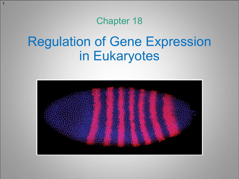
1 Chapter 18 Regulation of Gene Expression in Eukaryotes 2 Genetic Differences That Make Us Human While humans and chimpanzees diverged only 5 to 7 MEA, – Paradoxically, – – they differ in a large number of traits – anatomical, physiological, behavioral, etc. 96% of the DNA of humans and chimpanzees is identical How did humans and chimpanzees come to be so different? Where are the genes that make us human? Small number of genetic changes – in systems controlling gene expression produce large phenotypic differences between humans and chimpanzees Network of interacting genes for Transcription Factors differentially expressed in the brains of humans and chimpanzees. Red circles represent transcription factors that are more highly expressed in the human brain. Green circles represent transcription factors that are more highly expressed in the chimpanzee brain. 3 Different Patterns of Gene Expression Different cells in a multicellular organism – differ dramatically from one another both in structure and function While all cells contain the same genome and genes, – – – – the differences arise because cells have different sets of genes being actively expressed A typical human cell express 3060% of ~25,000 genes Some of them are common and some are tissue specific Each type of cells has a characteristic pattern of gene expression 1800 genes in 142 human tumor cell lines 4 Ways of Regulating Eukaryotic Gene Expression What level is the control of gene expression exercised at? Because of the way how eukaryotic genome is structured (chromosomes with several levels of chromatin packaging) and compartmentalized (separated from cytoplasm with the nuclear envelope), – the processes that regulate gene expression in eukaryotes are very diverse 5 Ways of Regulating Eukaryotic Gene Expression Expression of a gene in the eukaryotic cell includes multiple steps, each of which could in principle be a regulatory point 1. 2. 3. 4. 5. 6. Transcriptional control - when and where the gene is activated RNA processing control - determines the splicing pattern RNA transport control - determines which transcript to export Translational control - determines which RNA will be translated Degradation control - selectively targets RNAs for degradation Protein activity control - selectively activates/inactivates protein molecules 6 Ways of Regulating Eukaryotic Gene Expression 7 Alternative Splicing of RNA (Mammalian Troponin-T Gene) Alternative splicing of the rat troponin T gene, which leads to many cell-specific isoforms. The protein encoded by this gene is the tropomyosin-binding subunit of the troponin complex, which regulates muscle contraction in response to alterations in intracellular Ca2+ ion concentration. Phenomenon of splicing an RNA transcript in different ways is called alternative splicing Alternative splicing allows for a single gene to encode several polypeptides – It can serve many regulatory functions, e.g., control the variability of muscle cell contraction in mammals, or sex determination in flies 8 Alternative Splicing of RNA (Sex Determination in Drosophila) A cascade of alternative RNA splicing regulates sex determination – – – – – It includes three genes – doublesex (Dsx), transformer (tra), and Sex-lethal (Sxl) DsxF and DsxM are two isoforms of a transcription factor that activates femalespecific genes in females and represses the same female-specific genes in males DsxF and DsxM are produced by alternative splicing of the dsx pre-mRNA The Tra protein is an alternative splicing factor that regulates dsx splicing The tra gene expression is itself regulated by alternative splicing conducted by the Sxl protein (another splicing factor) 9 Alternative Splicing of RNA (Sex Determination in Drosophila) Sxl gene expression is governed by X-chromosome-encoded activators and autosome-encoded repressors – In females, Sxl activation prevails: the Sxl protein is produced and regulates tra RNA splicing, as well as, the splicing of Sxl RNA itself 10 RNA Stability Pathways of the degradation of eukaryotic mRNAs. The half-life of mRNA molecules can influence gene expression, – The stability of mRNA is extended by – as long-lived mRNAs can support multiple rounds of polypeptide synthesis the presence of the 5’-cap and the poly(A)-tail Among the factors decreasing stability are the AU-rich elements (AUUUA) repeated several times in the 3’UTR of some mRNAs – small noncoding RNA (snRNA) molecules – siRNAs and miRNAs – 11 Transcriptional Regulation For most genes, transcriptional regulation is the most important It is mediated through regulatory regions in DNA – They could be simple as a switch – responding to a particular signal, or could be complex (or modular) responding and integrating a variety of signals Transcription Factor Overall, transcriptional regulation consists of two components: – – Response Elements (RE) – short stretches of DNA Regulatory Proteins or Transcription Factors (TF) that recognize and bind RE Response Element 12 Key Points Eukaryotic gene transcripts may undergo alternative splicing to produce mRNAs that encode distinct, but related, polypeptides Alternative splicing is a possible point of regulation of gene expression The stability of mRNA can influence the amount of polypeptide being produced and correspondingly the level of gene expression Positive and negative regulatory proteins called transcription factors (TF) interact with specific DNA fragments (Response Elements or RE) to control the transcription of eukaryotic genes 13 Induction of Transcriptional Activity of Eukaryotic Genes Patterns of eukaryotic gene expression are constantly changing in response to either environmental cues (e.g., ToC increase) or in response to developmental needs communicated by signaling molecules (e.g., hormones and growth factors) – Both intracellular signaling and intercellular communication are important for transcriptional regulation in eukaryotes 14 The Heat-Shock Genes Exposure of cells to pulses of elevated temperature initiates the heat-shock response – – A restricted subset of genes – the Hsp genes (hsp22, hsp23, hsp26, hsp27, hsp68, hsp70, hsp83) – is activated and the majority of transcription and translation is shut-down The response may be elicited at all stages of the life cycle and in cultured cells The heat shock response (HSR) is a universal response to a large array of stresses (e.g., hypoxia or chemical stress) – – heat-shock proteins (HSP) play a role in the protection from these insults HSP stabilize the internal cellular environment 15 The Heat-Shock Genes The phenomenon of the heat-shock response was discovered in 1960 – – – – by Italian geneticist Ritossa, when he was studying polytene chromosomes Due to an accidental increase in ToC of the incubator where he was growing Drosophila, Ritossa observed a new pattern of “puffs” that occurs with increased temperature In salivary gland polytene chromosomes, existing puffs regress and a novel group quickly appears (33B; 63C; 64F; 67B; 70A; 87A; 87C; 93D; 95D) “Puffs” correspond to actively transcribed regions, as measured by radioactive uridine incorporation Ferruccio Ritossa (1936 – 2014) 16 Induction of the Drosophila hsp70 Gene by Heat Shock The heat shock response is very fast, within few minutes after ToC increase, the hsp gene encoded mRNAs can be detected – – – – Without the heat-shock, GAGA factor binds upstream of hsp70 gene and recruits NURF, which keeps the promoter free of nucleosomes and allows RNA polymerase binding Transcription starts, but at 25oC it stalls as the CTD of RNA Pol II is not phosphorylated enough With a sudden rise in ToC, Heat-Shock TF (HSTF) trimerizes and binds to heat shock element (HSE) It then interacts with Mediator and recruits a kinase, which phosphorylates the CTD and allows transcription to resume 17 Gene Expression and Signaling Molecules Regardless of the chemical nature, – the response to any signaling molecule starts with a specific receptor are two types of receptors – intracellular and cell-surface There – – Some signal molecules diffuse across the plasma membrane and activate intracellular receptors that directly regulate the transcription of specific genes In most cases, receptors are cell-surface or transmembrane proteins on the target cell surface; they act as signal transducers converting the extracellular binding into intracellular signals 18 Regulation of Gene Expression by Steroid Hormones The intracellular receptors could be activated by lipophilic signaling molecules: – – – steroid hormones (glucocorticoids and sex steroids); thyroid hormones; retinoids; vitamin D Although they differ greatly in both structure and function, they all act through a similar mechanism Lipophilic hormones diffuse directly across the plasma membrane to bind intracellular receptors 19 Regulation of Gene Expression by Steroid Hormones In early 1950s, while many steroid hormones have been characterized chemically and physiologically, their mode of action remained unknown – A breakthrough came from the collaboration of Ulrich Clever and Peter Karlson, when they studied the effect of ecdysone on the polytene chromosomes 20 Puffing of Giant Chromosomes and Steroid Hormones In 1960, Ulrich Clever analyzed developmental changes of the puffing pattern and found those that are characteristic for metamorphosis Peter Karlson at the same time purified the insect steroid hormone ecdysone, which regulates insect metamorphosis 21 Puffing of Giant Chromosomes and Steroid Hormones Together, Ulrich Clever and Peter Karlson found that ecdysone injection induces the appearance of metamorphic puffs These studies established a new paradigm of steroid hormone action – a direct interaction between the hormone and hormone-dependent genes 22 Regulation of Gene Expression by Steroid Hormones The steroid hormone enters the target cell and binds the nuclear receptor (NR) protein The hormone-receptor complex translocates into the nucleus and binds hormone response elements in DNA – Hormone response elements (HRE) are analogous to the heat-shock response elements (HSRE) The bound hormone-receptor complex (plays a role of the specific TF) stimulates gene expression (transcription) mRNA is processed and transported to the cytoplasm, where it is translated into a protein 23 Regulation of Gene Expression by Peptide Hormones A peptide hormone binds to a transmembrane receptor protein of the target cell The hormone-receptor complex activates a signal transduction pathway that brings the signal to the nucleus The signal induces a transcription factor (TF) to bind to a corresponding response element (RE) in the DNA The bound TF activates transcription mRNA is processed and transported to the cytoplasm, where it is translated into a protein 24 Key Points Eukaryotic gene expression is regulated by environmental cues or signaling molecules Transcription of the heat-shock genes in response to increased ToC is mediated by a heat-shock transcription factor Steroid hormones and their receptor proteins form complexes that act as transcription factors to regulate the expression of specific genes Peptide hormones interact with membrane-bound receptor proteins to activate a signal transduction pathway that regulates the expression of specific genes 25 Molecular Control of Transcription in Eukaryotes Transcription of eukaryotic genes is regulated by interactions between proteins and DNA sequences within or near the genes Three sets of regulatory DNA sequences are commonly involved in eukaryotic gene regulation – The core promoter region (immediately adjacent to the start of transcription) contains the TATA box and related sequences that bind RNA polymerase II and associated general transcription factors (TFIID, TFIIB, etc.) – Upstream of the core promoter region (approx. few hundred base pairs) are various proximal elements (GC box, CAAT box) that bind regulatory proteins essential for promoter activation – At greater distances from the core promoter are the enhancers 26 Properties of Enhancers Tissue-specific enhancers of the Drosophila yellow gene Unlike core promoter and proximal promoter elements, which are upstream and close to the gene they regulate, enhancers can be located close to or very far from the genes they regulate – Enhancers can be upstream, downstream, or within the genes (e.g., introns) – Enhancers bind regulatory proteins that interact with proteins bound to core and proximal promoter elements – Enhancers control the timing and location of gene transcription – Enhancers could be simple (like HSE) or modular (composed of multiple binding sites for different TFs) 27 Yeast Upstream Activator Sequences (Enhancers) In the yeast S. cerevisiae, transcription of genes in the galactose utilization pathway is regulated by enhancers – – When galactose is the only sugar available, yeast cells induce transcription of enzyme-encoding genes: GAL1, GAL2, GAL7, GAL10 Together these enzymes import and then break down galactose Each of the GAL genes has its own basal promoter and similar enhancers bound by a regulatory protein, Gal4 – The enhancer element is called the upstream activator sequence (UAS, or UASG) 28 Yeast Upstream Activator Sequences (Enhancers) Galactose absent: Gal4 is continuously present in cells; it interacts with the Gal80 protein (also continuously present) that binds Gal4 and keeps it inactive in the absence of galactose – – – Each UASG element contains two 17-bp repeat sequences that are binding sites for Gal4 Gal4 functions as a homodimer with two domains – the DNA-binding domain and the activation domain When Gal4 is bound to Gal80, the Gal4 DNA-binding domain is unable to bind UASG 29 Yeast Upstream Activator Sequences (Enhancers) Galactose present: When galactose is present, galactose and Gal3 (encoded by the GAL3 gene) form a complex and bind to Gal80 – – Gal80 releases Gal4, freeing the DNA-binding domain of Gal4 to recognize and bind to the UASG sites The transcriptional activation domain then activates transcription of the GAL genes 30 DNA Regulatory Elements Regulatory proteins are DNA-binding transcription factors (TF) – – – They typically recognize sequences less than 20 bp in length, referred to as response elements (RE) Each TF recognizes specific sequence of nts Upon binding they can change the level of gene expression 31 Structure of Transcription Factors Transcription factors have modular structure – DBD (DNA-binding domain) recognizes and binds specific response elements adjacent to regulated genes – TAD (transcriptional-activation domain) binds transcriptional coregulators and changes gene expression – SSD (signal-sensing domain) senses/binds external signals (e.g., ecdysone) and transmits the information to other domains (optional) TAD and SSD are often contained within the same domain 32 Modular Structure of Transcription Factors The yeast-bacterial Gal4-LexA fused protein activates expression of the reporter only when it has a corresponding response element (or binding site) 33 Structural Motifs of TFs Hundreds of DNA-binding regulatory proteins have been identified in eukaryotes They are categorized into four groups based on the structural motif that constitutes the DNA-binding domain – – – – the Zinc finger motif the helix-turn-helix motif (HTH) the basic-leucine zipper motif (bZIP) the basic helix-loop-helix motif (bHLH) 34 Structural Motifs of TFs * * Note: Homeodomain proteins constitute a subclass of the HTH (helix-turn-helix) group of proteins 35 Structural Motifs of TFs (Leucine Zipper Motif) Most of basic leucine zipper (bZIP) TFs are homodimers Each polypeptide of the bZIP protein – – – is an α-helix composed of two regions – the basic region that interacts with the major groove of DNA, and the hydrophobic region responsible for dimerization The hydrophobic region has a leucine residue at every 7th position; every second turn, leucine residues from two helices interact forming the “zipper” In the basic region, two α-helices separate to form a Y-shaped structure with two arms bound to a specific DNA sequence 36 Structural Motifs of TFs (Helix-loop-Helix Motif) A basic helix-loop-helix (bHLH) motif consists of two α-helices connected by a flexible loop In general, bHLH type TFs are dimeric Each polypeptide of the bHLH protein – has one α-helix containing basic AA (basic region) that facilitate DNA binding, and the second α-helix mediate dimerization 37 Key Points Unlike core and proximal promoter elements, enhancers act in an orientation-independent manner over considerable distances to regulate eukaryotic gene expression Transcription factors are regulatory proteins that recognize and bind to specific DNA sequences called response elements Transcription factors comprise four groups, each characterized by a structural motif in the DBD: the zinc finger, the helix-turn-helix, the leucine zipper, and the helix-loop-helix 38 Posttranscriptional Regulation of Gene Expression Short noncoding RNAs may regulate the expression of eukaryotic genes by interacting with the mRNAs produced by these genes – The phenomenon is called RNA interference (or RNAi) – RNAi occurs in plants and animals including humans 39 Posttranscriptional Regulation of Gene Expression Andrew Fire and Craig Mello were investigating – how gene expression is regulated in the nematode worm C. elegans Injecting ‘sense’ RNA molecules encoding a muscle protein led to no changes in the behaviour of the worms – Injecting ‘antisense’ RNA, which can pair with the mRNA, also had no effect – But when Fire and Mello injected sense and antisense RNA together, they observed that the worms displayed peculiar, twitching movements – Thus, a double-stranded RNA molecules can specifically silence genes whose code matches that of the injected RNA molecules – 40 Posttranscriptional Regulation of Gene Expression Andrew Fire (1959) Craig Mello (1960) Fire and Mello published their findings in the journal Nature on February 19, 1998 The Nobel Prize in Physiology or Medicine 2006 was awarded jointly to Andrew Fire and Craig Mello “for their discovery of RNA interference gene silencing by double-stranded RNA” 41 RNA Interference RNAi silences gene expression post-transcriptionally, but how? – – – Cells have a specific enzyme, Dicer that recognizes the double stranded RNA and chops it up into small fragments between 21-25 bp in length These short double stranded RNA fragments (small interfering RNA, or siRNA) bind to the RNA-Induced Silencing Complex (RISC) The RISC is activated when the siRNA unwinds and the sense strand is eliminated 42 RNA Interference 43 Double Stranded RNA Double stranded RNA originates either from genes, the transcription of other endogenous sequences, or from exogenous source – – In many eukaryotes, miRNA genes encode precursors of dsRNA that are cleaved into microRNAs of 21-24 nt Small interfering RNAs (siRNAs) are not encoded by genes; they originate from endogenous transcription or exogenous sources – Known eukaryotic siRNAs are mapped to transposons or other genomic regions that produce transcripts capable of forming dsRNA structures (e.g., inverted repeat structures, bidirectional transcription and antisense transcripts) – The exogenous source of dsRNA could be RNA viruses 44 microRNA Biogenesis miRNA genes are transcribed by RNA Pol II and produce transcripts that fold back upon themselves to produce dsRNA – The initial transcript is called primary microRNA (pri-miRNA); it forms a doublestranded stem of about 65-70 nucleotides, with free ends on one side and a singlestranded loop on the other The pri-miRNA is processed by Drosha enzyme complex – It cuts near the middle to produce a precursor miRNA (pre-miRNA), which is a 21-25 nt stem and the terminal loop – The pre-miRNA is transported to cytoplasm, where it is further processed by Dicer – RISC then binds to the remaining 21-25 bp and separates the strands to create miRNAs 45 microRNA Biogenesis (Summary) 46 Key Points siRNAs and miRNAs are produced from larger double-stranded precursors by the action of endonucleases Dicer siRNAs originate from transcription of sequences capable of forming dsRNA structures, e.g. transposons miRNAs originate from hundreds of genes present in eukaryotic genomes In RNA-Induced Silencing Complexes (RISC), siRNAs and miRNAs become single-stranded so they can target complementary sequences in mRNAs 47 Key Points RNA interference is used as a research tool to knock down the expression of genes in cells and organisms mRNA that has been targeted by siRNA is cleaved, and mRNA that has been targeted by miRNA is either cleaved or prevented from being translated 48 Chromatin Organization The defining feature of eukaryotic DNA is its packing into chromatin – Electron micrograph of a cell showing the heterochromatin localized to the nuclear periphery. Chromatin is a complex of DNA and proteins representing specific levels of DNA compactization Chromatin exists in two forms: – – Euchromatin (stains lighter; gene rich) is a less condensed form of chromatin that allows access of transcription machinery Heterochromatin (stains darker; gene poor) is a highly condensed form of chromatin that is typically not transcribed 49 Types of Heterochromatin Facultative (or intercalary) heterochromatin exhibits variable levels of condensation, reflecting activity of resident genes – Found in euchromatic environment Constitutive heterochromatin is permanently condensed, composed primarily of repetitive DNA sequences – Found in centromeres and telomeres, where it represses transposable element activity and thereby ensures genome stability and integrity 50 Gene Expression and Chromatin The structure of chromatin has a crucial role in eukaryotic gene transcription Changes in the level of compactization govern the accessibility of DNA by proteins (such as, TFs and enzymes) regulating and participating in gene expression – Position effect variegation (PEV) in Drosophila illustrates the effect of chromatin compaction on gene expression 51 Position effect variegation (PEV) in Drosophila The – PEV was first described by Herman Muller in 1930 Muller identified mutations in flies that resulted in variegated eye color – The X chromosomes of mutant flies had undergone inversion – The variability in the phenotype is caused by the relocation of the white+ gene from euchromatic into heterochromatic surrounding – The impaired ability of a gene to function at a new location is called Position Effect Variegation (PEV) 52 Chromatin Structure and PEV 53 Chromatin Structure and PEV The degree of heterochromatin spreading varies – In some cells euchromatic genes will not be affected by adjacent heterochromatin, and in others – certain genes will be silenced – Once heterochromatin-type structure and inactivation of neighboring euchromatin is established, it will be inherited by all cell progeny 54 Position effect variegation (PEV) in Drosophila Thus, the occurrence of PEV in Drosophila shows that – Gene expression can be silenced by its chromosomal position – Silencing is a feature of chromatin structure that can be transmitted from one cell generation to the next – This type of regulation is called epigenetic, as it is not due to changes in the DNA base sequence 55 Molecular Organization of Transcriptionally Active DNA Harold Weintraub (1945-1995) Mark Groudine Transcriptionally active DNA is more sensitive to DNase I than inactive This phenomenon was discovered in 1976 by Groudine and Weintraub – when they compared the β-globin (active) and ovalbumin (inactive) genes in chicken erythrocytes Gene expression is associated with “open” chromatin structure as it provides access to DNA by proteins involved in transcription 56 DNase I Hypersensitivity Dubrovsky et al. (1994), J Mol Biol 241, 353-362 Regions of chromatin that are sensitive to DNase I digestion are called DNase I hypersensitive sites (or DHS) – Today, mapping DNase I hypersensitive sites is a molecular approach to identify local changes in chromatin structure Mapping DHS is a highly accurate method for identifying promoters, enhancers, and other transcription control sequences 57 Key Points Heterochromatin is associated with the repression of transcription Position Effect Variegation is an example of the epigenetic regulation of gene expression Gene expression is associated with “open” (or loosely organized) chromatin Transcriptionally active DNA is more sensitive to digestion with DNase I 58 Chromatin Structure and Gene Expression So far, we have established that – The biological role of chromatin is not purely structural (storing and packing DNA), but it is quite a dynamic player in gene expression – Chromatin is not a static entity with defined regions of euchromatin and heterochromatin, instead there are dynamic changes in levels of compactization – As illustrated by PEV and DNase I hypersensitivity, dynamic changes in chromatin organization are directly associated with regulation of gene expression Still – questions to answer: What defines chromatin organization? How is it established? How is it maintained? How is it changed? 59 PEV Mutations Second-site mutations modifying PEV led to identification of proteins that play a role in establishing chromatin structures – – Two types of mutations have been identified – enhancers and suppressors of positioneffect variegation Several dozen E(var) and Su(var) mutations have been identified in Drosophila 60 E(var) Mutations E(var) mutations, enhancers of PEV, increase the appearance of the mutant white phenotype in flies with a variegating allele of the white gene – – E(var)s encourage the spread of heterochromatin further, and thus restrict w+ expression to even smaller patches of cells E(var)s produce a greater number of eye cells lacking pigment 61 Su(var) Mutations Su(var) mutations, suppressors of PEV, increase the appearance of the wild-type red phenotype in flies with a variegating allele of the white gene – – Su(var)s restrict the spread of heterochromatin or interfere with its function, and thus leave more cells with w+ expression Su(var)s produce a greater number of pigmented eye cells 62 Histone-Modifiers Analysis – genes encoding histone modifiers or proteins that make epigenetic marks on histones (e.g., adding methyl or acetyl group) These – – of PEV mutants led to covalent modifications can directly alter chromatin structure making it more open or reverse more condensed Thus, histone modifiers are critical in any process that involves accessing DNA (e.g., activation of gene expression) (a) Histone tails protrude from the nucleosome core. (b) Examples of histone tail modifications: ‘A’ represents acetylation; ‘M’ represents methylation. 63 HP-1 and HMT 64 Histone Acetylation Histone acetylation is another – post-translational modification of histones that can alter chromatin compaction and gene expression Covalent attachment of the acetyl (-COCH3) group is catalyzed by – histone acetyltransferase (HAT) Like any other histone modification, acetylation is reversible – Acetyl groups are removed by histone deacetylase (HDAC) Acetylation of lysine amino acids in histone tails opens the chromatin, exposing DNA to the activity of proteins that regulate transcription. 65 Histone Acetylation HDAC closes the chromatin structure HAT opens the chromatin structure Activity of these and other chromatin modifiers is under tight control 66 Histone Modifications Lysine (K) is the most frequently targeted residue for acetylation – Lysine and arginine (R) are the most frequent methylation targets Serine (S) and threonine (T) are targets for phosphorylation 67 Histone Modifications There are now known at least 150 histone modifications that utilize a wide variety of molecules: – acetyl, methyl, phosphoryl, ubiquitine, and ADP ribose Histone modifications can affect chromatin structure in different ways (and either activate or silence genes), but they are all reversible 68 Histone-Modifiers The modifications of histones are conducted by specific enzymes (histone-modifiers) 69 Histone Modifications Specific modifications are associated with different chromatin regions or cellular events E.g., acetylation of lysine residues at positions 8 and 16 of H4 is associated with chromatin regions where the start sites of the expressed genes are located 70 Histone Modifications Methylation of lysine residues at positions 4 and 36 of H3 typically is associated with the expressed genes 71 Histone Modifications On the other hand, methylation of lysine residues at positions 9 or 27 of H3 is associated with transcriptional repression
