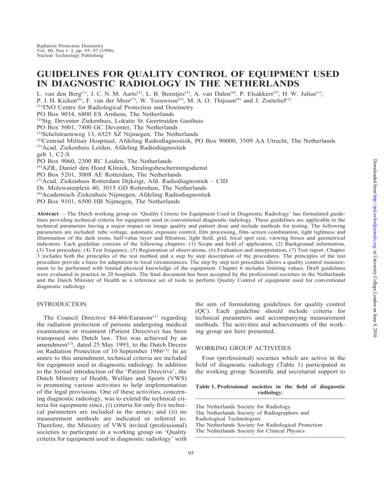
Radiation Protection Dosimetry Vol. 80, Nos 1–3, pp. 95–97 (1998) Nuclear Technology Publishing GUIDELINES FOR QUALITY CONTROL OF EQUIPMENT USED IN DIAGNOSTIC RADIOLOGY IN THE NETHERLANDS Abstract — The Dutch working group on ‘Quality Criteria for Equipment Used in Diagnostic Radiology’ has formulated guidelines providing technical criteria for equipment used in conventional diagnostic radiology. These guidelines are applicable to the technical parameters having a major impact on image quality and patient dose and include methods for testing. The following parameters are included: tube voltage, automatic exposure control, film processing, film–screen combination, light tightness and illumination of the dark room, half-value layer and filtration, light field, grid, focal spot size, viewing boxes and geometrical indicators. Each guideline consists of the following chapters: (1) Scope and field of application, (2) Background information, (3) Test procedure, (4) Test frequency, (5) Registration of observations, (6) Evaluation and interpretation, (7) Test report. Chapter 3 includes both the principles of the test method and a step by step description of the procedures. The principles of the test procedure provide a basis for adaptation to local circumstances. The step by step test procedure allows a quality control measurement to be performed with limited physical knowledge of the equipment. Chapter 6 includes limiting values. Draft guidelines were evaluated in practice in 20 hospitals. The final document has been accepted by the professional societies in the Netherlands and the Dutch Minister of Health as a reference set of tools to perform Quality Control of equipment used for conventional diagnostic radiology. INTRODUCTION the aim of formulating guidelines for quality control (QC). Each guideline should include criteria for technical parameters and accompanying measurement methods. The activities and achievements of the working group are here presented. The Council Directive 84/466/Euratom (1) regarding the radiation protection of persons undergoing medical examination or treatment (Patient Directive) has been transposed into Dutch law. This was achieved by an amendment (2), dated 25 May 1993, to the Dutch Decree on Radiation Protection of 10 September 1986 (3). In an annex to this amendment, technical criteria are included for equipment used in diagnostic radiology. In addition to the formal introduction of the ‘Patient Directive’, the Dutch Ministry of Health, Welfare and Sports (VWS) is promoting various activities to help implementation of the legal provisions. One of these activities, concerning diagnostic radiology, was to extend the technical criteria for equipment since, (i) criteria for only five technical parameters are included in the annex; and (ii) no measurement methods are indicated or referred to. Therefore, the Ministry of VWS invited (professional) societies to participate in a working group on ‘Quality criteria for equipment used in diagnostic radiology’ with WORKING GROUP ACTIVITIES Four (professional) societies which are active in the field of diagnostic radiology (Table 1) participated in the working group. Scientific and secretarial support to Table 1. Professional societies in the field of diagnostic radiology. The Netherlands Society for Radiology The Netherlands Society of Radiographers and Radiological Technologists The Netherlands Society for Radiological Protection The Netherlands Society for Clinical Physics 95 Downloaded from http://rpd.oxfordjournals.org/ at University College London on June 8, 2016 L. van den Berg(1), J. C. N. M. Aarts(2), L. B. Beentjes(3), A. van Dalen(4), P. Elsakkers(5), H. W. Julius(1), P. J. H. Kicken(6), F. van der Meer(7), W. Teeuwisse(5), M. A. O. Thijssen(8) and J. Zoetelief(1) (1) TNO Centre for Radiological Protection and Dosimetry PO Box 9034, 6800 ES Arnhem, The Netherlands (2) Stg. Deventer Ziekenhuis, Lokatie St. Geertruiden Gasthuis PO Box 5001, 7400 GC Deventer, The Netherlands (3) Schelstraeteweg 13, 6525 SZ Nijmegen, The Netherlands (4) Centraal Militair Hospitaal, Afdeling Radiodiagnostiek, PO Box 90000, 3509 AA Utrecht, The Netherlands (5) Acad. Ziekenhuis Leiden, Afdeling Radiodiagnostiek geb 1, C2-S PO Box 9060, 2300 RC Leiden, The Netherlands (6) AZR, Daniel den Hoed Kliniek, Stralingsbeschermingsdienst PO Box 5201, 3008 AE Rotterdam, The Netherlands (7) Acad. Ziekenhuis Rotterdam Dijkzigt, Afd. Radiodiagnostiek – CID Dr. Molewaterplein 40, 3015 GD Rotterdam, The Netherlands (8) Academisch Ziekenhuis Nijmegen, Afdeling Radiodiagnostiek PO Box 9101, 6500 HB Nijmegen, The Netherlands L. VAN DEN BERG et al STRUCTURE AND CONTENTS OF THE GUIDELINES The guidelines can be used in different ways, dependent on the experience of the reader. By following the successive chapters (Table 3) a rather inexperienced user obtains information on: (i) technical and physical aspects of the parameter(s), (ii) the impact of the parameter(s) on image quality and patient dose, (iii) the principles of the measurement method and a step by step procedure for performing the test, (iv) registration of the results of measurements and calculations, (v) evaluation of the results and corrective actions where necessary. A more experienced user can easily apply the guidelines by using the measurement equipment, the step by step method and the registration forms. The most experienced users, e.g. persons responsible for QC, can adapt the step by step method to the local situation. If the adaptation is in accordance with the principles of the measurement method, the accompanying limiting values are still applicable. The most essential aspects of the guidelines are the limiting values and the measurement methods for the eleven parameters (Table 2). When the measurement results are in compliance with the limiting values no noticeable deviation in image quality or in patient dose is to be expected. The guidelines also Table 2. Technical parameters influencing the performance of X ray equipment with the accompanying limiting values and measurement methods. Parameter Limiting values Tube voltage Automatic exposure control 5% accuracy; 2.5% precision. Optical density of the film between 1.10 OD and 1.50 OD. Maximal deviation fog ± 0.05 OD; speed and contrast ± 0.15 OD. Maximal deviation of the same speed class ± 0.10 OD; no artefacts. Film processing Film–screen combination Light tightness and illumination of the darkroom Half-value layer and filtration Light beam alignment Grid Focal spot size Viewing boxes Geometrical indicators of the X ray unit Measurement method or equipment Electronic kVp meter. Exposing film at selected tube voltages and thickness of the PMMA phantom. Constancy test using film exposed with a sensitometer. Exposure of 4 films simultaneously to a given radiation quality, e.g. 80 kV and 25 mm Al filter. Maximal increment 0.10 OD after 4 min Film, pre-exposed with a sensitometer, kept to exposure of a pre-exposed film. various darkroom conditions. Inherent filtration ⬎2.5 mm Alequivalent. It is Measurement of the HVL at 80 kV and use of recommended to add Cu filter for look-up diagrams for filtration. ⬎100 kV. Edges light and X ray beam within 1% Use of a specific alignment phantom. focus–film distance. Transmission factor ⬎64%; ratio 6:1, grid Dose measurements of primary and scattered factor ⬍3; no artefacts. radiation in the presence and the absence of the grid. In compliance with the specification of the Star pattern phantom according to the IECmanufacturer. 336. cd.m−2 meter for brightness of the viewing ⬎ 3000 cd.m−2 and 25% homogeneity; ambient light sources ⬍25 lux. box; lux meter for ambient light. Readouts ⬍1 cm; ⬍1°; orthogonality Centimetre, angle meter and orthogonality X ray beam to table ⬍1°. phantom. 96 Downloaded from http://rpd.oxfordjournals.org/ at University College London on June 8, 2016 the working group was given by the TNO Centre for Radiological Protection and Dosimetry. The working group aimed at the establishment of guidelines including limiting values and measurement methods for conventional installations in the Dutch language. Based on the literature and the experience of the members of the working group, eleven technical parameters having a major impact on image quality and patient dose were selected (Table 2). The limiting values for these parameters are based on values recommended in internationally accepted standards, for instance those from the International Electrotechnical Commission (IEC) (4). The measurement methods were selected on the basis of simplicity, time and cost effectiveness. Most measurement methods, as described in international guidelines, are appropriated in a technical setting but were not considered to be easily applicable in a clinical situation. Drafts of the guidelines were tested by radiographers, instrumental engineers and medical physicists in 20 departments of radiology of university and peripheral hospitals. These tests resulted in all types of comment which were implemented in the guidelines where appropriate. In September 1997 about 400 copies of the final version of the guidelines were distributed among the boards of directors of health institutes having a diagnostic X ray installation, and among all diagnostic radiology departments in the Netherlands. GUIDELINES FOR QUALITY CONTROL IN THE NETHERLANDS include annexes on definition on terms, measurement equipment, phantoms and registration forms. an acceptance test is carried out and appropriate QC programmes are implemented by the holder of the radiological installation. The professional societies and the Minister of Health in the Netherlands consider these guidelines to be a valuable instrument for QC of equipment used in diagnostic radiology. The Minister of Health recommends that holders of radiological installations gain experience with these guidelines to be able to meet future requirements. (5) As mentioned before, the guidelines are restricted to conventional X ray installations. For other types of radiological equipment, such as computed tomography systems, fluoroscopy and digital imaging systems additional guidelines should also be formulated. CONCLUSIONS AND FUTURE DEVELOPMENTS The working group has formulated 11 guidelines for Quality Control of conventional diagnostic radiology installations and accessories (film–screen combination, dark room, film processor and viewing boxes). The guidelines include limiting values and measurement methods. The new European patient directive (97/43/ Euratom) (5) states that Member States shall ensure that Chapter Contents 1. Scope and field of application 2. Background information Aims and types of apparatus Information on physical and technical aspects and impact on image quality and patient dose Principles of the test method; a step by step description of the method, including a list of instrumentation Time interval between tests Registration of measurement results Calculations; comparison of results with limiting values; corrective actions; trend analysis Test report with a completed registration form 3. Test procedures 4. Test frequency 5. Registration of observations 6. Evaluation and interpretation 7. Test report REFERENCES 1. European Commission. Council Directive of 3 September 1984 (84/466/Euratom) laying down the Basic Measures for the Radiation Protection of Persons undergoing Medical Examination or Treatment. Official Journal of the European Communities No L 265 (1984). 2. Decree of 25 May 1993. Amendment of Decree on Radiation Protection. Official Journal of the Netherlands 317 (1993). 3. Decree of 10 September 1986. Enforcement of the Articles 28 up to and including 32 and the Application of Article 34 of the Atomic Energy Act (Decree on Radiation Protection Atomic Energy Act). Official Journal of the Netherlands 465 (1986). 4. Technical Committee No 62B of the International Electrotechnical Commission. X-ray equipment operating up to 400 kV and accessories. Quality Assurance in Diagnostic X-ray Departments, Evaluation and Routine Testing in Medical Imaging Departments; Part 2–11: Constancy Tests — Equipment for General Direct Radiography. IEC Publications 1223-1-11 (1995). 5. European Commission. Council Directive 97/43/Euratom of 30 June 1997 on Health Protection of Individuals against the Dangers of Ionizing Radiation in Relation to Medical Exposure, and Repealing Directive 84/466/Euratom. Official Journal of the European Communities No L 180/22 (1997). 97 Downloaded from http://rpd.oxfordjournals.org/ at University College London on June 8, 2016 Table 3. Structure of the guidelines.
