593
Glycoside hydrolases and glycosyltransferases: families and
functional modules
Yves Bourne and Bernard Henrissat*
The past year has witnessed the expected increase in the
number of solved structures of glycoside hydrolases and
glycosyltransferases, and their constitutive modules. These
structures show that, while glycoside hydrolases display an
extraordinary variety of folds, glycosyltransferases and
carbohydrate-binding modules appear to belong to a much
smaller number of folding families.
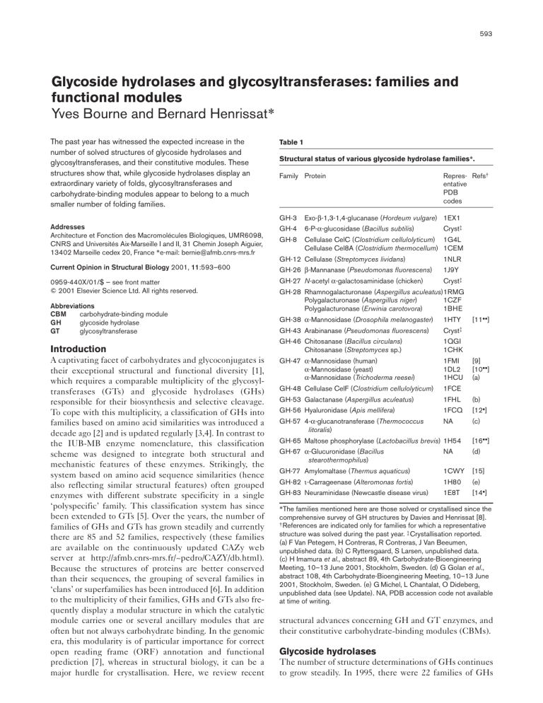
Table 1
GH-3
Exo-β-1,3-1,4-glucanase (Hordeum vulgare) 1EX1
Addresses
Architecture et Fonction des Macromolécules Biologiques, UMR6098,
CNRS and Universités Aix-Marseille I and II, 31 Chemin Joseph Aiguier,
13402 Marseille cedex 20, France *e-mail: bernie@afmb.cnrs-mrs.fr
GH-4
6-P-α-glucosidase (Bacillus subtilis)
GH-8
Cellulase CelC (Clostridium cellulolyticum) 1G4L
Cellulase Cel8A (Clostridium thermocellum) 1CEM
Structural status of various glycoside hydrolase families*.
Family Protein
GH-12 Cellulase (Streptomyces lividans)
Repres- Refs†
entative
PDB
codes
Cryst‡
1NLR
Current Opinion in Structural Biology 2001, 11:593–600
GH-26 β-Mannanase (Pseudomonas fluorescens)
1J9Y
0959-440X/01/$ — see front matter
© 2001 Elsevier Science Ltd. All rights reserved.
GH-27 N-acetyl α-galactosaminidase (chicken)
Cryst‡
GH-28 Rhamnogalacturonase (Aspergillus aculeatus)1RMG
Polygalacturonase (Aspergillus niger)
1CZF
Polygalacturonase (Erwinia carotovora)
1BHE
Abbreviations
CBM
carbohydrate-binding module
GH
glycoside hydrolase
GT
glycosyltransferase
GH-38 α-Mannosidase (Drosophila melanogaster)
1HTY
GH-43 Arabinanase (Pseudomonas fluorescens)
Cryst‡
Introduction
GH-46 Chitosanase (Bacillus circulans)
Chitosanase (Streptomyces sp.)
1QGI
1CHK
GH-47 α-Mannosidase (human)
α-Mannosidase (yeast)
α-Mannosidase (Trichoderma reesei)
1FMI
1DL2
1HCU
GH-48 Cellulase CelF (Clostridium cellulolyticum)
1FCE
A captivating facet of carbohydrates and glycoconjugates is
their exceptional structural and functional diversity [1],
which requires a comparable multiplicity of the glycosyltransferases (GTs) and glycoside hydrolases (GHs)
responsible for their biosynthesis and selective cleavage.
To cope with this multiplicity, a classification of GHs into
families based on amino acid similarities was introduced a
decade ago [2] and is updated regularly [3,4]. In contrast to
the IUB-MB enzyme nomenclature, this classification
scheme was designed to integrate both structural and
mechanistic features of these enzymes. Strikingly, the
system based on amino acid sequence similarities (hence
also reflecting similar structural features) often grouped
enzymes with different substrate specificity in a single
‘polyspecific’ family. This classification system has since
been extended to GTs [5]. Over the years, the number of
families of GHs and GTs has grown steadily and currently
there are 85 and 52 families, respectively (these families
are available on the continuously updated CAZy web
server at http://afmb.cnrs-mrs.fr/~pedro/CAZY/db.html).
Because the structures of proteins are better conserved
than their sequences, the grouping of several families in
‘clans’ or superfamilies has been introduced [6]. In addition
to the multiplicity of their families, GHs and GTs also frequently display a modular structure in which the catalytic
module carries one or several ancillary modules that are
often but not always carbohydrate binding. In the genomic
era, this modularity is of particular importance for correct
open reading frame (ORF) annotation and functional
prediction [7], whereas in structural biology, it can be a
major hurdle for crystallisation. Here, we review recent
[11••]
[9]
[10••]
(a)
GH-53 Galactanase (Aspergillus aculeatus)
1FHL
(b)
GH-56 Hyaluronidase (Apis mellifera)
1FCQ
[12•]
GH-57 4-α-glucanotransferase (Thermococcus
litoralis)
NA
(c)
GH-65 Maltose phosphorylase (Lactobacillus brevis) 1H54
[16••]
GH-67 α-Glucuronidase (Bacillus
stearothermophilus)
NA
(d)
GH-77 Amylomaltase (Thermus aquaticus)
1CWY
[15]
GH-82 ι-Carrageenase (Alteromonas fortis)
1H80
(e)
GH-83 Neuraminidase (Newcastle disease virus)
1E8T
[14•]
*The families mentioned here are those solved or crystallised since the
comprehensive survey of GH structures by Davies and Henrissat [8].
† References are indicated only for families for which a representative
structure was solved during the past year. ‡ Crystallisation reported.
(a) F Van Petegem, H Contreras, R Contreras, J Van Beeumen,
unpublished data. (b) C Ryttersgaard, S Larsen, unpublished data.
(c) H Imamura et al., abstract 89, 4th Carbohydrate-Bioengineering
Meeting, 10–13 June 2001, Stockholm, Sweden. (d) G Golan et al.,
abstract 108, 4th Carbohydrate-Bioengineering Meeting, 10–13 June
2001, Stockholm, Sweden. (e) G Michel, L Chantalat, O Dideberg,
unpublished data (see Update). NA, PDB accession code not available
at time of writing.
structural advances concerning GH and GT enzymes, and
their constitutive carbohydrate-binding modules (CBMs).
Glycoside hydrolases
The number of structure determinations of GHs continues
to grow steadily. In 1995, there were 22 families of GHs
594
Carbohydrates and glycoconjugates
Figure 1
Ribbon diagrams of (a) Saccharomyces
cerevisiae α-mannosidase I (PDB accession
code 1DL2) and (b) Drosophila
α-mannosidase II (PDB accession code
1HTY) — representative of GH families GH-47
and GH-38, respectively. β Sheets are
coloured in cyan and α helices are in red.
Figure prepared with SPOCK [45] and
Raster3D [46].
with known representative structures [8]. The number of
families with known structures has almost doubled since to
reach 38 at the time of writing, with no less than 9 families
reported since January 2000 (see Table 1). These new 3D
structures have further expanded the extraordinary variety
of folds exhibited by these enzymes (for a review, see [8])
and suggest that yet more folds might exist for families
that are still awaiting structural determination. A good
example is the α-mannosidases, which are commonly
found in GH families GH-38 and GH-47 [9,10••,11••].
Remarkably, the representative structures of each family
revealed a novel fold (Figure 1)!
In addition, several other structures determined in 2000
have reinforced the families of folds already known for
GH enzymes. For example, bee venom hyaluronidase,
the first structure reported for family GH-56, resembles a
classical triose phosphate isomerase, except that the barrel is composed of only seven strands [12•]. The structure
of the complex of the hyaluronidase with a substrate
analogue suggests a molecular mechanism involving
anchimeric assistance of the N-acetyl group of the substrate in catalysis. Such a mechanism has been confirmed
for family GH-20 by the structure of Streptomyces plicatus
β-N-acetylhexosaminidase in complex with N-acetylglucosamine-thiazoline [13]. Despite insignificant sequence
similarity with bacterial and influenza virus neuraminidases, the structure of the family GH-83
hemagglutinin-neuraminidase from Newcastle disease
virus revealed a typical neuraminidase active site within
a β-propeller fold [14•]. This clear resemblance allows
the inclusion of family GH-83 in a superfamily called
‘clan GH-E’, which already contained families GH-33
and GH-34 (for more on clans, see [6]).
In a similar vein, the first structural description of a family
GH-77 member has also been accomplished with the
resolution of the structure of Thermus aquaticus amylomaltase, a transglycosylating enzyme that produces amylose
macrocycles. The (β/α)8 structure reveals a clear resemblance to family GH-13 (also known as the α-amylase
superfamily) members [15].
Family GH-65 groups trehalases together with maltose
phosphorylases. The structure of maltose phosphorylase
from Lactobacillus brevis shows a striking resemblance to
the (α/α)6-barrel structure of family GH-15 glucoamylases
[16••]. This resemblance allows the creation of a new clan
of GHs (‘clan GH-L’, comprising families GH-65 and
GH-15) and provides a remarkable illustration of how little
it takes to evolve an inverting GH into a phosphorylase by
recruiting phosphate instead of water as the nucleophile in
the single displacement mechanism (see also Update).
Structures have also been reported for families that already
had a known structural representative, but these are too
numerous to all be reported here. Amongst the most
notable of these structures, one can mention a family GH-1
plant β-glucosidase, whose narrow substrate specificity for
aromatic aglycones is dictated by a slot-like aglycone-binding
subsite [17]. In the same family, Burmeister et al. [18•] have
reported several high-resolution structures of the plant
defence protein myrosinase in complex with inhibitors
and ascorbate. Another interesting structure published
Glycoside hydrolases and glycosyltransferases Bourne and Henrissat
595
Table 2
Glycosyltransferases: families and structures (May 2001).
Family
Protein
Fold
Representative
PDB codes
References*
GT-1
GtfB (Amycolatopsis orientalis)
GT-2
SpsA (Bacillus subtilis)
GT-B
1IIR
[29]
GT-A
1QG8
GT-6
α-1,3-galactosyltransferase (bovine)
GT-A
1FG5
[27•]
GT-7
β-1,4-galactosyltransferase β4GalT1 (bovine)
GT-A
1FGX
GT-8
α-1,4-galactosyltransferase LgtC (Neisseria meningitidis)
Glycogenin (rabbit)
GT-A
1GA8
NA
[26••]
(a)
GT-13
β-1,2-N-acetylglucosaminyltransferase GnT1 (rabbit)
GT-A
1FO8
[24]
GT-28
β-1,4-N-acetylglucosaminyltransferase MurG
GT-B
1FOK
[47]
GT-35
Maltodextrin phosphorylase (E. coli)
Glycogen phosphorylase (human)
Glycogen phosphorylase (rabbit)
Glycogen phosphorylase (yeast)
GT-B
1AHP
1EM6
1ABB
1YGP
GT-43
β-1,3-glucuronyltransferase (human)
GT-A
1FGG
NC
β-Glucosyltransferase (bacteriophage T4)
GT-B
1BGT
[25]
*References are indicated only for families for which a representative structure was solved during the past year. (a) BJ Gibbons, PJ Roach, TD
Hurley, abstract W0269, Annual Meeting of the American Crystallographic Association, 21–26 July 2001, Los Angeles, CA. NA, PDB accession
code not available at time of writing; NC, nonclassified.
this year was that of family GH-18 endo-β-N-acetylglucosaminidase F3 from Flavobacterium meningosepticum [19].
The structure of Bacillus agaradhaerens cellulase Cel5A
(family GH-5) in complex with a substrate analogue in
which a single α-1,4 glycosidic bond was incorporated into
an otherwise all β-1,4-linked oligosaccharide has led to the
discovery of a whole new class of cellulase inhibitors.
These inhibitors have affinities that are 150 times better
than that observed for an all β-linked compound [20•].
Finally, and very recently, the mechanism of hen eggwhite lysozyme has been revisited by a clever alliance of
mutagenesis, organic chemistry, mass spectrometry and
X-ray crystallography. The outcome, that hen egg-white
lysozyme does form a covalent glycosyl–enzyme intermediate using Asp52, puts an end to a long-lived controversy
and discards the ion-pair intermediate hypothesis found in
all textbooks [21••].
Glycosyltransferases
While only two GT structures were solved by 1995 (rabbit
muscle glycogen phosphorylase and bacteriophage T4
β-glucosyltransferase), structures of representatives of nine
families of GTs have now been solved in total, with structures reported for six families since January 2000 (Table 2).
The folds of glycosyltransferases
In marked contrast to the wide variety of folds displayed
by the GHs, GTs are less ‘exciting’ if one considers only
their folds. So far, all GT structures adopt only two folds
[22]. By analogy to the GH clans, we name these two
folds ‘GT-A’ and ‘GT-B’ (Table 2). The GT-A fold, best
represented by family GT-2 SpsA from Bacillus subtilis [23],
comprises two dissimilar domains, one involved in
nucleotide binding (called the SGC domain in [22]) and
the other binding the acceptor. The structures of several
GTs sharing this fold have been solved recently, including
rabbit N-acetylglucosaminyltransferase GnT1 (family
GT-13) [24] and human β-1,3-glucuronyltransferase I
(family GT-43) [25].
While all previously determined GT structures were
‘inverting’ enzymes (e.g. those that produce β-bonds from
α-linked nucleotide sugars), Persson et al. [26••] have
accomplished remarkable work with the structural resolution
of a ‘retaining’ enzyme from family GT-8, the bacterial
α-1,4-galactosyltransferase LgtC from Neisseria meningitidis.
Shortly after, the structure of bovine α-1,3-galactosyltransferase (family GT-6), another retaining enzyme, was also
reported [27•]. Both structures adopt the GT-A fold.
The GT-B fold, originally found in phage T4 DNA-glucosyltransferase [28] and characterised by two similar
Rossmann fold subdomains, is also found in families
GT-28 and GT-35 (Table 2). Mulichak et al. [29] have
completed this folding family with the crystal structure of
the family GT-1 UDP-glucosyltransferase GtfB, which is
involved in the biosynthesis of the vancomycin group
of antibiotics.
As only two large folding superfamilies (GT-A and GT-B)
have emerged so far for GTs, we have submitted the
remaining, unsolved families to threading analyses [30,31].
The results suggest that the other families will fold either
like GT-A (examples include GT-12, GT-21 and GT-27) or
like GT-B (for instance GT-4, GT-5, GT-9, GT-19, GT-20,
GT-30 and GT-33). A number of families resist the
threading analyses, suggesting that other folds perhaps
exist. This optimism should be moderated by the fact that
596
Carbohydrates and glycoconjugates
Figure 2
Stereo view of the superimposition of four
structures from the GT-A fold family around
the active site: SpsA in cyan (family GT-2),
GnT1 in orange (family GT-13), β4GalT1 in
green (family GT-7 [48]) and
glucuronyltransferase 1 in magenta (family
GT-43). The catalytic base appears at the top
in the same colour code. The sugar-nucleotide
donor around the manganese-binding region
is shown at the bottom. For clarity, the ligands
of the manganese ion (amino acid sidechains,
solvent and nucleotide-sugar) are shown only
for GnT1 (PDB accession code 1FOA).
nucleotide binding may constitute an important constraint,
which might prevent the proliferation of folds seen with
the glycosidases. Finally, the high sensitivity of threading
analyses has an unexpected drawback: the fold might be
conserved, but not the function. Each of the two GT
folding superfamilies has counterparts with significant
structural similarity detectable by these threading analyses.
Thus, a bacterial UDP-N-acetylglucosamine 2-epimerase
clearly belongs to the GT-B fold [29,32]. In a similar
fashion, the bacterial glucosamine-1-phosphate pyrophosphorylase GlmU shows substantial resemblance to the
GT-A fold [33].
The mechanism of glycosyltransferases
In contrast to the GH ‘clans’, in which the catalytic mechanism is strictly conserved, the two folding superfamilies
of GTs group families of retaining and inverting enzymes
together. Whereas the mechanism of inverting GTs
appears reasonably well understood (a single displacement reaction with base activation of the acceptor), that of
retaining enzymes remains poorly understood. Although a
double displacement is deemed necessary, no evidence of
a glycosyl–enzyme intermediate has yet been found for
retaining GTs and the nature of the enzymatic nucleophile is the subject of debate [34]. By direct analogy with
the GHs, aspartic acid or glutamic acid groups are excellent candidates for this task. Because these residues are
sometimes not conserved in certain GT families or are
sometimes not located appropriately in the active site,
however, alternative nucleophiles have to be identified.
The carbonyl oxygen in an amide sidechain or even
perhaps in the mainchain could, in principle, also play
the role of the nucleophile, as shown by the retaining
N-acetylglucosaminidases, in which the enzymatic nucleophile is replaced by the acetamido group of the
substrate. More structural work and mechanism-based
inhibitors of retaining GTs are needed to resolve this
important issue.
Another important mechanistic feature emerging from 3D
structures of GTs is the interplay between the donor and
acceptor subsites: in some cases, it is only upon binding
the nucleotide-sugar that the acceptor-binding site
becomes fully functional (for instance, GnT1 and LgtC),
whereas the reverse happens in other cases (for instance,
GtfB). Perhaps important is the presence of a hinge region,
typical of the GT-B fold, that separates the two constitutive subdomains and whose flexibility might be critical for
specificity and catalysis. A beautiful example of the conformational restraints required within the acceptor-binding
site should soon be demonstrated by the expected structure of rabbit glycogenin from family GT-8 (BJ Gibbons, PJ
Roach, TD Hurley, abstract W0269, Annual Meeting of
the American Crystallographic Association, 21–26 July
2001, Los Angeles, CA). This enzyme has the unique
property of self-transferring several glucose moieties
from a nucleotide-glucose donor to a tyrosine residue, forming a 10-residue α-1,4-glucan chain sufficient to initiate
glycogen biosynthesis.
The two main superfamilies of GTs also diverge in the
utilisation of divalent cations. In all characterised members
Glycoside hydrolases and glycosyltransferases Bourne and Henrissat
of the GT-A fold superfamily, there is a strong structural
restraint on the coordination of a metal ion by two phosphate
oxygens of the nucleotide and sidechain residues from the
protein (Figure 2). These sidechains are often called the
‘DXD motif’ [35], although the aspartic acid residues in
this motif can be replaced by other residues. In contrast,
members of the GT-B fold superfamily do not have such a
motif and no metal has been identified clearly in the 3D
structures solved so far, even though some (but not all) of
these enzymes have been reported to be metal-dependent
(see also Update).
Structures of carbohydrate-binding modules
and entire multimodular enzymes
The CBMs also form sequence-based families (27 at the
time of writing) and they have also witnessed a recent
acceleration in the number of structural determinations,
with three families structurally depicted by 1995 and thirteen now, with five families described since January 2000
(Table 3). Because of their frequent small size, a significant number of CBM structures have been determined
by NMR.
Isolated carbohydrate-binding modules
Five families of CBMs have seen their first structural
characterisation recently. The xylan-binding module of
xylanase A from Pseudomonas fluorescens ssp cellulosa
(family CBM-10) consists of two antiparallel β sheets, one
with two strands and one with three, with a short α helix
across one face of the three-stranded sheet [36]. The 3D
structure of the family CBM-12 module of chitinase A1
from Bacillus circulans WL-12 was determined by NMR
[37]. This module, which binds chitin, has a compact
twisted β-sandwich structure reminiscent of that found in
family CBM-5 [38]. Family CBM-14 contains chitin-binding
modules either borne by chitinases or existing in isolation,
such as the chitin-binding protein tachycitin, a 73-residue
antimicrobial polypeptide from Tachypleus tridentatus. The
3D structure of tachycitin shares some similarity with the
chitin-binding modules of family CBM-18; this has been
proposed to have arisen by convergent evolution [39]. In
an elegant piece of work, Charnock and co-workers [40]
have reported both the function and the X-ray structure
of a family CBM-22 module that binds xylan. This work
also showed that some CBM-22 family members have
lost their polysaccharide-binding function. Finally, the
β-sandwich structure of the C-terminal CBM-9 module of
Thermotoga maritima xylanase 10A has been determined
alone and in complex with cellobiose by X-ray crystallography [41•]. The stunning result is that the T. maritima
CBM appears to bind selectively to the reducing ends
of cellulose.
Because of their relative small size, CBMs fold predominantly as all-β proteins. Like the hydrolases and transferases,
superfamilies are also beginning to emerge for the CBMs.
For instance, families CBM-4 and CBM-22 are clearly
related and, based on sequence motifs, it has been proposed
597
Table 3
Carbohydrate-binding modules: families and structures
(May 2001).
Family
Protein
Representative
PDB
codes
Refs*
CBM-1
Cellulase Cel7A (Trichoderma reesei)
1CBH
CBM-2
Xylanase Xyn10A (Cellulomonas fimi)
Xylanase Xyn11A (Cellulomonas fimi)
1EXG
2XBD
CBM-3
Scaffoldin (Clostridium cellulolyticum)
Scaffoldin (Clostridium thermocellum)
Cellulase Cel9A (Thermobifida fusca)
1G43
1NBC
1TF4
CBM-4
Cellulase Cel9B (Cellulomonas fimi)
1ULO
CBM-5
Cellulase Cel5A (Erwinia chrysanthemi)
1AIW
CBM-6
Xylanase U (Clostridium thermocellum)
NA
(a)
CBM-9
Xylanase Xyn10A (Thermotoga maritima)
1I8A
[41•]
CBM-10 Xylanase Xyn10A (Pseudomonas
fluorescens)
1QLD
[36]
CBM-12 Chitinase A1 (Bacillus circulans)
Chitinase B (Serratia marcescens)
1ED7
1E15
[37]
[44]
CBM-13 Xylanase (Streptomyces olivaceoviridis)
Ricin (Ricinus communis)
Ebulin (Sambucus ebulus)
1XYF
2AAI
1HWM
[43•]
[49]
CBM-14 Tachycitin (Tachypleus tridentatus)
1DQC
[39]
CBM-17 Endoglucanase EngF (Clostridium
cellulovorans)
1J83
(b)
CBM-18 Hevein (Hevea brasiliensis)
Antimicrobial peptide 2 (Amaranthus
caudatus)
Agglutinin (Triticum aestivum)
1HEV
1MMC
1WGC
CBM-20 Glucoamylase (Aspergillus niger)
β-Amylase (Bacillus cereus)
1KUM
1CQY
CBM-22 Xylanase Xyn10B (Clostridium
thermocellum)
1DYO
[40]
*References are indicated only for families for which a representative
structure was solved during the past year. (a) M Czjzek et al., abstract
131, 4th Carbohydrate-Bioengineering Meeting, 10–13 June 2001,
Stockholm, Sweden. (b) V Notenboom, B Boraston, A Freelove,
D Kilburn, DR Rose, unpublished data. NA, PDB accession code not
available at time of writing.
that they form a superfamily with families CBM-16,
CBM-17 and CBM-27 [42].
Entire multimodular enzymes
The flexibility of the linker peptides connecting the various modules makes intact modular enzymes with a
catalytic module and a CBM particularly recalcitrant to
crystallisation. As a consequence, there are only a very few
solved structures of intact modular GHs and none of a
modular GT. In this respect, the two entire modular GHs
solved this year probably represent a tour de force. The
first was a xylanase from Streptomyces olivaceoviridis featuring a catalytic module from family GH-10 carrying a
xylan-binding module from family CBM-13 [43•]. The
other is chitinase B from Serratia marcescens comprising a
family GH-18 catalytic module and a C-terminal chitinbinding module from family CBM-12 [44] (Figure 3).
598
Carbohydrates and glycoconjugates
Figure 3
Ribbon diagrams of (a) S. olivaceoviridis
family GH-10 xylanase (yellow) linked to a
CBM from family CBM-13 (cyan) (PDB
accession code 1XYF) and (b) S. marcescens
family GH-18 chitinase B (yellow) linked to a
CBM from family CBM-12 (cyan) (PDB
accession code 1E15) via a long ordered
linker (orange).
Conclusions
References and recommended reading
While the number of enzyme families (GH or GTs) will
grow relatively slowly now, a systematic analysis of protein
modularity should reveal novel families of noncatalytic
modules (PM Coutinho, B Henrissat, unpublished data),
some of which might turn out to be CBMs. A major challenge remains the structural elucidation of multimodular
enzymes: some have over ten different modules! Indeed,
the adjunction of multiple CBMs to the catalytic modules
is probably a convenient way to build larger active sites
from pre-existing scaffolds and these extended sites allow
the perception of ligand structures remote from the site of
catalysis itself.
Papers of particular interest, published within the annual period of review,
have been highlighted as:
Update
The crystal structure of the muramidase from Streptomyces
coelicolor has recently been determined [50]. This structure
(PBD accession code 1JFX) is the first reported for a
family GH-25 member. The structure of the mannanase
from Pseudomonas cellulosa (family GH-26; Table 1; PBD
accession code 1J9Y) has now been published [51]. Recent
work has demonstrated that T4 phage β-glucosyltransferase, which adopts the GT-B fold, can bind metal ions
near the β-phosphate of the nucleotide [52].
• of special interest
•• of outstanding interest
1.
Laine RA: A calculation of all possible oligosaccharide isomers
both branched and linear yields 1.05 x 10¹² structures for a
reducing hexasaccharide: the Isomer Barrier to development of
single-method saccharide sequencing or synthesis systems.
Glycobiology 1994, 4:759-767.
2.
Henrissat B: A classification of glycosyl hydrolases based on
amino acid sequence similarities. Biochem J 1991,
280:309-316.
3.
Henrissat B, Bairoch A: New families in the classification of
glycosyl hydrolases based on amino acid sequence similarities.
Biochem J 1993, 293:781-788.
4.
Henrissat B, Bairoch A: Updating the sequence-based
classification of glycosyl hydrolases. Biochem J 1996,
316:695-696.
5.
Campbell JA, Davies GJ, Bulone V, Henrissat B: A classification
of nucleotide-diphospho-sugar glycosyltransferases
based on amino acid sequence similarities. Biochem J 1997,
326:929-939.
6.
Henrissat B, Davies G: Structural and sequence-based
classification of glycoside hydrolases. Curr Opin Struct Biol 1997,
7:637-644.
7.
Henrissat B, Davies GJ: Glycoside hydrolases and
glycosyltransferases: families, modules and implications for
genomics. Plant Physiol 2000, 124:1515-1519.
8.
Davies G, Henrissat B: Structures and mechanisms of glycosyl
hydrolases. Structure 1995, 3:853-859.
9.
Vallée F, Karaveg K, Herscovics A, Moremen KW, Howell PL:
Structural basis for catalysis and inhibition of N-glycan
processing class I α1,2-mannosidases. J Biol Chem 2000,
275:41287-41298.
Acknowledgements
We would like to thank Jim Rini, Michael Garavito, David Rose and
Herman van Tilbeurgh for providing the coordinates and preprints for
GnT1, GtfB, α-mannosidase II and maltose phosphorylase, respectively,
before release.
Glycoside hydrolases and glycosyltransferases Bourne and Henrissat
10. Vallée F, Lipari F, Yip P, Sleno B, Herscovics A, Howell PL: Crystal
•• structure of a class I α1,2-mannosidase involved in N-glycan
processing and endoplasmic reticulum quality control. EMBO J
2000, 19:581-588.
The first crystal structure of S. cerevisiae α-1,2-mannosidase I reveals a novel
(α/α)7-barrel fold for family GH-47. The unexpected presence of a fully ordered
N-glycan within the active site of a neighbouring molecule in the crystal provides
a detailed description of the catalytic mechanism involving a calcium ion
11. van den Elsen JMH, Kuntz DA, Rose DR: Structure of Golgi α
•• mannosidase II: a target for inhibition of growth and metastasis
of cancer cells. EMBO J 2001, 20:3008-3017.
The structure of the enormous (1108 residues) Drosophila Golgi α-mannosidase II has been solved in the presence of the anticancer agent swainsonine and the inhibitor deoxymannojirimicin. The structure reveals a novel
protein fold for family GH-38, consisting of an N-terminal α/β domain, a
three-helical bundle and an all-β C-terminal domain forming a single compact
entity. A zinc atom appears to be involved both in the substrate specificity of
the enzyme and directly in the catalytic mechanism.
12. Markovic-Housley Z, Miglierini G, Soldatova L, Rizkallah PJ, Muller U,
•
Schirmer T: Crystal structure of hyaluronidase, a major allergen of
bee venom. Structure 2000, 8:1025-1035.
This first structural determination for family GH-56 not only reveals the overall
topology of bee venom hyaluronidase but also suggests a molecular
mechanism involving anchimeric assistance of the N-acetyl group of the
substrate for catalysis.
13. Mark BL, Vocadlo DJ, Knapp S, Triggs-Raine BL, Withers SG, James MN:
Crystallographic evidence for substrate-assisted catalysis in a
bacterial β-hexosaminidase. J Biol Chem 2001, 276:10330-10337.
599
22. Ünligil UM, Rini JM: Glycosyltransferase structure and mechanism.
Curr Opin Struct Biol 2000, 10:510-517.
23. Charnock SJ, Davies GJ: Structure of the nucleotide-diphosphosugar transferase, SpsA from Bacillus subtilis, in native and
nucleotide-complexed forms. Biochemistry 1999, 38:6380-6385.
24. Ünligil U, Zhou S, Yuwaraj S, Sarkar M, Schachter H, Rini J: X-ray
crystal structure of rabbit N-acetylglucosaminyltransferase I:
catalytic mechanism and a new protein superfamily. EMBO J
2000, 19:5269-5280.
25. Pedersen LC, Tsuchida K, Kitagawa H, Sugahara K, Darden TA,
Negishi M: Heparan/chondroitin sulfate biosynthesis: structure
and mechanism of human glucuronyltransferase I. J Biol Chem
2000, 275:34580-34585.
26. Persson K, Ly HD, Dieckelmann M, Wakarchuk WW, Withers SG,
•• Strynadka NC: Crystal structure of the retaining galactosyltransferase
LgtC from Neisseria meningitidis in complex with donor and acceptor
sugar analogs. Nat Struct Biol 2001, 8:166-175.
The first structure of a retaining GT. This beautiful piece of work, which
combines X-ray crystallography, carbohydrate chemistry and mutagenesis,
also reports the first structure of a ternary complex containing both the
sugar-nucleotide donor and an acceptor analogue. Although these combined approaches have not uncovered all the details of the catalytic mechanism, they have revealed that the enzyme does not have a carboxylic
nucleophile equivalent to that of the retaining GHs.
14. Crennell S, Takimoto T, Portner A, Taylor G: Crystal structure of the
•
multifunctional paramyxovirus hemagglutinin-neuraminidase. Nat
Struct Biol 2000, 7:1068-1074.
The crystal structure of the family GH-83 hemagglutinin-neuraminidase from
Newcastle disease virus bound to either an inhibitor or the β-anomer of sialic
acid reveals a typical neuraminidase active site within a β-propeller fold.
Gastinel LN, Bignon C, Misra AK, Hindsgaul O, Shaper JH,
Joziasse DH: Bovine α1,3-galactosyltransferase catalytic domain
structure and its relationship with ABO histo-blood group and
glycosphingolipid glycosyltransferases. EMBO J 2001, 20:638-649.
This enzyme synthesises the epitope that causes hyperacute rejection
observed in pig-to-human xenotransplantation. The crystal structure of this
retaining glycosyltransferase in complex with a modified UDP-Gal sugar
donor allowed the authors to describe a catalytic mechanism that, unlike
LgtC, involves the formation of a covalent glycosyl–enzyme intermediate.
15. Przylas I, Tomoo K, Terada Y, Takaha T, Fujii K, Saenger W, Strater N:
Crystal structure of amylomaltase from Thermus aquaticus, a
glycosyltransferase catalysing the production of large cyclic
glucans. J Mol Biol 2000, 296:873-886.
28. Vrielink A, Ruger W, Driessen HP, Freemont PS: Crystal structure of
the DNA modifying enzyme β-glucosyltransferase in the presence
and absence of the substrate uridine diphosphoglucose. EMBO J
1994, 13:3413-3422.
16. Egloff MP, Uppenberg J, Haalck L, van Tilbeurgh H: Crystal structure
•• of maltose phosphorylase from Lactobacillus brevis: unexpected
evolutionary relationship with glucoamylases. Structure 2001,
9:689-697.
Maltose phosphorylase is a family GH-65 enzyme that catalyses the conversion
of maltose and inorganic phosphate into β-D-glucose-1-phosphate and glucose,
without any cofactor. The 3D structure strongly suggests that this enzyme,
which has evolved from family GH-14 glucoamylase, has conserved one
carboxylate group for acid catalysis and has exchanged the catalytic base for
a phosphate-binding pocket.
29. Mulichak AM, Losey HC, Walsh CT, Garavito RM: Structure of the
UDP-glucosyltransferase GtfB that modifies the heptapeptide
aglycone in biosynthesis of the vancomycin group of antibiotics.
Structure 2001, 9:547-557.
17.
Czjzek M, Cicek M, Zamboni V, Bevan DR, Henrissat B, Esen A: The
mechanism of substrate (aglycone) specificity in β-glucosidases
is revealed by crystal structures of mutant maize β-glucosidaseDIMBOA, -DIMBOAGlc, and -dhurrin complexes. Proc Natl Acad
Sci USA 2000, 97:13555-13560.
18. Burmeister WP, Cottaz S, Rollin P, Vasella A, Henrissat B: High
•
resolution X-ray crystallography shows that ascorbate is a
cofactor for myrosinase and substitutes for the function of the
catalytic base. J Biol Chem 2000, 275:39385-39393.
Several high-resolution structures of myrosinase in complex with inhibitors
and/or L-ascorbate have brought a final answer to the question of the missing
acid/base catalyst and the particular activation of the enzyme by ascorbate.
19. Waddling CA, Plummer TH, Tarentino AL, Van Roey P: Structural
β-Nbasis for the substrate specificity of endo-β
acetylglucosaminidase F3. Biochemistry 2000, 39:7878-7885.
20. Fort S, Varrot A, Schülein M, Cottaz S, Driguez H, Davies GJ: Mixed
•
linkage cellooligosaccharides: a new class of glycoside hydrolase
inhibitors. Chem Biochem 2001, 2:319-325.
A beautiful example of unexpected synergy between chemistry (and its pitfalls) X-ray crystallography has led the authors to design a clever class of
inhibitors that ‘by-pass’ the catalytic subsite while maintaining binding to the
surrounding subsites of endoglucanase Cel5A from B. agaradhaerens.
21. Vocadlo DJ, Davies GJ, Laine R, Withers SG: Catalysis by hen egg
•• white lysozyme proceeds via a covalent intermediate. Nature
2001, 412:835-838.
This paper brings the long debate on the nature of the reaction intermediate
in egg-white lysozyme catalysis to an end. Most textbooks will need to be
updated, with the covalent glycosyl–enzyme intermediate replacing the
famous (but erroneous) ion pair.
27.
•
30. Jones DT: GenTHREADER: an efficient and reliable protein fold
recognition method for genomic sequences. J Mol Biol 1999,
287:797-815.
31. Kelley LA, MacCallum RM, Sternberg MJ: Enhanced genome
annotation using structural profiles in the program 3D-PSSM.
J Mol Biol 2000, 299:499-520.
32. Campbell RE, Mosimann SC, Tanner ME, Strynadka NC: The
structure of UDP-N-acetylglucosamine 2-epimerase reveals
homology to phosphoglycosyl transferases. Biochemistry 2000,
39:14993-15001.
33. Brown K, Pompeo F, Dixon S, Mengin-Lecreulx D, Cambillau C,
Bourne Y: Crystal structure of the bifunctional
N-acetylglucosamine 1-phosphate uridyltransferase from
Escherichia coli: a paradigm for the related pyrophosphorylase
superfamily. EMBO J 1999, 18:4096-4107.
34. Davies GJ: Sweet secrets of synthesis. Nat Struct Biol 2001,
8:98-100.
35. Wiggins CA, Munro S: Activity of the yeast MNN1 α-1,3mannosyltransferase requires a motif conserved in many other
families of glycosyltransferases. Proc Natl Acad Sci USA 1998,
95:7945-7950.
36. Raghothama S, Simpson PJ, Szabo L, Nagy T, Gilbert HJ, Williamson MP:
Solution structure of the CBM10 cellulose binding module from
Pseudomonas xylanase A. Biochemistry 2000, 39:978-984.
37.
Ikegami T, Okada T, Hashimoto M, Seino S, Watanabe T,
Shirakawa M: Solution structure of the chitin-binding domain of
Bacillus circulans WL-12 chitinase A1. J Biol Chem 2000,
275:13654-13661.
38. Brun E, Moriaud F, Gans P, Blackledge MJ, Barras F, Marion D:
Solution structure of the cellulose-binding domain of the
endoglucanase Z secreted by Erwinia chrysanthemi. Biochemistry
1997, 36:16074-16086.
600
Carbohydrates and glycoconjugates
39. Suetake T, Tsuda S, Kawabata S, Miura K, Iwanaga S, Hikichi K,
Nitta K, Kawano K: Chitin-binding proteins in invertebrates and
plants comprise a common chitin-binding structural motif. J Biol
Chem 2000, 275:17929-17932.
40. Charnock SJ, Bolam DN, Turkenburg JP, Gilbert HJ, Ferreira LM,
Davies GJ, Fontes CM: The X6 ‘thermostabilizing’ domains of
xylanases are carbohydrate-binding modules: structure and
biochemistry of the Clostridium thermocellum X6b domain.
Biochemistry 2000, 39:5013-5021.
41. Notenboom V, Boraston AB, Kilburn DG, Rose DR: Crystal
•
structures of the family 9 carbohydrate-binding module from
Thermotoga maritima xylanase 10A in native and ligand-bound
forms. Biochemistry 2001, 40:6248-6256.
The crystal structure of the C-terminal module of T. maritima xylanase 10A is
the first to be reported for family CBM-9. This work also reveals the first complex of a cellulose-binding CBM bound to cellobiose. The structure suggests
that this CBM binds selectively to the reducing ends of cellulose.
42. Sunna A, Gibbs MD, Bergquist PL: Identification of novel β-mannanand β-glucan binding modules: evidence for a superfamily of
carbohydrate-binding modules. Biochem J 2001, 356:791-798.
43. Fujimoto Z, Kuno A, Kaneko S, Yoshida S, Kobayashi H, Kusakabe I,
•
Mizuno H: Crystal structure of Streptomyces olivaceoviridis E-86
β-xylanase containing xylan-binding domain. J Mol Biol 2000,
300:575-585.
The crystal structure of S. olivaceoviridis xylanase A provides a first view of
a multimodular enzyme from family GH-10 carrying a xylan-binding CBM
from family 13. This structure offers a preview of the overall architecture of
other CBM-13-containing modular enzymes, such as GTs from family GT-27.
44. van Aalten DM, Synstad B, Brurberg MB, Hough E, Riise BW,
Eijsink VG, Wierenga RK: Structure of a two-domain
chitotriosidase from Serratia marcescens at 1.9-Å resolution. Proc
Natl Acad Sci USA 2000, 97:5842-5847.
45. Christopher JA: SPOCK: the Structural Properties Observation and
Calculation Kit Program Manual. Texas: The Center for
Macromolecular Design, Texas A&M University, College Station; 1998.
46. Merritt EA, Bacon DJ: Raster3D: photorealistic molecular graphics.
Methods Enzymol 1997, 277:505-524.
47.
Ha S, Walker D, Shi Y, Walker S: The 1.9 Å crystal structure of
Escherichia coli MurG, a membrane-associated
glycosyltransferase involved in peptidoglycan biosynthesis.
Protein Sci 2000, 9:1045-1052.
48. Gastinel LN, Cambillau C, Bourne Y: Crystal structures of the
bovine β4galactosyltransferase catalytic domain and its complex
with uridine diphosphogalactose. EMBO J 1999, 18:3546-3557.
49. Pascal JM, Day PJ, Monzingo AF, Ernst SR, Robertus JD, Iglesias R,
Perez Y, Ferreras JM, Citores L, Girbes T: 2.8-Å crystal structure of a
nontoxic type-II ribosome-inactivating protein, ebulin l. Proteins
2001, 43:319-326.
50. Rau A, Hogg T, Marquardt R, Hilgenfeld R: A new lysozyme fold.
Crystal structure of the muramidase from Streptomyces coelicolor
at 1.65 Å resolution. J Biol Chem 2001, 276:31994-31999.
51. Hogg D, Woo EJ, Bolam DN, McKie VA, Gilbert HJ, Pickersgill RW:
Crystal structure of mannanase 26A from Pseudomonas cellulosa
and analysis of residues involved in substrate binding. J Biol
Chem 2001, 276:31186-31192.
52. Moréra S, Larivière L, Kurzeck J, Aschke-Sonnenborn U,
Freedmont PS, Janin J, Rüger W: High resolution crystal
structures of T4 phage β-glucosyltransferase: induced fit
and effect on substrate and metal binding. J Mol Biol 2001,
311:569-577.
Now in press
The work referred to in Table 1 as (G Michel, L Chantalat, O Dideberg,
unpublished data) is now in press:
53. Michel G, Chantalat L, Fanchon E, Henrissat B, Kloareg B,
Dideberg O: The iota-carrageenase of Alteromonas fortis:
a β-helix fold-containing enzyme for the degradation of a highly
polyanionic polysaccharide. J Biol Chem 2001, in press.


