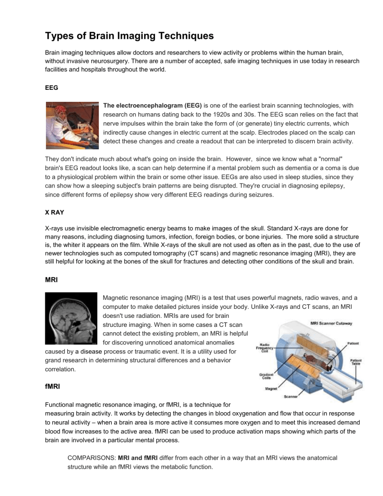
Types of Brain Imaging Techniques Brain imaging techniques allow doctors and researchers to view activity or problems within the human brain, without invasive neurosurgery. There are a number of accepted, safe imaging techniques in use today in research facilities and hospitals throughout the world. EEG The electroencephalogram (EEG) is one of the earliest brain scanning technologies, with research on humans dating back to the 1920s and 30s. The EEG scan relies on the fact that nerve impulses within the brain take the form of (or generate) tiny electric currents, which indirectly cause changes in electric current at the scalp. Electrodes placed on the scalp can detect these changes and create a readout that can be interpreted to discern brain activity. They don't indicate much about what's going on inside the brain. However, since we know what a "normal" brain's EEG readout looks like, a scan can help determine if a mental problem such as dementia or a coma is due to a physiological problem within the brain or some other issue. EEGs are also used in sleep studies, since they can show how a sleeping subject's brain patterns are being disrupted. They're crucial in diagnosing epilepsy, since different forms of epilepsy show very different EEG readings during seizures. X RAY X-rays use invisible electromagnetic energy beams to make images of the skull. Standard X-rays are done for many reasons, including diagnosing tumors, infection, foreign bodies, or bone injuries. The more solid a structure is, the whiter it appears on the film. While X-rays of the skull are not used as often as in the past, due to the use of newer technologies such as computed tomography (CT scans) and magnetic resonance imaging (MRI), they are still helpful for looking at the bones of the skull for fractures and detecting other conditions of the skull and brain. MRI Magnetic resonance imaging (MRI) is a test that uses powerful magnets, radio waves, and a computer to make detailed pictures inside your body. Unlike X-rays and CT scans, an MRI doesn't use radiation. MRIs are used for brain structure imaging. When in some cases a CT scan cannot detect the existing problem, an MRI is helpful for discovering unnoticed anatomical anomalies caused by a disease process or traumatic event. It is a utility used for grand research in determining structural differences and a behavior correlation. fMRI Functional magnetic resonance imaging, or fMRI, is a technique for measuring brain activity. It works by detecting the changes in blood oxygenation and flow that occur in response to neural activity – when a brain area is more active it consumes more oxygen and to meet this increased demand blood flow increases to the active area. fMRI can be used to produce activation maps showing which parts of the brain are involved in a particular mental process. COMPARISONS: MRI and fMRI differ from each other in a way that an MRI views the anatomical structure while an fMRI views the metabolic function. CT or CAT CT, or CAT scans, are special X-ray tests that produce cross-sectional images of the body using X-rays and a computer. CT scans are also referred to as computerized axial tomography. CT scan can help doctors to visualize small nodules or tumors, which they cannot see with a plain film X-ray. CT scan images allow the doctor to look at the inside of the body just as one would look at the inside of a loaf of bread by slicing it. This type of special X-ray, in a sense, takes "pictures" of slices of the body so doctors can look right at the area of interest. CT scans are frequently used to evaluate the brain, neck, spine, chest, abdomen, pelvis, and sinuses. CT is a commonly performed procedure. Scanners are found not only in hospital X-ray departments, but also in outpatient offices. COMPARISONS: CT and MRI are similar to each other, but provide a much different view of the body than an X-ray does. CT and MRI produce cross-sectional images that appear to open the body up, allowing the doctor to look at it from the inside. MRI uses a magnetic field and radio waves to produce images, while CT uses X-rays to produce images. Plain X-rays are an inexpensive, quick test and are accurate at diagnosing things such as pneumonia, arthritis, and fractures. CT and MRI better to evaluate soft tissues such as the brain, liver, and abdominal organs, as well as to visualize subtle abnormalities that may not be apparent on regular X-ray tests. PET Positron Emission Tomography (PET) uses trace amounts of short-lived radioactive material to map functional processes in the brain. When the material undergoes radioactive decay a positron is emitted, which can be picked up by the detector. Areas of high radioactivity are associated with brain activity, because that is where the blood flows to. EEG Electroencephalography (EEG) is the measurement of the electrical activity of the brain by recording from electrodes placed on the scalp. The resulting traces are known as an electroencephalogram (EEG) and represent an electrical signal from a large number of neurons. EEGs are frequently used in experimentation because the process is non-invasive to the research subject. The EEG is capable of detecting changes in electrical activity in the brain on a millisecond-level. It is one of the few techniques available that has such high temporal resolution. MEG Magnetoencephalography (MEG) is an imaging technique used to measure the magnetic fields produced by electrical activity in the brain via extremely sensitive devices known as SQUIDs (which need liquid helium and are very expensive). These measurements are commonly used in both research and clinical settings. There are many uses for the MEG, including assisting surgeons in localizing a pathology, assisting researchers in determining the function of various parts of the brain, neurofeedback, and others. NIRS Near infrared spectroscopy is an optical technique for measuring blood oxygenation in the brain. It works by shining light in the near infrared part of the spectrum (700-900nm) through the skull and detecting how much the remerging light is attenuated. How much the light is attenuated depends on blood oxygenation and thus NIRS can provide an indirect measure of brain activity. Sites Cites: psychcentral.comhttp://psychcentral.com/lib/types-of-brain-imaging-techniques/0001057 https://io9.gizmodo.com/5495712/six-ways-science-can-see-into-your-brain https://www.webmd.com/a-to-z-guides/what-is-an-mri#1 https://www.emedicinehealth.com/ct_scan/article_em.htm http://www.differencebetween.net/technology/difference-between-mri-and-fmri/


