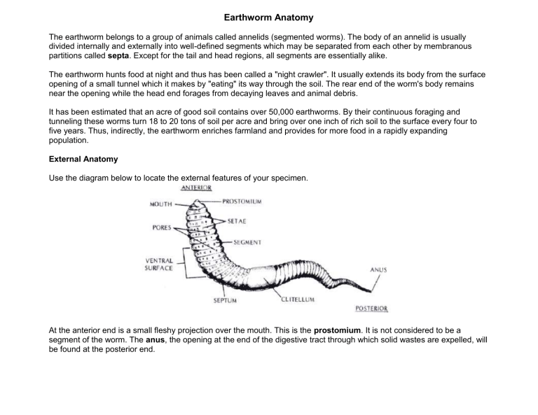
Earthworm Anatomy The earthworm belongs to a group of animals called annelids (segmented worms). The body of an annelid is usually divided internally and externally into well-defined segments which may be separated from each other by membranous partitions called septa. Except for the tail and head regions, all segments are essentially alike. The earthworm hunts food at night and thus has been called a "night crawler". It usually extends its body from the surface opening of a small tunnel which it makes by "eating" its way through the soil. The rear end of the worm's body remains near the opening while the head end forages from decaying leaves and animal debris. It has been estimated that an acre of good soil contains over 50,000 earthworms. By their continuous foraging and tunneling these worms turn 18 to 20 tons of soil per acre and bring over one inch of rich soil to the surface every four to five years. Thus, indirectly, the earthworm enriches farmland and provides for more food in a rapidly expanding population. External Anatomy Use the diagram below to locate the external features of your specimen. At the anterior end is a small fleshy projection over the mouth. This is the prostomium. It is not considered to be a segment of the worm. The anus, the opening at the end of the digestive tract through which solid wastes are expelled, will be found at the posterior end. About one-third of the way back from the mouth is a thick cylindrical collar - the clitellum. This structure is involved in reproduction. Place the worm so that the ventral (lower) side is uppermost. In living worms the ventral surface is a lighter color than the dorsal (upper) surface. With your finger, lightly stroke the ventral surface in an anterior direction. The bristles you feel are the setae and are used by the worm in movement. 1. How many segments does your worm have? _______________________ 2. How many pairs of setae are there in each segment of the worm? _______________________ 3. Which segments of the worm do not have setae? ____________________________________ 4. Which segments are covered by the clitellum? _______________________ Each segment (except the first three and the last one) have pores. The small openings connect with the metanephridia which are the primitive kidneys of the earthworm. Liquid wastes, which collect in the body cavity, are excreted through these nephridial openings. Opening the Body Place the earthworm on the dissecting tray, dorsal side up. Place a pin through the prostomium and the anal segment, as in the diagram below. Use the diagram below as a guide. With forceps, lift the dorsal skin. Insert scissors at the base of the forceps and cut a line (slightly off center) through to the anus. BE CAREFUL to cut only as deep as the skin to avoid damaging the internal organs. Use the diagram below as a guide. Beginning at the anal end, hold the body wall with forceps and with a scalpel, cut through the septa on both sides of the intestine. Cut to within 1/2 to 1 inch of the clitellum. Pin the body wall to the dissecting tray as shown below. Cut forward through the clitellum. BE CAREFUL to cut only as deep as the skin to avoid damaging the internal organs. Sever the septa and pin the body wall to the tray as in the diagram below. Digestive System The diagram below shows the general location and structure of the digestive system. Locate the mouth, just below the overlapping prostomium. The mouth leads to a slightly expanded and muscular pharynx which is usually covered by three pairs of whitish seminal vesicles. Food is passed on by muscular contractions in the pharynx through the exophagus to the crop where it is temporarily stored. The crop opens into a thick-walled, and highly muscular gizzard where food, with the aid of samll soil particles taken in during feeding, is ground up. Food then goes into the intestine where digestion and absorption occur. Solid waste products of digestion are passed to the exterior through the anus. Circulatory System The closed circulatory system of the earthworm moves blood through a series of blood vessels. The blood is red because it contains hemoglobin, the same pigment that gives the red color to human blood. The major vessels of the circulatory system are a dorsal blood vessel laying on top of the digestive tract and a ventral blood vessel lying below the digestive tract. These two vessels are connected with each other by a number of vessels passing around the digestive tract. Five pairs of these (located in segments 7 to 11) are larger than the others and comprise the "hearts". Pulsation of the hearts cause circulation of the blood. Reproductive System Cut through the intestine near the clitellum. Carefully lift it and ease it free as far forward as the posterior end of the pharynx. Then cut it out. Use the diagram below as a guide to locate the reproductive organs. Although earthworms are monoecious, possess a complete set of both male and female reproductive organs, they undergo cross-fertilization during copulation. Two worms come tigether along their ventral sides and release sperm cells from the seminal vesicles into the seminal receptacles of the opposite worm. The animals separate and the clitellum of each worm then secretes a mucus ring. The worm then "backs" out of this ring. As it slides over the anterior segments, it picks up eggs from the oviducts in the 14th segment and sperm then slides off the anterior end of the worm to form theegg cocoon, from which the young eventually hatch. The seminal vesicles are found lying alongside the crop and gizzard. The testes are too small to be seen. Near the seminal vesicles (in segments 9 and 10) are the four small, spherical seminal receptacles. Nervous System The nervous system in the worm is difficult to study. Its major component is the ventral nerve cord, which runs the lingth of the worm on the inner ventral surface. At its anterior end the cord divides and passes around the front part of the pharynx where it enlarges to form two swellings - the cerebral ganglia. These might be considered primitive "brains". Along the length of the cord, lateral nerves are given off which go to the muscles of the body wall. Excretory System Each segment of the body, except the first three and the last one, contains a pair of excretory structures called metanephridia. These coiled tubular structures, lying next to the body wall, open to the exterior by a pore called a nephridiopore. Internally they are connected to the septum of the segment just anterior to them. Each nephridium collects fluid wastes from the segment anterior to the segment in which it is located.
