ARTICLES
PUBLISHED ONLINE: 2 MAY 2016 | DOI: 10.1038/NNANO.2016.62
Spatiotemporal dynamics of the nuclear pore
complex transport barrier resolved by high-speed
atomic force microscopy
Yusuke Sakiyama1, Adam Mazur2, Larisa E. Kapinos1 and Roderick Y. H. Lim1*
Nuclear pore complexes (NPCs) are biological nanomachines that mediate the bidirectional traffic of macromolecules
between the cytoplasm and nucleus in eukaryotic cells. This process involves numerous intrinsically disordered, barrierforming proteins known as phenylalanine-glycine nucleoporins (FG Nups) that are tethered inside each pore. The selective
barrier mechanism has so far remained unresolved because the FG Nups have eluded direct structural analysis within NPCs.
Here, high-speed atomic force microscopy is used to visualize the nanoscopic spatiotemporal dynamics of FG Nups inside
Xenopus laevis oocyte NPCs at timescales of ∼100 ms. Our results show that the cytoplasmic orifice is circumscribed by
highly flexible, dynamically fluctuating FG Nups that rapidly elongate and retract, consistent with the diffusive motion of
tethered polypeptide chains. On this basis, intermingling FG Nups exhibit transient entanglements in the central channel,
but do not cohere into a tightly crosslinked meshwork. Therefore, the basic functional form of the NPC barrier is comprised
of highly dynamic FG Nups that manifest as a central plug or transporter when averaged in space and time.
N
uclear pore complexes mediate the traffic of mRNA, preribosomal proteins and other essential cargoes between the
cytoplasm and nucleus in eukaryotic cells1. After two
decades of work, it is still debated how NPCs promote the selective
transport of cargo-carrying transport receptors known as karyopherins but delay the passage of large nonspecific molecules2.
This problem stems from the fact that the intrinsically disordered,
barrier-forming FG Nups3 have never been directly visualized
inside the pore. In marked contrast, the overall NPC structure has
been refined by cryo-electron tomography (CET)4,5 to a resolution
of ∼20 Å (ref. 6). Inevitably, in vitro experimentation has led to
barrier models that postulate different spatial FG Nup arrangements
in the NPC, but however remain unverified. Briefly, the virtual
gating7,8/polymer brush model9,10 predicts that incoming cargoes
encounter an entropic barrier consisting of rapidly fluctuating
FG Nups that extend away from their tether sites due to confinement and crowding. The selective phase model claims that the FG
Nups span the pore as a tightly crosslinked meshwork based on
cohesive hydrophobic interactions between FG repeats. The size
limit to passive diffusion is then determined by the static mesh
size11,12. Finally, the Forest model3 proposes that cohesive and noncohesive FG Nup regions can assemble into a structure known as
the ‘central plug’ or ‘transporter’4,5,13 (abbreviated to CP/T).
To be precise, nucleocytoplasmic transport in vivo proceeds
through NPCs in a matter of milliseconds14. Therefore, it is the
dynamic spatiotemporal behaviour of the FG Nups rather than
their static time-independent properties that governs the NPC
selective barrier. Yet very little is known about FG Nup dynamics
given the lack of techniques that can first visualize and then
probe their time-dependent behaviour inside NPCs. Electron
microscopy-based approaches require vacuum conditions and
sample freezing/fixation which precludes dynamic observation
although immunogold labels15 can provide static snapshots of
FG Nup localization. Atomic force microscopy (AFM) can provide
time-lapse imaging of native NPCs16 and stiffness measurements17
by invasively indenting into the pore. However, AFM data are typically slow to acquire (on the order of minutes) and lack the spatial
resolution required to resolve the FG Nups18. On the other hand,
FG Nup motion can be studied using fluorescent protein tags19, or
inferred from the localization of fluorescently labelled FG segments,
karyopherins and other transport substrates20,21. However, it is
important to bear in mind that the FG Nups themselves remain structurally invisible in fluorescence-based studies.
Several key features make high-speed AFM (HS-AFM) advantageous for observing NPCs in action. First, stable tapping-mode
imaging is possible in aqueous environments at low piconewton
forces using short cantilevers and nonlinear feedback22. Second,
the tapping force is applied in microsecond pulses, which minimizes
disturbances to the FG Nups as the transfer momentum or impulse
is very small23. Third, at the most rapid scanning speeds, HS-AFM
movies are able to capture dynamic nanoscale movements at ∼100 ms
frame rates, as has been shown for myosin V24, the F1-ATPase25 and
intrinsically disordered proteins26. Fourth, each cantilever presents a
high-aspect-ratio, electron beam-deposited (EBD) amorphous
carbon nanofibre tip23 that is ideal for probing the NPC. In this
work, each homegrown EBD tip is 420 ± 141 nm long and has a tip
radius of 5.5 ± 0.9 nm (see Supplementary Information).
Examining native NPCs directly
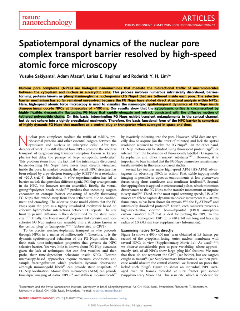
Figure 1a shows a 400 × 400 nm2 scan obtained at 1.8 frames per
second of the cytoplasm-facing, outer nuclear membrane with
several NPCs in view (Supplementary Movie 1a). As usual4–6,16,
we observe considerable pore-to-pore variability, where approximately 40% of all NPCs show large ‘plug-like’ features. We note
that these do not represent the CP/T (see below), but are cargoes
caught in transit16 (see Supplementary Information). As their presence would obscure the central channel, we focused on pores that
lacked such ‘plugs’. Figure 1b shows an individual NPC averaged over 68 frames recorded at 0.74 frames per second
(Supplementary Movie 1b). This scan rate, which is moderate for
1
Biozentrum and the Swiss Nanoscience Institute, University of Basel, Klingelbergstrasse 70, CH-4056 Basel, Switzerland. 2 Research IT, Biozentrum,
University of Basel, CH-4056 Basel, Switzerland. * e-mail: roderick.lim@unibas.ch
NATURE NANOTECHNOLOGY | VOL 11 | AUGUST 2016 | www.nature.com/naturenanotechnology
© 2016 Macmillan Publishers Limited. All rights reserved
719
ARTICLES
NATURE NANOTECHNOLOGY
b
c
1
8
z height (nm)
2
7
*
*
*
10
Cytoplasmic
filament
0
3
Central channel
6
−60
*
d
Pore diameter
20
−40 −20
0
20
40
Distance from pore axis (nm)
4
5
e
60
f
1
Along nuclear filaments
Between nuclear filaments
8
2
3
7
4
6
5
z height (nm)
40
30
20
Nu
cle
ar
fila
me
10
nt
Distal ring
a
DOI: 10.1038/NNANO.2016.62
0
−60
−40 −20
0
20
40
Distance from pore axis (nm)
60
Figure 1 | Observing native NPCs by HS-AFM. a, Numerous NPCs decorate the cytoplasm-facing outer nuclear membrane. Pore-to-pore variability shows
vacant NPCs as well as those that are clogged with large cargoes-in-transit (indicated by white stars). Scale bar, 100 nm. b, Average projected structure of a
vacant NPC showing eight cytoplasmic filaments (numbered) that surround a central pore. Scale bar, 25 nm. c, Average cross-sectional height profile of
b showing that the height of a single cytoplasmic filament is ∼13 nm. Error bars denote the standard deviation. The overall pore diameter is ∼80 nm when
measured from the maxima of opposing filaments, whereas the central channel diameter is ∼40 nm. d, Nuclear baskets protrude away from the inner
nuclear membrane (same scale as a). e, Average structure of a nuclear basket showing eight distinct nuclear filaments (numbered) that fuse into a distal
ring (same scale as b). f, Average cross-sectional height profile of e, showing that the nuclear basket is ∼40-nm tall and ∼120-nm wide. At the bottom of
the structure, ∼45-nm long nuclear filaments fuse into a distal ring that is ∼40-nm wide and ∼20-nm thick. In a,b,d,e brightness corresponds to
feature height. Note: The nuclear basket cross-section in f is inverted with respect to e so as to conform to the orientation of the NPC as defined by c.
HS-AFM but still far exceeds conventional AFM speeds, facilitates structural averaging by the successive capture of several
image frames. This reveals eight cytoplasmic filaments that are
13.7 ± 2.9 nm high, denoting that the eight-fold rotational symmetry of the NPC is consistent with CET measurements6
(Fig. 1c). From here the central channel diameter measures
∼40 nm from the full-width at half-maximum (FWHM) of opposite-facing filaments, whereas the overall NPC diameter is 80 nm
when measured from their maxima.
Separate recordings of the inner nuclear membrane reveal the
presence of nuclear baskets that decorate the NPCs on their nucleoplasmic end (Fig. 1d; Supplementary Movie 1c). A single nuclear
basket structure, averaged over eight images recorded at
1.5 frames per second, shows eight clearly resolved nuclear filaments
that assemble into the so-called distal ring4,5 (Fig. 1e; Supplementary
Movie 1d). We note that dynamic movements were observed in the
nuclear filaments and distal ring, however, because this sample was
pre-fixed with 2.5% gluteraldehyde only the overall structure of the
nuclear basket is analysed. The entire nuclear basket has a diameter
of ∼120 nm whereas the distal ring is ∼40 nm in diameter5 and ∼20 nm
thick, judging from where individual nuclear filaments separate (Fig. 1f).
Dynamic disorder underpins FG Nup barrier function
To resolve FG Nup behaviour we focused a 50 × 50 nm2 scan area
squarely on the entrance of the aqueous central channel surrounded
by the cytoplasmic filaments (same NPC as Fig. 1b), and increased
the scan rate to 5.6 frames per second (180 ms per frame; fast). A
post-experiment image registration algorithm was then used to
align consecutive frames in the x–y plane and to correct for drift
in the z direction (see Supplementary Information). Subsequent
playback shows remarkable dynamic behaviour within the pore
720
(Supplementary Movie 2a). Further implementing basic image
filtering (see Supplementary Information) reveals the flailing
motion of polypeptide chains being the FG Nups that repeatedly
extend into and retract from the central channel (Supplementary
Movie 2b). This is similar to the diffusive motion of a different
intrinsically disordered protein previously observed by HS-AFM26
and evokes the characteristics of virtual gating7,8, where the FG Nups
collectively bristle, whip and writhe in a brush-like manner from
their tethering points9.
Figure 2a shows successive snapshots of the same region, highlighting the sequential changes in FG Nup motion under the
elapsed time of 180 ms per frame. First and foremost, the
FG Nups emanate from eight apparent tether points that seem to
be unchanged from one frame to the next, although their positions
deviate from an eightfold rotational symmetry. Typically, not all
eight FG Nups are present in a single frame as HS-AFM has
difficulty resolving the ones that protrude into or out of the x–y
plane—this is consistent with the dynamics of Nup153 at the
nuclear basket19. Yet, their dynamic behaviour is unmistakable in
that no two frames share the same features and the pore is never
devoid of FG Nups for more than the elapsed time between
frames. Although their exact identity is unclear, we speculate that
the FG Nups represent either Nup214 or Nup62 due to their
location close to the cytoplasmic entrance27. The average extension
length of the FG Nups is 15.1 ± 3.9 nm, which exceeds the insolution hydrodynamic diameter (∼9 nm) of several metazoan
FG Nups including Nup214, Nup62, Nup98 and Nup15328.
Cross-sectional height analyses further show that the average
FG Nup thickness is 0.48 ± 0.12 nm (see Supplementary
Information), which is consistent with the persistence length of an
individual FG Nup9. The lateral width substantially exceeds this
NATURE NANOTECHNOLOGY | VOL 11 | AUGUST 2016 | www.nature.com/naturenanotechnology
© 2016 Macmillan Publishers Limited. All rights reserved
NATURE NANOTECHNOLOGY
ARTICLES
DOI: 10.1038/NNANO.2016.62
a
4
360 ms
540 ms
720 ms
900 ms
3
1,080 ms
2
1
Relative height (nm)
180 ms
0
1,260 ms
1,440 ms
1,620 ms
1,800 ms
1,980 ms
FG Nup
2,160 ms
Tether point
Entanglement
b
Extended
Entangled
2
1
Radial
1
2
1
3
3
6
2
0.5
0.0
1
0
2
3
10
4
20
5
6
30
Distance along path (nm)
1
40
1.5
1.0
0.5
0.0
1
2
0
10
4
5
Height (nm)
1.5
1.0
Height (nm)
Height (nm)
3
4
5
3
20
1
30
1.5
1.0
0.5
0.0
40
Distance along path (nm)
1
0
2
3
10
4
20
5
1
30
40
Distance along path (nm)
Figure 2 | HS-AFM resolves dynamic FG Nup behaviour inside an individual NPC. a, Successive frames reveal the spatiotemporal behaviour of the FG Nups.
The accompanying illustrations and legend represent the key features in each frame corresponding to the FG Nups, including eight apparent tethering points
that remain unchanged even though no two frames are the same. The time elapsed between frames is 180 ms. A representative FG Nup is highlighted
(red oval) at 540 ms. Scale bar, 10 nm. b, Dynamic FG Nups that coincide and intermingle in the central channel can manifest transient arrangements.
The height profiles correspond to the cross-sectional features encountered along the path demarcated by a polygon (yellow). Each number corresponds to
the corner of a polygon. The accompanying illustrations highlight the observed FG Nup behaviour in each frame. Scale bar, 10 nm.
value due to tip convolution effects, which is well-known in AFM29
(see Supplementary Information).
Interestingly, the FG Nups can adopt dynamic spatial conformations
that recall static descriptions of the NPC barrier3,7–13 (Fig. 2b). In particular, extended FG Nups that coincide and intermingle in the
central channel give the appearance of a sieve-like conformation.
Nevertheless, this entanglement is short-lived as the FG Nups detach
within two or three frames (less than 500 ms). Therefore any
resemblance to the formation of a tightly crosslinked, pore-spanning
meshwork11,12 is coincidental, although charged or hydrophobic
inter-FG repeat interactions might factor into this behaviour. Still, it is
striking that the FG Nups can radiate inwardly from their tether
points to form a radial arrangement of polypeptide chains, even
appearing to straighten under tension9. Overall, these transient entanglements are considerably smaller in size (1.71 ± 0.39 nm) than the
hydrodynamic diameters of a karyopherin (karyopherinβ1 ∼10 nm)28
or large cargo (>5 nm) and should not be mistaken for cargoes
caught in transit4,5,16 (see Supplementary Information).
Exposing the identity of the CP/T
Molecular fluctuations occur on microsecond timescales30, therefore
the exact trajectories of individual FG Nups remain inaccessible at
current HS-AFM scan speeds. To gain insight into collective
FG Nup motion, we plot their z-directional mean-square-displacement (MSDz) as a function of time in Fig. 3a (see also
Supplementary Information) comparing values obtained at the
pore edge and centre. In contrast to free diffusion, within one
second both MSDs reach saturation, in agreement with the
restricted diffusion of a tethered polypeptide chain31. This gives a
maximum z-diffusional limit32 Lz (where MSDz ∝ L2z /6) of 0.9 nm
NATURE NANOTECHNOLOGY | VOL 11 | AUGUST 2016 | www.nature.com/naturenanotechnology
© 2016 Macmillan Publishers Limited. All rights reserved
721
ARTICLES
a
NATURE NANOTECHNOLOGY
MSDz (nm2)
simulations. However, a consensus view of transport-relevant phenomena has yet to emerge. This is due in part to differences in parameterization, structural detail and simulation time. Our findings indicate
that different cargoes probably experience varying degrees of entropic
exclusion depending on their size, (size-dependent) diffusion times
and biochemical interactions with the FG Nups. This alludes to the
presence of a spatiotemporal barrier as shown in Fig. 4, defined as
the number of FG Nup fluctuations that collide with a particle in
space to delay it in time. Thus, a small cargo with a short diffusion
time (fast) encounters a low barrier and has a high probability of
entry. In contrast, a large particle with a long diffusion time (slow)
experiences a high barrier and is more effectively hindered from
entering the pore. Indeed, the resilience of the FG Nups against the
HS-AFM tip—which oscillates with a mechanical energy of ∼20kBT
(ref. 42; kB is the Boltzmann constant and T temperature)—further
underscores the effectiveness of entropic exclusion against macromolecular diffusion (kBT), contrary to recent claims12,43. This barrier
is lowered36 for large cargo-carrying karyopherins that exert fast
binding kinetics with the FG Nups28,44,45 that retract dynamically10,28
into a more malleable form. Importantly, these scenarios are consistent
with the finding that entry into the NPC does not pose a rate-limiting
step for selective transport35. More generally, this underscores the
‘fuzzy’ dynamics46 that intrinsically disordered proteins or domains
might confer to regulating protein–protein interactions47.
0.5
Lz (center) =
1.5 ± 0.1 nm
0.4
0.3
0.2
Lz (edge) =
0.8 ± 0.1 nm
0.1
0.0
0
1
2
DOI: 10.1038/NNANO.2016.62
3
Time (s)
b
Watching molecular machines at work
Figure 3 | Spatiotemporal averaging of FG Nup behaviour. a, A plot of
MSDz as a function of time shows saturating behaviour consistent with the
restricted diffusion of tethered polypeptide chains. The z-diffusional limit Lz
shows that FG Nup diffusion is more constrained close to the tethering
points at the pore edge then their fluctuations at the pore centre. b, The
average projection of ten successive frames taken 180 ms apart simulates
the time-independent outcome of an ensemble-averaged measurement.
Effectively all dynamic FG Nup structure is lost and replaced by a central
condensate-like structure resembling the CP/T. Scale bar, 10 nm.
and 1.6 nm at the pore edge and centre, respectively (see
Supplementary Information), providing that larger fluctuations are
not being supressed or undetected by the HS-AFM tip. Evidently
surface tethering plays a key role in constraining the FG Nups to the
NPC wall whereupon their intrinsically disordered domains
emanate and diffuse into the central channel. Hence, a lack of such
physical tethers could facilitate the unconstrained aggregation of
free-floating FG Nups in solution33 because their mobility would
no longer be spatially restricted to their anchoring sites. This calls
into question the static properties of the FG Nups at equilibrium
timescales inside the NPC. Figure 3b simulates the effect of scanning
slowly at 1,800 ms per frame by averaging over every ten successive
frames 180 ms apart. Effectively all dynamic motion is lost and
replaced by a static structure resembling the CP/T3,13,17,34
(Supplementary Movie 3).
Entropic exclusion operates in space and time
a
Small molecule
non-specific
Fast
Small t, low barrier
Based on the above analysis, static ‘time-averaged’ interpretations of
the NPC barrier mechanism provide only limited insight, bearing in
mind that karyopherins import in ∼5 ms14, whereas larger mRNAs
export in ∼200 ms (ref. 35). Clearly, transport-relevant FG Nup
behaviour is underpinned by constraints in both space (for example,
surface tethering) and time (such as translocation speed). In this
respect, aspects of their spatial arrangement34,36–38 and chain
dynamics39–41 have been explored theoretically and in computational
studies ranging from atomistic to coarse-grained molecular dynamics
722
To conclude, we have used HS-AFM to resolve FG Nup behaviour
approaching the spatiotemporal resolution of coarse-grained computer simulations. This uniquely addresses the spatiotemporal
‘no-man’s-land’ that lies between time-averaged measurements
and atom-resolved molecular dynamics. In surpassing static
measurements of ‘averaged-out’ behaviour, HS-AFM could be of
particular use in studying highly dynamic and disordered systems.
By observing the pore at transport-relevant timescales, we have
obtained the first direct physical proof of the NPC barrier mechanism, which comprises highly dynamic FG Nups. Importantly, this
brings consensus and clarity to barrier models, which mainly disagree on their static arrangements in the pore. Nevertheless,
whether and how such dynamic FG Nup behaviour changes
under authentic trafficking conditions within a cellular milieu
remains to be ascertained. In particular, the questions of how
cargo-carrying karyopherins rapidly negotiate the central channel
and whether large cargoes perturb the overall NPC structure await
further study.
b
Large molecule
non-specific
Slow
Large t, high barrier
c
Karyopherin
specific
Slow
FG Nup binding,
barrier penetration
Figure 4 | Entropic exclusion works in both space and time. a, A small
molecule undergoing fast diffusion is more likely to enter the NPC because
it ‘sees’ the FG Nups moving in ‘slow motion’. b, When diffusion is slow,
large particles are effectively hindered by repeated collisions with the rapidly
fluctuating FG Nups that envelop more space. c, Cargo-carrying
karyopherins penetrate the spatiotemporal barrier by exerting fast binding
kinetics with the FG Nups. t, time. Darker shading emphasizes
spatiotemporal motion.
NATURE NANOTECHNOLOGY | VOL 11 | AUGUST 2016 | www.nature.com/naturenanotechnology
© 2016 Macmillan Publishers Limited. All rights reserved
NATURE NANOTECHNOLOGY
DOI: 10.1038/NNANO.2016.62
Methods
Methods and any associated references are available in the online
version of the paper.
Received 21 October 2015; accepted 15 March 2016;
published online 2 May 2016
References
1. Grünwald, D., Singer, R. H. & Rout, M. Nuclear export dynamics of RNAprotein complexes. Nature 475, 333–341 (2011).
2. Popken, P., Ghavami, A., Onck, P. R., Poolman, B. & Veenhoff, L. M. Sizedependent leak of soluble and membrane proteins through the yeast nuclear
pore complex. Mol. Biol. Cell 26, 1386–1394 (2015).
3. Yamada, J. et al. A bimodal distribution of two distinct categories of intrinsically
disordered structures with separate functions in FG nucleoporins. Mol. Cell.
Proteomics 9, 2205–2224 (2010).
4. Stoffler, D. et al. Cryo-electron tomography provides novel insights into nuclear
pore architecture Implications for nucleocytoplasmic transport. J. Mol. Biol. 328,
119–130 (2003).
5. Beck, M. et al. Nuclear pore complex structure and dynamics revealed by
cryoelectron tomography. Science 306, 1387–1390 (2004).
6. Eibauer, M. et al. Structure and gating of the nuclear pore complex. Nature
Commun. 6, 7532 (2015).
7. Rout, M. P. et al. The yeast nuclear pore complex: composition, architecture, and
transport mechanism. J. Cell Biol. 148, 635–651 (2000).
8. Rout, M. P., Aitchison, J. D., Magnasco, M. O. & Chait, B. T. Virtual gating and
nuclear transport: the hole picture. Trends Cell Biol. 13, 622–628 (2003).
9. Lim, R. Y. H. et al. Flexible phenylalanine-glycine nucleoporins as entropic
barriers to nucleocytoplasmic transport. Proc. Natl Acad. Sci. USA 103,
9512–9517 (2006).
10. Lim, R. Y. H. et al. Nanomechanical basis of selective gating by the nuclear pore
complex. Science 318, 640–643 (2007).
11. Frey, S. & Görlich, D. A saturated FG-repeat hydrogel can reproduce the
permeability properties of nuclear pore complexes. Cell 130, 512–523 (2007).
12. Hülsmann, B. B., Labokha, A. A. & Görlich, D. The permeability of reconstituted
nuclear pores provides direct evidence for the selective phase model. Cell 150,
738–751 (2012).
13. Akey, C. W. Visualization of transport-related configurations of the nuclear-pore
transporter. Biophys. J. 58, 341–355 (1990).
14. Dange, T., Grünwald, D., Grünwald, A., Peters, R. & Kubitscheck, U. Autonomy
and robustness of translocation through the nuclear pore complex A singlemolecule study. J. Cell Biol. 183, 77–86 (2008).
15. Fahrenkrog, B. et al. Domain-specific antibodies reveal multiple-site topology of
Nup153 within the nuclear pore complex. J. Struct. Biol. 140, 254–267 (2002).
16. Stoffler, D., Goldie, K. N., Feja, B. & Aebi, U. Calcium-mediated structural
changes of native nuclear pore complexes monitored by time-lapse atomic force
microscopy. J. Mol. Biol. 287, 741–752 (1999).
17. Bestembayeva, A. et al. Nanoscale stiffness topography reveals structure and
mechanics of the transport barrier in intact nuclear pore complexes. Nature
Nanotech. 10, 60–64 (2015).
18. Kramer, A., Liashkovich, I., Ludwig, Y. & Shahin, V. Atomic force microscopy
visualises a hydrophobic meshwork in the central channel of the nuclear pore.
Pflugers Arch. 456, 155–162 (2008).
19. Cardarelli, F., Lanzano, L. & Gratton, E. Capturing directed molecular motion
in the nuclear pore complex of live cells. Proc. Natl Acad. Sci. USA 109,
9863–9868 (2012).
20. Ma, J., Goryaynov, A., Sarma, A. & Yang, W. Self-regulated viscous channel in
the nuclear pore complex. Proc. Natl Acad. Sci. USA 109, 7326–7331 (2012).
21. Ma, J., Goryaynov, A. & Yang, W. Super-resolution 3D tomography of
interactions and competition in the nuclear pore complex. Nature Struct. Mol.
Biol. 23, 239–247 (2016).
22. Ando, T., Uchihashi, T. & Fukuma, T. High-speed atomic force microscopy for
nano-visualization of dynamic biomolecular processes. Prog. Surf. Sci. 83,
337–437 (2008).
23. Uchihashi, T., Kodera, N. & Ando, T. Guide to video recording of structure
dynamics and dynamic processes of proteins by high-speed atomic force
microscopy. Nature Protoc. 7, 1193–1206 (2012).
24. Kodera, N., Yamamoto, D., Ishikawa, R. & Ando, T. Video imaging of
walking myosin V by high-speed atomic force microscopy. Nature 468,
72–76 (2010).
25. Uchihashi, T., Iino, R., Ando, T. & Noji, H. High-speed atomic force microscopy
reveals rotary catalysis of rotorless F-1-ATPase. Science 333, 755–758 (2011).
26. Miyagi, A. et al. Visualization of intrinsically disordered regions of proteins by
high-speed atomic force microscopy. ChemPhysChem 9, 1859–1866 (2008).
27. Chatel, G., Desai, S. H., Mattheyses, A. L., Powers, M. A. & Fahrenkrog, B.
Domain topology of nucleoporin Nup98 within the nuclear pore complex.
J. Struct. Biol. 177, 81–89 (2012).
ARTICLES
28. Kapinos, L. E., Schoch, R. L., Wagner, R. S., Schleicher, K. D. & Lim, R. Y. H.
Karyopherin-centric control of nuclear pores based on molecular occupancy and
kinetic analysis of multivalent binding with FG nucleoporins. Biophys. J. 106,
1751–1762 (2014).
29. Vesenka, J., Manne, S., Giberson, R., Marsh, T. & Henderson, E. Colloidal gold
particles as an incompressible atomic-force microscope imaging standard for
assessing the compressibility of biomolecules. Biophys. J. 65, 992–997 (1993).
30. Chattopadhyay, K., Elson, E. L. & Frieden, C. The kinetics of conformational
fluctuations in an unfolded protein measured by fluorescence methods.
Proc. Natl Acad. Sci. USA 102, 2385–2389 (2005).
31. Windisch, B., Bray, D. & Duke, T. Balls and chains - A mesoscopic approach to
tethered protein domains. Biophys. J. 91, 2383–2392 (2006).
32. Kusumi, A., Sako, Y. & Yamamoto, M. Confined lateral diffusion of membrane
receptors as studied by single particle tracking (nanovid microscopy). Effects of
calcium-induced differentiation in cultured epithelial cells. Biophys. J. 65,
2021–2040 (1993).
33. Schmidt, H. B. & Görlich, D. Nup98 FG domains from diverse species
spontaneously phase-separate into particles with nuclear pore-like
permselectivity. eLife 4, e04251 (2015).
34. Osmanovic, D. et al. Bistable collective behavior of polymers tethered in a
nanopore. Phys. Rev. E 85, 061917 (2012).
35. Grünwald, D. & Singer, R. H. In vivo imaging of labelled endogenous b-actin
mRNA during nucleocytoplasmic transport. Nature 467, 604–609 (2010).
36. Tagliazucchi, M., Peleg, O., Kroger, M., Rabin, Y. & Szleifer, I. Effect of charge,
hydrophobicity, and sequence of nucleoporins on the translocation of model
particles through the nuclear pore complex. Proc. Natl Acad. Sci. USA 110,
3363–3368 (2013).
37. Ando, D. et al. Nuclear pore complex protein sequences determine overall
copolymer brush structure and function. Biophys. J. 106, 1997–2007 (2014).
38. Ghavami, A., Veenhoff, L. M., van der Giessen, E. & Onck, P. R. Probing the
disordered domain of the nuclear pore complex through coarse-grained
molecular dynamics simulations. Biophys. J. 107, 1393–1402 (2014).
39. Mincer, J. S. & Simon, S. M. Simulations of nuclear pore transport yield
mechanistic insights and quantitative predictions. Proc. Natl Acad. Sci. USA 108,
E351–E358 (2011).
40. Gamini, R., Han, W., Stone, J. E. & Schulten, K. Assembly of Nsp1 nucleoporins
provides insight into nuclear pore complex gating. PLoS Comp. Biol. 10,
e1003488 (2014).
41. Peyro, M., Soheilypour, M., Ghavami, A. & Mofrad, M. R. K. Nucleoporin’s like
charge regions are major regulators of FG coverage and dynamics inside the
nuclear pore complex. PLoS ONE 10, e0143745 (2015).
42. Ando, T. High-speed atomic force microscopy. Microscopy 62, 81–93 (2013).
43. Schmidt, H. B. & Görlich, D. Transport selectivity of nuclear pores, phase
separation, and membraneless organelles. Trends Biochem. Sci. 41, 46–61 (2016).
44. Hough, L. E. et al. The molecular mechanism of nuclear transport revealed by
atomic-scale measurements. eLife 4, e10027 (2015).
45. Milles, S. et al. Plasticity of an ultrafast interaction between nucleoporins and
nuclear transport receptors. Cell 163, 734–745 (2015).
46. Hoh, J. H. Functional protein domains from the thermally driven motion of
polypeptide chains A proposal. Proteins 32, 223–228 (1998).
47. Fuxreiter, M. et al. Disordered proteinaceous machines. Chem. Rev. 114,
6806–6843 (2014).
Acknowledgements
We are grateful to D. Mathys, M. Dueggelin and M. Dürrenberger for EM and FIB; T. Ando,
N. Kodera and T. Uchihashi for HS-AFM support; R. Strittmatter and S. Saner for
machining and electronics, respectively; N. Ehrenfeucter and H. Stahlberg for advice on
image processing; and Ch. Gerber for stimulating discussions. Y.S. is supported by a PhD
Fellowship from the Swiss Nanoscience Institute. R.Y.H.L. is funded by the Swiss
Nanoscience Institute and the Biozentrum at the University of Basel, as well as the Swiss
National Science Foundation as part of the NCCR in Molecular Systems Engineering.
Author contributions
Y.S. and R.Y.H.L. conceived the study and designed the experiments. Y.S. performed the
HS-AFM experiments. A.M. wrote customized software for data analysis and image
processing. L.E.K. extracted Xenopus laevis oocytes, contributed materials and conducted
dynamic light scattering experiments. R.Y.H.L. wrote the paper with input from Y.S. and
A.M.. All authors analysed data, discussed the results and commented on the manuscript.
Additional information
Supplementary information is available in the online version of the paper. Reprints and
permissions information is available online at www.nature.com/reprints. Correspondence and
requests for materials should be addressed to R.Y.H.L.
Competing financial interests
The authors declare no competing financial interests.
NATURE NANOTECHNOLOGY | VOL 11 | AUGUST 2016 | www.nature.com/naturenanotechnology
© 2016 Macmillan Publishers Limited. All rights reserved
723
ARTICLES
Methods
NATURE NANOTECHNOLOGY
Nuclear envelope preparation. All samples were prepared according to previously
described methods16. Mature oocytes were surgically removed from female
Xenopus laevis and kept in modified Barth`s saline (MBS) (88 mM NaCl, 1 mM
KCl, 0.82 mM MgSO4 , 0.33 mM Ca(NO3)2 , 0.41 mM CaCl2 , 10 mM Hepes; pH 7.5,
and 100 U ml–1 of penicillin streptmycin) for up to 3 days. Nuclei were ejected from
needle-punctured oocytes in a low salt buffer (LSB) (1 mM KCL, 0.5 mM MgCl2 ,
10 mM Hepes; pH 7.5) within 20 min. Yolk particles and other debris were removed
by pipette aspiration. Nuclei were transferred to a clean Petri dish filled with LSB and
adsorbed onto a poly-L-lysine (PLL)-coated HS-AFM glass sample stage without
further chemical treatments. Glass capillaries were used to spread the nuclear
envelope onto the sample stage, followed by a 2 h incubation in LSB to remove
chromatin and other debris. HS-AFM experiments proceeded in 60 µl LSB to
facilitate structural comparisons with CET studies4,6. Local salt concentrations
increased over time due to evaporation at room temperature. Subsequent nuclear
basket experiments included a prefixation step using 2.5% gluteraldehyde in LSB
before HS-AFM. Dynamic light scattering (Zetasizer Nano, Malvern Instruments
Limited) shows that FG Nup hydrodynamic size is not significantly different in LSB
compared to other buffers (see Supplementary Information).
DOI: 10.1038/NNANO.2016.62
High-speed atomic force microscopy. All experiments were conducted using a
commercial HS-AFM 1.0 system (RIBM, Japan) featuring a standard scanner with a
maximum scan speed of 80 ms per frame. Electron-beam deposited (EBD) carbon
tips were grown onto commercial BL-AC10DS-A2 (Olympus) cantilever tips.
Supplier specifications indicate that these cantilevers have a nominal spring constant
of 0.1 N m−1, a resonance frequency of 300–700 kHz and a quality factor of ∼2 in
water. Overall, more than 100 nuclear envelopes were scanned and visualizing NPCs
was routine. However, image resolution can vary depending on the tip quality,
stability and sample roughness. Hence, the FG Nup data shown here are
representative of our most highly resolved data. Typical experiments commenced
with a coarse ‘zoom out’ scan of the nuclear envelope, followed by zooming into an
individual NPC and finally a high-speed scan of the central channel. HS-AFM
movies of the FG Nups were recorded using setpoints that corresponded to a
maximum tapping force of ∼100 pN. The 2.5 µs duration of each tap ensures that the
impulse22 or momentum transferred to the FG Nups is small in order to minimize
any disturbance to the FG Nups (see Supplementary Information).
Image and data analysis. All data were analysed using custom-written code in Python
and Fiji image analysis packages as described in the Supplementary Information.
NATURE NANOTECHNOLOGY | www.nature.com/naturenanotechnology
© 2016 Macmillan Publishers Limited. All rights reserved

