Reporting - Clinical Neurophysiology
advertisement

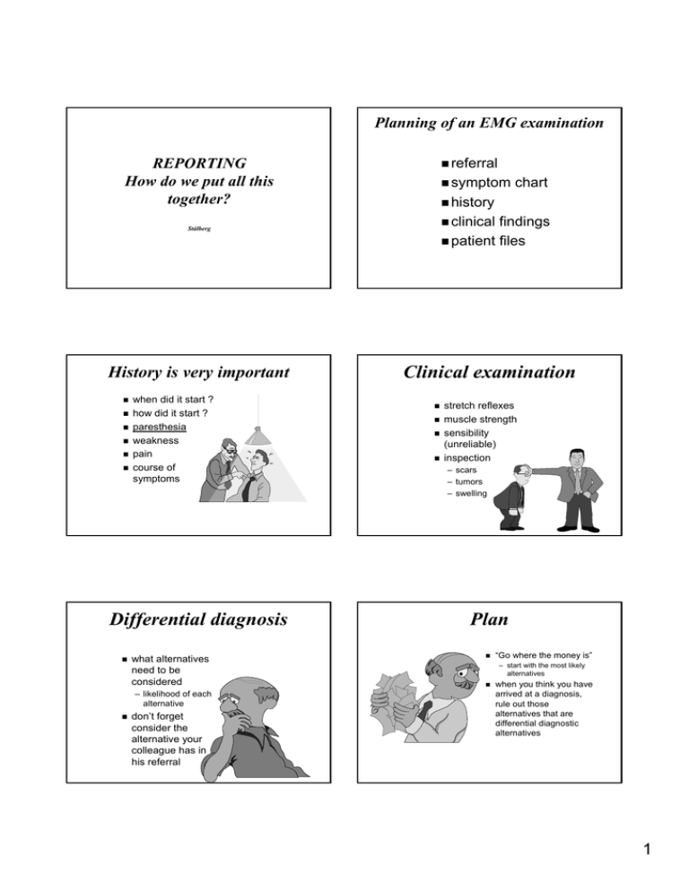
Planning of an EMG examination REPORTING How do we put all this together? referral symptom history clinical findings patient files Stålberg History is very important when did it start ? how did it start ? paresthesia weakness pain course of symptoms Differential diagnosis what alternatives need to be considered – likelihood of each alternative don’ don’t forget consider the alternative your colleague has in his referral chart Clinical examination stretch reflexes muscle strength sensibility (unreliable) inspection – scars – tumors – swelling Plan “Go where the money is” is” – start with the most likely alternatives when you think you have arrived at a diagnosis, rule out those alternatives that are differential diagnostic alternatives 1 Tricks to use when you do not know what to do When nothing else helps - think! – logical thinking – lateral thinking – consult literature/internet Check to opposite side and other limbs Are the electrodes and equipment working? Consult a colleague before the patient leaves Severe unilateral carpal tunnel syndrome Ulnar conduction block at the elbow, right side low sens ampl dig IV and V without motor involvement, left side Conduction block, radial nerve Bilateral carpal tunnel syndrome, left severe, right moderate Bilateral moderate carpal tunnel syndrome 2 Practical approach Often we start with neurography – sometimes this is enough – supplement with inching, centimetering – autonomic tests – TMS – quantitative sensory testing Strategy that may change dynamically as findings evolve Second step is EMG Successive steps depend on findings – RNS – SFEMG,Macro EMG NOTE Be open to the unexpected Results must harmonize ampl decay and normal # F prox ampl higher than dist jitter/ blocking but no weakness good strength - low CMAP - techn anomal.inn techn bad stim or inexcitabilty good strength - low CMAP - inexcitable nerve high F waves - normal MUPs - central hyperexcitability biopsy type grouping - normal FD - cong myopathy high MUPs in GBS day 3, - no reinnervation (FD normal) - loss of small MUs NOTE Be open for the unexpected Practical hints - the examiner Practical hints - the patient inform the patient about reason for EMG explain expected discomfort do not display the electrode term ”pin” pin” (or similar) better than needle keep bloody tissues away do not state number of remaining muscles inform about soreness for 11-2 days inform the patient about next step medical consultation read referral before you see the patient check history, phys exam formulate strategy inform the patient about the progress have all supplies ready before exam use gloves 3 Reporting Practical hints - the investigation Summary of clinical situation no skin preparation is necessary support your hand on the area of needle insertion electrode perpendicular to the skin small but brisk insertion through the skin do not go very deep, just beneath the fascia investigate the muscle at – rest (denervation), – slight contraction (MUP) and – strong contraction (IP) Results Conclusion AANEM 2006 Demographic data Reason for referral, symptoms, signs, possible diagnosis AANEM 2006 Neurography report - concise 2. NCS neurophysiological conclusion clinical interpretation 1. Pat data and Clinical problem Report (AANEM 2006) 1. Clinical problems 2. NCS, specific details 3. EMG, specific details 4. Summary Section: summarize NCS and EMG - integrate 5. Diagnostic interpretation Section 6. Option: clinical diagnosis, differential diagnostic alternative graphic reports tables signals brief summary of essential findings Methods,anti/orthoMethods,anti/ortho-dromic, dromic, ampl methods SCS sites, segments, ampl, ampl, CV, MCS sites, segments, data F-waves nerve, #, latency H refl nerve, physiological state, ampl ratio RNS nerve, phys state, Hz, ampl, ampl, decr, decr, facilitation AANEM 2006 4 Graphic reporting of EMG findings 3. EMG Optimal # muscles describe spont activity, MUP shape, IP in tabular for report limitations (pain, ..) AANEM 2006 Summary of findings Conclusion report only relevant findings on which you base your conclusion use simple terminology that your colleagues understand words like “fibrillation potentials” potentials” or “polyphasic MUPs” MUPs” must be avoided sometimes it important to underline that some findings are normal or Neurophysiological conclusion localization severity pathophysiology time course distribution When in doubt, doubt, say SO When in doubt, doubt, say NO 5. Diagnostic Interpretation Describe if the study over all is abnormal, and why provide electrophysiological diagnosis, and level describe limitations that make the study incomplete compare to earlier tests AANEM 2006 5 Examples of neurophysiological conclusion moderate subacute axonal sensory and motor polyneuropathy slight conduction block of the ulnar nerve in the cubital tunnel severe axonal lesion of the suprascapular nerve in incisura scapulae Example of a report summary EMG: There are slight subacute neurogenic findings in the first dorsal interosseus and flexor carpi ulnaris muscles. Nerve conduction studies: The motor conduction velocity of the ulnar nerve was slightly reduced in the retroepiconylar region at at the elbow. Conclusion. The findings are compatible with a slight axonal and demyelinating ulnar nerve lesion in the retroepicondylar region at the elbow. The symptoms have progressed slowly over a two years and the patient has clinically arthrosis of the elbow. This ulnar neuropathy neuropathy is probably a so called “ tardy ulnar palsy” palsy” (entrapment due to arthrosis of the elbow). Clinical interpretation “beware of weak ice” ice” suite the clinical conclusion to you clinical experience be clear provide differential diagnosis if indicated recommend a control EMG only if there is a specific reason for it Factors determining the usefulness of Neurophysiology quality of examination – (technique, technique, concept, concept, interpretation) – (experience and training of staff) staff) clinical material availability waiting list reporting patient acceptance cost / reimbursement Stålberg 6


