Brain Research, 566 (1991) 308-315
~) 1991 Elsevier Science Publishers B.V. All rights reserved. 0006-8993/91/$03.50
308
BITES 24940
A glycinergic projection from the ventromedial lower brainstem to spinal
motoneurons. An ultrastructural double labeling study in rat
J.C. H o l s t e g e and C . M . H . B o n g e r s
Department of Anatomy, Erasmus University Medical School, Rotterdam (The Netherlands)
(Accepted 27 August 1991)
Key words: Glycine; Motoneuron; Spinal cord; Brainstem; Sleep; Electron microscopy; Immunogold; Locomotion
In the present study it was determined whether glycine was present in the descending brainstem projections to spinal motoneurons in the
rat. For this purpose injections of wheatgerm agglutinin-horseradish peroxidase (WGA-HRP) were made in the ventromedial part of the
lower brainstem at the levels of the rostral inferior olive and the caudal facial nucleus. After perfusion, WGA-HRP histochemistry was
performed, fo!lowed by the postembedding immunogold technique with an antibody against glycine. Electron microscopical examination of
the lumbar motoneuronal cell groups showed that 15% of the WGA-HRP labeled terminals, derived from the ventromedial reticular formation, were also labeled for glycine. The majority (91%) of these double labeled terminals were of the F-type (containing many flattened
vesicles), while the remaining 9% were of the S-type (containing mostly spherical vesicles). Many of the double labeled terminals established
a synapse, mostly with proximal and distal dendrites. The present data, combined with our previous findings that 40% of the projections from
the same ventromedial brainstem area to lumbar motoneurons contained y-aminobutyric acid (GABA), indicate that over 50% of these
brainstem projections contain GABA and/or glycine, exerting a direct inhibitory effect on spinal motoneurons. The possibility that the glycinergic fibers within these projections play an important role in producing muscle atonia during rapid eye movement (REM) sleep is discussed.
The projections from the lower brainstem to the spinal cord have been extensively investigated, both with
retrograde and anterograde tracing techniques (for a review see ref. 14). It was demonstrated that the projection fibers from the ventromedial reticular formation and
the adjacent raphe nuclei of the caudal brainstem terminated in all laminae, including the spinal motoneuronal
cell groups, throughout the spinal cord. Ultrastructural
studies on these projections t°,t3 showed that their terminals within the motoneuronal cell groups could be sub.
divided in morphologically different types: F-type terminals (containing many flattened vesicles), S-type terminals (containing many spherical vesicles) and G-type
terminals (containing many dense cored (granular) vesicles). In addition it was found t3, by combining the anterograde transport of [3H]leucine from the ventro.medial reticular formation with the retrograde transport of
horseradish peroxidase (HRP) from the hindleg muscles,
that over 50% of the radioactively labeled terminals derived from the lower brainstem actually contacted HRP
labeled dendrites of motoneurons in the lumbar spinal
cord.
Recently we have started to investigate whether the
different types of terminals, which were labeled from the
ventromedial brainstem, could be correlated with different transmitters. Dahlstrgm and Fuxe~ showed that there
exists a prominent serotonergic projection from the ventromedial brainstem to the spinal cord, including the motoneuronal cell groups. These findings were confirmed in
studies='as combining retrograde tracing from the spinal
cord with immunocytochemistry for serotonin. These
studies also showed the existence of a prominent nonserotonergic spinal projection from the ventromedial
brainstem, suggesting that, apart from serotonin, one or
more other transmitters were present in those projections. In the motoneuronal cell groups, serotonin is contained within G-type terminals3t which originate mainly
in the raphe nuclei of the lower brainstem and to a much
lesser extent in the adjacent ventromedial reticular formation t3. The latter area gives rise mainly to (non-serotonergic) F-type terminals m't3. By using the combination
of wheat- germ agglutinin coupled to HRP (WGA-HRP)
anterograde tracing with the postembedding immunogold
technique for y-aminobutyric acid (GABA) 7, we have
recently established" that GABA is present in many of
the F-type terminals derived from the ventro-medial reticular formation at the level of the rostral inferior olive and
the caudal facial nucleus. However, it was also found that
Correspondence: J.C. Holstege, Dept. of Anatomy, Erasmus University Medical School, P.O. Box 1738, 3000 DR Rotterdam, The Neth-
erlands.
309
action product was stabilized with DAB-Cobalt TM. The
slabs were postfixed with osmium tetroxide (1.5% in
phosphate buffer (pH 7.3) containing 8% glucose), chemically dehydrated z~ and flat-embedded in Araldite. The
plastic embedded vibratome sections were examined in
the light microscope and from each rat the two sections
with the highest amount of WGA-HRP labeling were
selected• They were glued to plastic blocks and pyramids
were made, containing the entire lateral motoneuronal
cell group. Ultrathin sections were cut and collected on
formvar coated nickel one hole grids. These were incubated overnight at 4 °C with a polyclonal glycine antibody (kindly provided by Dr. R.L Wenthold, for specificity tests, see ref. 33), diluted 1:250 in Tris buffered
saline (0.05 M, pH 7.6), containing 0.1% Triton X-100,
followed by a 2 h incubation with a goat anti-rabbit antibody, labeled with 15 nm gold particles (Amersham).
The sections were counterstained with lead citrate and
uranyl acetate and examined in a Philips 300 electron
microscope. From each rat two blocks were used for
analysis and from each block two sections (at least 1/~m
apart) were analyzed, i.e. a total of 12 sections. The data
obtained in the various sections were grouped together
and the results were expressed as percentages.
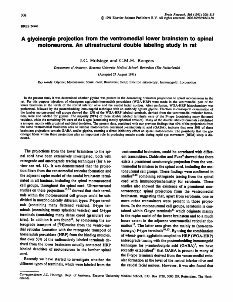
The light microscopic sections of the lower brainstem
showed that the WGA-HRP injection site was located in
the ventromedial reticular formation at the level of the
rostral inferior olive and the caudal part of the facial
nucleus (Fig. 1). This area included the gigantocellular
reticular nucleus pars ventralis and pars a and part of
the paragigantocellular reticular nucleus ~, The iajection
some of the F-type terminals derived from the ventromedial reticular formation were not labeled for GABA,
suggesting that they contained another transmitter. In
the motoneuronal cell groups, F-type terminals were
found to contain either G A B A n2'~9'23 o r glycine ~9'22'32.
We have initiated the present study to determine whether
glycine was present in the non-GABAergic F-type terminals derived from the ventromedial reticular formation. The findings presented in this paper show that in
lumbar motoneuronal cell groups approximately 15% of
the terminals labeled with WGA-HRP from the ventromedial reticular formation contained glycine.
Three rats were used for ultrastructural analysis. In
short the following steps were taken (for details of the
procedure see ref. 11). The rats were deeply anaesthetized with pentobarbital (60 mg/kg) and injected with
0.15/d WGA-HRP (5% in saline) in the ventromedial
part of the reticular formation at the level of the rostral
inferior olive and the caudal part of the facial nucleus.
After 3 days survival, the animals were re-anaesthetized
with pentobarbitai and perfused transcardially with 40 ml
saline in phosphate buffer (0.1 M, pH 7.3) immediately
followed by I ! of 5% glutaraldehyde in phosphate buffer
(0.1 M, pH 7.3). After perfusion, the brainstem and
lumbar spinal cord were dissected. From the brainstem
injection sites, frozen sections were cut, incubated with
diaminobenzidine (DAB) 9, counterstained with Cresyl
violet and coverslipped. The 5th and 6th lumbar segments were cut in slabs (75/~m) on a vibratome slicer.
These slabs were then reacted for WGA-HRP using tetramethylbenzidine (TMB) as a chromogen u and the re-
.~0,
:"
.
..
II
.;~ '
• . , ,,~,~,..".~' ,
•
'cii
;~ ,~,'~
t
,
,.-.,
,
,
', i I i'f'
?' ,,.!
., ,'
, .
'i '~i' '~"
,~
~o
','";!!"~"ii
"~...,.
.,.,..
.
t~,
~,,,
,
~ ~,
'*'
•
.,~,
,,,
I ~, .~ ~q~
~.'.~
•.
..',,,:~
,~
,,
.
,~'~,.
~',~,~,
,~',,.
.
,
,
~'~'~"~"~I;~,,,:...-',
'~,~ "'~
~
'
.
~.,
,
,.~,~
,,,~,g/~,' ........
'
',. ",'- 'C
~
, ~
• .
,
,.,
..
.
:'
.
.
"
,,
~
.
•
~,.
~
..
• .
,
.
.
'~
~".
.
,
, ..~
. q?,.
~
,~,,~,:,;,,;
• ,.
~,
".,
'
'., ,, ,,,,o.,:,~;
,.'
.,.....
@
',
,;,,:'.,'.,,
%~,'
,, ~"~."
,
•,~,,
'~;
,
"
"
• '.~
Fig. 1. Light micrograph of the wheat germ agglutinin-horseradishperoxidase (WGA-HRP) injection site in the ventromedia!part of the rat
lower brainstem at the level of the caudal facial nucleus. Bar - 0.5 mm.
310
site also included part bf the rostral inferior olive and
the nucleus raphe magnus.
Light microscopic examination of the semithin sections from the 5th and 6th lumbar segments showed anterograde WGA-HRP labeling bilaterally, with a clear
ipsilateral predominance. Most labeling was seen in the
motoneuronal cell groups in the ventral horn (c.f. refs.
10,11). Several retrogradely labeled cells were present,
mostly in the contralateral intermediate zone, and only
a few on the ipsilateral side, but not in the direct vicinity of the lateral motoneuronai cell groups. A more detailed description of the light microscopic findings has
been given previously t~. In the ultrathin sections several
WGA-HRP labeled structures were present in addition
to profiles which carried several gold particles, indicating the presence of glycine. A structure was considered
to be labeled for glycine if the number of gold particles
overlying the terminal was at least 8 times the number
of gold particles overlying nearby unlabeled structures
(terminals and dendrites). Although a detailed study of
the glycinergic structures was not performed, it was noted
that most of the glycinergic terminals were of the F-type
and that (mostly myelinated) axonal profiles were often
labeled with a larger number of gold particles than the
terminal profiles. Axo-axonic (presynaptic) terminal arrangements involving glycinergic terminals were not encountered. The latter finding emphasizes the specificity
of the glycine antibody, since presynaptic terminals in
the motoneuronal cell groups are strongly immunoreactive for GABA n. In this study, attention was focused
on WGA.HRP labeled terminals, therefore other WGA.
HRP labeled structures were discarded. A total of 448
WGA-HRP labeled terminal profiles were encountered.
Three types of terminals were labeled with the WGAHRP reaction product: F-type terminals (70%), S-type
terminals (19%) and G-type terminals (11%). These results are in general agreement with the findings of previous studies t°'tt't3. For each WGA-HRP labeled terminal profile it was determined whether it was also labeled
for glycine (double labeled). It was found that 15% of
the WGA-HRP labeled terminals were double labeled
for glycine (Fig, 2). When considering the different types
of terminals it was found that 19% of the WGA-HRP
labeled F-type terminals and 7% of the S-type terminals
were double labeled. G-type terminalb were never double labeled. When considering only the douole labeled
terminal profiles, it was found that 91% were F-type
(Figs. 3-5) and 9% were S-type, The double labeled terminals established a synapse in 68% of the cases, These
synapses were made with proximal dendrites (containing
ribosomes) (44%) and with distal dendrites (36%) (Figs,
3 and 5) and to a lesser extent (20%) with cell somata
(Fig. 4C). Axo-axonic (pre-synaptic) contacts were never
established by ally of the labeled terminals.
The present experiment showed that in the lumbar
motoneuronal cell groups approximately 15% of the terminals, which were labeled with WGA-HRP from the
ventromedial reticular formation, contained glycine. In
a similar experiment 1~ in which WGA-HRP anterograde
tracing was combined with postembedding immunocytochemistry for GABA, it was found that 40% of the
descending brainstem projections to lumbar motoneurons contained GABA. This percentage was considered
as rather accurate since the same technique applied to
other systems resulted in nearly 100% double labeling7'
34. Whether such a high yield can also be obtained with
the glycine antibody is not known. However, it seems
likely that the properties of the glycine antibody are similar to those of the GABA antibody since (1) glycine and
GABA are both small amino acid transmitters, (2) the
glycine and GABA antibodies were raised in a similar
manner (conjugated to glutaraldehyde) and in the same
species (rabbit) and (3) both antibodies were used in a
similar postembedding procedure. This would mean that
the 15% double labeling should also be considered as a
rather accurate figure. If the glycine antibody would be
less sensitive as compared to the GABA antibody, the
15% of double labeling found in the present experiment
should be regarded as an underestimate.
In a previous study t3 we have shown that the majority of the terminals, autoradiographically labeled from
the ventromedial reticular formation, contacted HRP-Ia-
% 80
60
40
20
WGA-HRP
WGA-HRP
+
Glycine
Fig. 2, Histogram showing the percentages of all terminals, labeled
with WGA-HRP from the ventromedial lower brainstem, which
were glycinergic (right column) or non-glycinergic(left column).
The frequency of the F-, G- and S-types of terminals is indicated.
311
.,,~ .~
•
.-~.~.
ta
. . . .
~L
,
,
, .~
~ ~
.
~ ~'
.
/
,
/*f
o
Fig, 3. Electronmicrographsof two adjacent sections from the rat Ls lateral motoneuronal cell groups, showingF-type terminal profiles which
were double labeled with a small amount of WGA-HRP reaction product (large arrows) and many gold particles, indicating that the terminals originated from the ventromedial reticular formation and contained glycine as a transmitter. Small arrows indicate a possible synaptic
junction with part of a proximal dendrite. Asterisks indicate unlabeled terminal profiles. Bar = 0.5 ~ m .
beled motoneurons in the lumbar spinal cord. It is assumed, therefore, that also in the present study on the
same system, the majority of the WGA-HRP labeled
terminals, including the glycinergic ones, contacted motoneurons (for a more detailed discussion, see ref. 11).
When the present finding on the glycinergic brainstem
projections to lumbar motoneurons are combined with
the previous finding 11 that 40% of these projections contained GABA, it may be concluded that over 50% of the
descending brainstem projections to spinal motoneurons
contain either GABA or glycine. Since glycine and
GABA are the main inhibitory transmitters in the spinal
cord (for review see ref. 16), it seems likely that over
50% of the projections descending from the ventromedial brainstem exert an inhibitory effect on spinal motoneurons. A further comparison of the data obtained in
the two experiments show that 55% of the F-type terminals originating in the ventromedial reticiTlar formation
contained GABA, while the present data showed that
19% contained glycine. Thus 74% (55% + 19%) of the
F.type terminals contain either glycine or GABA, leaving approximately 26% still unaccounted for. Since F-type
.'~
J~
.~. -
• u
~mu
"2--~~'~4
IxJ
313
Fig. 4. Electronmicrographs from the rat 1.5 (a,b) and I..6 (c) lateral motoneuronal cell groups. The terminal profiles were labeled with
WGA-HRP reaction product from the ventro-medial reticular formation and contained glycine as indicated by the many gold p~rticles o,~erlying the terminals. Bar ffi 0.5/~m. a,b: two sections taken from a row of 5 serial sections. The WGA-HRP reaction product is clearly visible
on the left and at the bottom of the terminal. An unlabeled terminal is situated very close to the double labeled terminal (top right). The
electron dense material in the areas of contact (arrowheads in a) show the characteristics of puncta adhaerentia rather than synaptic junctions, c: a double labeled terminal of the F-type, establishing a symmetrical synapse with a cell soma, containing a Oolgi apparatus (asterisks). Large arrows indicate the WGA-HRP reaction product.
terminals are generally associated with an inhibitory
transmitter 3°, it is difficult to suggest which transmitter,
apart from G A B A and glycine would be present in the
remaining 26% of the F-type terminals. However, it
should be kept in mind that it is increasingly difficult to
reliably categorize a terminal, when the tissue has been
extensively processed as in the present experiment.
Moreover, the various data were obtained from separate
studies and should not be treated as one experiment.
Therefore final conclusions cannot be drawn at this point
and additional experiments, combining G A B A and glycine immunocytochemistry within the same experiment
are needed to resolve these problems and to determine
whether co-existence of G A B A and glycine may occur
in the descending brainstem projections.
An important indication of the functional significance
of the descending brainstem projections was provided by
Magoun and Rhines ~ in 1946. They were the first to
show that electrical stimulation of the cat medial lower
brainstem resulted in inhibition of spinal reflexes, a finding confirmed in later studies ~s'2°. This electrophysiologicaUy identified 'inhibitory center of Magoun' in the cat
may well correspond to the area in the rat from which
the G A B A - and glycinergic spinal projections, which we
have identified anatomically, are derived. In this respect
it is interesting to note the differential effects obtained
after electrical or chemical stimulation of neurons in different parts of the cat lower brainstem ~. Stimulation of
the ventromedial reticular formation resulted in inhibition of spinal reflexes, which is in agreement with forementioned studies. However, after stimulation of the adjacent raphe, spinal reflexes were mainly facilitated (c.f.
i ~•
~:~,
......
~ti~
:~:.?
',~
1
Fig. 5. Electronmicrograph from the rat L5 lateral motoneuronai cell groups, showing an F-type terminal with WGA-HRP reaction product
(large arrows) and many gold particles. Small arrows indicate a symmetricalsynaptic junction with a proximal dendrite, containing ribosomes
(not visible on this part of the micrograph). Bar - 0.5 ~m.
314
shown 29 to be the main transmitter responsible for the
strong inhibition of lumbar motoneurons after electrical
stimulation in the ventromedial lower brainstem during
R E M sleep (see also ref. 5). However, these studies also
suggested that the connection between the lower brainstem and the motoneurons were not monosynaptic and
involved one or more interneurons in the spinal cord,
but so far these interneurons have not been identified.
In contrast, it was found 25 that one of the interneuronal
candidates for this inhibition, the Renshaw cell, was itself inhibited during R E M sleep. This indicated that not
only motoneurons but also interncurons, involved in motor control, are inhibited during R E M sleep. It may well
be that the glycinergic projection from the lower brainstem is responsible for the simultaneous inhibition of
both spinal motoneurons and interneurons during R E M
sleep.
~fs. 3, 8, 27). This difference can be explained by our
anatomical data: the majority of the terminals in the spinal motoneuronal cell groups, which are derived from
the raphe nuclei, are of the G-type and contain serotonin 3t (for review see ref. 14), which has been shown to
exert a facilitatory effect on spinal motoneurons 35. In
contrast the majority of the terminals on spinal motoneurons, which are derived from the adjacent ventro-medial
reticular formation are of the F-type and contain the inhibitory transmitters G A B A and glycine. Thus it seems
likely that the descending facilitory and inhibitory projections from the lower brainstem to spinal motoneurons
are, at least in part, mediated through direct monosynaptic connections with those motoneurons.
It is still unclear under which circumstances the projections from the ventromedial lower brainstem to spinal motoneurons are functionally active. In respect to
the glycinergic fibers within these projections, it is tempting to assume that they are involved in producing muscle atonia during rapid eye movement (REM)sleep. The
main reasons for this assumption are that (1) muscle atonia can be produced by chemically stimulating the area
in the lower brainstem from which the descending glycinergic projections originate t7 and (2) glycine was
The authors would like to thank Dr. J. Voogd for reading the
manuscript, Mr. E. Dalm and Mr. H. van den Burg for their help
with the surgery and Mrs. H. Goedknegt for her technical assistance. This investigation was supported by Grant 900-550-072 from
the Foundation for Medical Research MEDIGON, which is subsidized by the Netherlands Research Organization (NWO).
1 Andrezik, J.A. and Beitz, A.J., Reticular formation, central
grey and related tegmcntal nuclei. In G. Paxinos (Ed.), The Rat
Nervous System, Vol 2, Hindbrain and Spinal Cord, Academic
Press, Australia, 1985, pp. 1-28.
2 Bowker, R.M. and Abbott, L.C., Quantitative re-evaluation of
descending serotonergic and non.serotoncrgic projections from
the medulla of the rodent: evidence for extensive to.existence
of serotonin and peptides in the same spinally projecting neurons, but not from the nucleus raphe magnus, Brain Research,
512 (1990) 15-25.
3 Cardona, A. and Rudomin, E, Activation of brainstem sere.
tonergle pathways decreases homosynaptic depression of mona.
synaptic responses of frog spinal motoneurons, Brain Research,
280 (1983) 373-378.
4 Chai, C.Y., Lin, Y.F., Wang, H.Y., Wu, W.C., Yen, C.T., Kuo,
J.S. and Wayner, M.J., Inhibition of spinal reflexes by paramedian reticular nucleus, Brain Res. Bull., 25 (1990) 581-588.
5 Chase, M.H., Soja, P.J. and Morales, F.R., Evidence that glycine mediates the postsynaptic potentials that inhibit lumbar
motoneurons during the atonia of active sleep, J. Neurosci., 9
(1989) 743-751.
6 DahlstrOm, A. and Fuxe, K., Evidence for the existence of
monoamine-containing neurons in the central nervous system.
II. Experimentally induced changes in the intraneuronal amine
levels of bulbospinal neuron systems, Acta Physiol. Scand., 64
(1965) 6-36.
7 De Zeeuw, C.I., Holstege, J.C., Calkoen, F., Ruigrok, T.J,H.
and Voogd, J., A new combination of WGA-HRP anterograde
tracing and GABA-immunocytochemistry applied to afferents
of the cat inferior olive at the ultrastructural level, Brain Research, 447 (1988) 369-375.
8 Fang, S.J. and Barnes, C.D., Raph~-produced excitation of spinal cord motoneurons in the cat, Neurosci. Lett., 103 (1989)
185-190.
9 Graham, R.C. and Karnovsky, M.J., The early stages of absorption of injected horseradish peroxidase in the proximal tu-
bules of the mouse kidney. Ultrastructural cytochemistry by a
new technique, J. Histochem, Cytochem,, 14 (1966) 291-302.
10 Holstege, J.C,, Brainstem projections to lumbar motoneurons
in rat. I1. An ultrastructural study by means of the anterograde
transport of wheat.germ agslutlntn coupled to honeradlsh peroxidase and using the tetramethyl benzldine reaction, Neuro.
science, 21 (1987) 369-376,
11 Holstege, J.C., Ultrastructural evidence for GABAergle brain
stem projections to spinal motoneurons in the rat, J. IVeuroscl.,
11 (1991) 159-167.
12 Holstege, J,C, and Calkoen, F,, The distdbutlon of GABA in
lumbar motoneuronal cell groups, A quantitative ultrastruetural
study in rat, Brain Research, 530 (1990) 130-137.
13 Holstege, J.C, and Kuypers, H,G.J.M., Brainstem projections
to lumbar motoneurons in rat. I. An ultrastructural study using
autoradiography and the combination of autoradiography and
HRP histochemistry, Neuroscience, 21 (1987) 345-367.
14 Holstege, J.C, and Kuypers, H.GJ,M., Brainstem projections
to spinal motoneurons' an update, Neumscience, 23 (1987) 809821.
15 Jankowska, E,, Land, S., Lundberg, A. and Pompeiano, O.,
Inhibitory effects evoked through ventral reticolospinai pathways, Arch. Ital. Biol., 106 (1968) 124-140,
16 Krnjdvic, K., Transmitters in motor systems, In J,M. Brookhart,
V.B. Mountcastle, V.B. Brooks and S.R. Geiger (Eds.), Handbook of Physiology, the Nervous System Vol. !!, Motor Control
part L American Physiol. Society, Bethesda, 1981, pp. 107-155.
17 Lai, Y.Y, and Siegel, J.M,, Medullary regions mediating atonia, J. Neurosci., 8 (1988) 4790.-4796.
18 Lemann, W., Saper, C.B., Rye, D.B. and Wainer, B.H., Stabilization of TMB reaction product for electron microscopic retrograde and anterograde fiber tracing, Brain Res. Bull., 14
(1985) 277-281.
i9 Ljungdahl, A. and H6kfelt, T., Autoradiographic uptake patterns of [3H]GABA and [3H]glycine in central nervous tissues
with special reference to the cat spinal cord, Brain Research, 62
315
(1973) 587-595.
20 Llinas, R. and Terzuolo, C.A., Mechanisms of supraspinal actions upon spin~l cord activities. Reticular inhibitory mechanisms on alpha-extensor motoneuroas, J. Neurophysiol,, 27
(1964) 579-591.
21 Magoun, H.W. and Rhines, R., An inhibitory mechanism in the
bulbar reticular formation, J. Neurophysiol., 9 (1946) 165-171.
22 Matus, A.I. and Dennison, M.E., Autoradiographic localisation
of tritiated glycine at 'fiat-vesicle" synapses in spinal cord, Brain
Research, 32 (1971) 195-197.
23 MeLaughlin, B.J., Barber, R., Saito, K., Roberts, E. and Wu,
J.-Y., Immunocytochemical localization of glutamate decarboxylase in the rat spinal cord, Y. Comp. Neurol., 164 (1975) 305322.
24 Mesulam, M.-M., Tetramethyl benzidine for horseradish peroxidase neurohistochemistry: a non-carcinogenic blue reactionproduct with superior sensitivity for visualizing neural afferents
and efferents, Z Histochem. Cytochem., 26 (1978) 106-117.
25 Morales, ER., Engelhardt, J.K., Pereda, E.P., Yamuy, J. and
Chase, M.H., Renshaw cells are inactive during motor inhibition elicited by pontine microinjection of carbachol, Neurosci.
Leu., 86 (1988) 289-295.
26 Muller, L.L. and Jacks, T.J., Rapid chemical dehydration of
sampler for electron microscopic examinations, Y. Histochem.
Cytochem., 23 (1975) 107-110.
27 Roberts, M.H., Davies, M., Girdlestone, D. and Foster, G.A.,
Effects of 5.hydroxytryptamine agonists and antagonists on the
responses of rat spinal motoneurons to raphe obscurus stimulation, Br. J. Pharmacol., 95 (1988) 437-448,
28 Skagerberg, G. and Bj6rklund, A., Topographic principles in
the spinal projections of serotonergic and non-serotonergic
brainstem neurons in the rat, Neuroscience, 15 (1985) 445-480.
29 Soja, P.J., Morales, F.R., Baranyi, A. and Chase, M.H., Effect
of inhibitory amino acid antagonists on IPSPs induced in lumbar motoneurons upon stimulation of the nucleus reticularis gigantocellularis during active sleep, Brain Research, 423 (1987)
353-358.
30 Uchizono, K., Excitatory and inhibitory synapses in the cat spinal cord, Jpn. J. Physiol., 16 (1966) 570-575.
31 Ulfhake, B., Arvidsson, U., Cullheim, S., H6kfelt, T., Brodin,
E., Verhofstad, A. and Visser, T., An uitrastructural study of
5-hydroxytryptamine-, thyrotropin-releasing hormone- and substance P-immunoreactive axonal boutons in the motor nucleus
of spinal cord segments L7-$1 in the adult cat, Neuroscience,
23 (1987) 917-929.
32 Van den Pol, A.N. and Gores, T., Glycine and glycine receptor
immunoreactivity in brain and spinal cord, Y. Neurosci., 8 (1988)
472-492.
33 Wenthold, R.J., Huie, D., Altschuler, R.A. and Reeks, K.A.,
Glycine immunoreactivity localized in the cochlear nucleus and
superior olivary complex, Neuroscience, 22 (1987) 897-912.
34 Wentzei, P.R., Holstege, J.C., Thunnissen, I.E. and Voogd, J.,
Ultrastructural evidence for GABA as inhibitory transmitter in
the vestibulo-oculomotor projection in the rabbit, Fur. J. Neurosci., 2 (1989) 33.
35 White, S.R. and Neuman, R.S., Facilitation of spinal motoneurone excitability by 5-hydroxytryptamine and noradrenaline,
Brain Research, 188 (1980) 199-127.

