Application Note
IV. Materials
Worldwide Offices
ITEM
MANUFACTURER
CATALOG NUMBER
LabChip 3000 Drug Discovery System
Caliper Life Sciences
Chip Module TC
Caliper Life Sciences
Off-Chip Mobility-Shift Chip,
12-sipper, with coating reagent 3
Caliper Life Sciences
761037-0372R
bRAF (D1-415), active, Lot # 25491AU
Upstate
14-530
MEK1, unactive, Lot # 25557AU
Upstate
14-420
ERK2, unactive, Lot # 25760AU
Upstate
14-536
MEK1, active, Lot # 26355BU
Upstate
14-429
ERK2, active, Lot # 23529BU
Upstate
14-173
5-FAM-IPTSPITTTYFFFKKK-COOH
Caliper Life Sciences
760352
HEPES, Free Acid ULTROL
Calbiochem
391338
HEPES, Sodium Salt ULTROL
Calbiochem
391333
MOPS, Free Acid ULTROL
Calbiochem
475898
Magnesium Chloride, hexahydrate
Sigma
M2670
Triton X-100
Sigma
T9284
Brij-35 Solution
Sigma
B4184
DTT
EMD
3860
ATP, disodium salt
Sigma
A7699
EDTA, disodium salt, 0.5 M, pH 8.0
Ambion
9260G
DMSO
JT Baker
9224-33
Staurosporine 1mM in DMSO
Calbiochem
569396
18 MΩ water
Benelux
Caliper Life Sciences N.V.
Klapstraat 13
B-1790 Teralfene, Belgium
Telephone: +32-53-66-26-70
Fax: +32-53-66-27-32
France
Caliper Life Sciences S.A.
ZAC PARIS-NORD II
13 rue de la Perdrix
BP 48016 Tremblay en France
95911 Roissy CDG Cedex, France
Telephone: +33-1-48-63-71-35
Fax: +33-1-48-63-71-53
Germany
Caliper Life Sciences GmbH
Eisenstrasse 9c
DE-65428 Rüsselsheim, Germany
Telephone: +49-6142-834-93-0
Fax: +49-6142-162-821
Japan
Caliper Life Sciences Japan
Saito-Bldg. 2F
Yushima 2-17-15, Bunkyo-ku
Tokyo 113-0034, Japan
Telephone: +81-3-5840-6551
Fax: +81-3-5840-6554
Switzerland
Caliper Life Sciences AG
Nordstrasse 17
CH-4665 Oftringen, Switzerland
Telephone: +41-62-788-7000
Fax: +41-62-788-7017
United Kingdom
Caliper Life Sciences Ltd.
1 Wellfield
Preston Brook
Runcorn, Cheshire WA7 3AZ
United Kingdom
Telephone: +44-1928-711448
Fax: +44-1928-791228
Caliper Life Sciences has
representative offices worldwide.
Please visit www.caliperLS.com
for locations and contact
information.
4
©2007 Caliper Life Sciences, Inc.
All rights reserved.
Caliper and LabChip are registered trademarks, the Caliper Logo is the trademark
of Caliper Life Sciences, Inc.
LC3000-AP-209 02/07
Off Chip Incubation, Mobility Shift
jáíçÖÉåJ^Åíáî~íÉÇ=mêçíÉáå=háå~ëÉ=`~ëÅ~ÇÉ
I. Introduction
II. Methods
One of the most conserved signal transduction pathways in
eukaryotes is the mitogen-activated protein kinase (MAPK)
cascade, which is involved in the control of cellular growth,
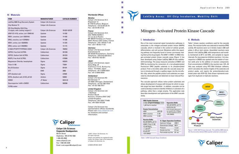
differentiation, and cell survival. Mutations within this signaling pathway are frequently found in human carcinomas making them prime drug targets. The bRAF = MEK1 = ERK2 mitogen-activated protein kinase cascade assay (Figure 1) has
been developed using Caliper LabChip 3000 off-chip mobilityshift technology. The assay measures activation of MEK1 and
ERK2 by monitoring the conversion of a non-phosphorylated
fluorescent ERK2 peptide substrate to its phosphorylated
product. From a microtiter plate well, the cascade assay mixture is introduced through a capillary sipper into the microfluidic chip, where the peptide product and substrate are separated by electrophoresis and detected via laser-induced fluorescence.
Table 1 shows reaction conditions used for the cascade
assay. The reaction buffer was selected to maximize ERK2
activity. All reactions were run in kinetic mode in 384-well
plates in 60 μL total volume. Assembled reactions were
placed in the LabChip 3000, with temperature and humidity maintained at 20o C and 50%, and sampled at regular
intervals. For staurosporine inhibition reactions, 1 mL staurosporine in DMSO was spotted onto the bottom of reaction wells prior to the addition of reaction components.
Final concentration of DMSO in the reaction was 1.6%. All
data was analyzed using HTS Well Analyzer software,
which calculates the relative heights of the substrate and
product peaks, and reports the product/(product+substrate) peak ratio (P/(P+S)). Data shown represents averages from duplicate or triplicate reactions.
The cascade approach utilizes native protein substrates and
allows development of assays with kinases for which no peptide target has been identified. In addition, cascades can be
used to develop screens to identify inhibitors or activators of a
pathway, rather than a single enzyme. This application note
describes development and optimization of the bRAF cascade
assay.
ERK2 Peptide Substrate
5-FAM-IPTSPITTTYFFFKKK-COOH
1 μM final in reaction
Enzyme Concentrations
bRAF
0.05 nM
MEK1 unactive
2 nM
ERK2 unactive
2 nM
Reaction Buffer
50 mM MOPS, pH 7.0
0.004% Triton X-100
10 mM MgCl2
Caliper Life Sciences
Corporate Headquarters
68 Elm Street
Hopkinton, MA 01748-1668
1-508-435-9500
www.caliperLS.com
Fax: 1-508-435-3439
Email: cust.support@caliperLS.com
LabChip Assay:
209
2 mM DTT
35 μM ATP
1.6% DMSO
bRAF
MEK1
unactive
MEK1
active
PO3
Separation Buffer
100 mM HEPES, pH 7.5
0.015% Brij-35
0.1% Coating Reagent 3
10 mM Disodium EDTA
1.6% DMSO
12- Sipper Separation Conditions
Upstream Voltage
-2200 V
Downstream Voltage -500 V
Pressure
-1.2 psi
Sample sip time
0.2 sec
Post sample sip time 30 sec
Table 1. bRAF Cascade Assay Conditions.
ERK2
unactive
ERK2
active
PO3
PO3
ERK2 Peptide
Substrate
ERK2 Peptide
Product
Figure 1. bRAF MAP Kinase Cascade. The LabChip 3000 detects
the flourescently labeled ERK2 peptide substrate and product.
209
Mitogen-Activated Protein Kinase Cascade
III. Results
Cascade Proof of Principle
Figure 1 illustrates the bRAF MAP kinase cascade. Activated
ERK2 adds a phosphate group to the fluorescently labeled ERK2
peptide. The phosphorylated peptide product migrates faster
through the chip than the non-phosphorylated substrate
(Figure 2). The presence of multiple enzymes and phosphorylation steps did not impede the sampling, separation or detection
of non-phosphorylated and phosphorylated ERK2 peptide.
substrate
only
Mitogen-Activated Protein Kinase Cascade
These results verify that data from assays containing active
bRAF, unactive MEK1, and unactive ERK2 (the full cascade)
represent independent activities of all three enzymes.
209
For the full and partial cascade reactions, staurosporine IC50
values increased with increasing ATP concentration (Figure 7
and Table 2). This is consistent with an ATP-competitive mode
of inhibition. At all ATP concentrations, the IC50 values for the
full cascade increased relative to those for the partial cascade
(Table 2), indicating that bRAF was less sensitive to staurosporine than MEK1. The change in IC50 values between the
full cascade and half cascade reflected the difference in the
final molar concentration of MEK1 in the two reactions (2 nM
vs. 0.4 nM).
bRAF Enzyme Titration
Increasing concentrations of bRAF were added to reactions
containing 2 nM unactive MEK1 and 2 nM unactive ERK2 (Figure
4). The concentration of bRAF affected both the lag time before
phosphorylated product was observed, and the linear rate of
product formation. This showed that the downstream targets of
bRAF were sufficiently in excess to act as indicators of bRAF
activity. The bRAF concentration resulting in approximately
30% conversion of ERK2 peptide substrate to product in 45 min
(0.05 nM) was chosen for further studies.
substrate
Figure 5. ATP Km for ERK2 active.
Figure 6 shows product accumulation in cascade reactions
containing 0.05 nM bRAF, 2 nM MEK1 unactive, 2 nM ERK2
unactive, and increasing concentrations of ATP. ATP concentration affected both the lag time before phosphorylated product was observed, and the rate of product formation. The ATP
concentration giving the half-maximal rate during the linear
phase of the full cascade reaction (27 μM) was consistent with
the measured ERK2 ATP Km (32 μM).
product
Figure 2. LabChip 3000 Data Signature. The signal from 6 channels of a
12-sipper chip are shown. The differing product peak heights reflect
differences in product accumulation due to variations in ATP concentration in different reaction wells.
Reactions were assembled containing various combinations of
active and unactive enzymes, and the accumulation of ERK2
peptide product was monitored for 75 minutes (Figure 3).
Product accumulation was observed only in reactions containing either active or activated ERK2. For reactions showing
product formation, reaction kinetics varied based on the concentration of ERK2 and the time required for activation of ERK2
by its upstream activator(s).
Figure 4. Effect of bRAF enzyme concentration on rate of phosphorylated peptide product accumulation.
ATP Titrations
ATP Km for ERK2 was measured by adding increasing amounts
of ATP to reactions containing 0.4 nM ERK2 and 1 μL peptide
substrate. Initial reaction rates were plotted vs. ATP concentration, and the Vmax and Km (32 μL) were determined via nonlinear regression analysis using the Michaelis-Menten Equation
(Figure 5).
Inhibition Assays
Staurosporine, a known ATP competitive inhibitor, was selected
to demonstrate use of the cascade assay to identify a pathway
inhibitor and determine its mechanism of action. Staurosporine
titrations were run at 4 different ATP concentrations with reactions containing:
Full Cascade
0.05 nM bRAF, 2 nM unactive MEK1, 2 nM unactive ERK2
Partial Cascade 0.4 nM active MEK1, 2 nM unactive ERK2
ERK2 Only
0.4 nM
Reactions containing ERK2 only progressed at the same rate
across all staurosporine concentrations (data not shown) indicating that ERK2 is not sensitive to this inhibitor.
Figure 7. Staurosporine inhibition curves for reactions containing the
full cascade (A) or the partial cascade (B).
[ATP]
IC50
Full Cascade
Partial Cascade
ERK2 Only
1000 mM
236 nM
67.2 nM
no effect
333 mM
44.6 nM
12.3 nM
no effect
111 mM
12.0 nM
3.8 nM
no effect
37 mM
2.9 nM
1.3 nM
no effect
Table 2. Staurosporine IC50 values at varying ATP concentrations.
2
Figure 3. Accumulation of phosphorylated peptide product is dependent upon the activity of ERK2. The legend summarizes which combinations of
active enzyme at 0.4 nM (A) and unactive enzyme at 2 nM (U) resulted in peptide product formation.
Figure 6. Effect of ATP concentration on rate of phosphorylated
product accumulation in cascade reactions.
3
209
Mitogen-Activated Protein Kinase Cascade
III. Results
Cascade Proof of Principle
Figure 1 illustrates the bRAF MAP kinase cascade. Activated
ERK2 adds a phosphate group to the fluorescently labeled ERK2
peptide. The phosphorylated peptide product migrates faster
through the chip than the non-phosphorylated substrate
(Figure 2). The presence of multiple enzymes and phosphorylation steps did not impede the sampling, separation or detection
of non-phosphorylated and phosphorylated ERK2 peptide.
substrate
only
Mitogen-Activated Protein Kinase Cascade
These results verify that data from assays containing active
bRAF, unactive MEK1, and unactive ERK2 (the full cascade)
represent independent activities of all three enzymes.
209
For the full and partial cascade reactions, staurosporine IC50
values increased with increasing ATP concentration (Figure 7
and Table 2). This is consistent with an ATP-competitive mode
of inhibition. At all ATP concentrations, the IC50 values for the
full cascade increased relative to those for the partial cascade
(Table 2), indicating that bRAF was less sensitive to staurosporine than MEK1. The change in IC50 values between the
full cascade and half cascade reflected the difference in the
final molar concentration of MEK1 in the two reactions (2 nM
vs. 0.4 nM).
bRAF Enzyme Titration
Increasing concentrations of bRAF were added to reactions
containing 2 nM unactive MEK1 and 2 nM unactive ERK2 (Figure
4). The concentration of bRAF affected both the lag time before
phosphorylated product was observed, and the linear rate of
product formation. This showed that the downstream targets of
bRAF were sufficiently in excess to act as indicators of bRAF
activity. The bRAF concentration resulting in approximately
30% conversion of ERK2 peptide substrate to product in 45 min
(0.05 nM) was chosen for further studies.
substrate
Figure 5. ATP Km for ERK2 active.
Figure 6 shows product accumulation in cascade reactions
containing 0.05 nM bRAF, 2 nM MEK1 unactive, 2 nM ERK2
unactive, and increasing concentrations of ATP. ATP concentration affected both the lag time before phosphorylated product was observed, and the rate of product formation. The ATP
concentration giving the half-maximal rate during the linear
phase of the full cascade reaction (27 μM) was consistent with
the measured ERK2 ATP Km (32 μM).
product
Figure 2. LabChip 3000 Data Signature. The signal from 6 channels of a
12-sipper chip are shown. The differing product peak heights reflect
differences in product accumulation due to variations in ATP concentration in different reaction wells.
Reactions were assembled containing various combinations of
active and unactive enzymes, and the accumulation of ERK2
peptide product was monitored for 75 minutes (Figure 3).
Product accumulation was observed only in reactions containing either active or activated ERK2. For reactions showing
product formation, reaction kinetics varied based on the concentration of ERK2 and the time required for activation of ERK2
by its upstream activator(s).
Figure 4. Effect of bRAF enzyme concentration on rate of phosphorylated peptide product accumulation.
ATP Titrations
ATP Km for ERK2 was measured by adding increasing amounts
of ATP to reactions containing 0.4 nM ERK2 and 1 μL peptide
substrate. Initial reaction rates were plotted vs. ATP concentration, and the Vmax and Km (32 μL) were determined via nonlinear regression analysis using the Michaelis-Menten Equation
(Figure 5).
Inhibition Assays
Staurosporine, a known ATP competitive inhibitor, was selected
to demonstrate use of the cascade assay to identify a pathway
inhibitor and determine its mechanism of action. Staurosporine
titrations were run at 4 different ATP concentrations with reactions containing:
Full Cascade
0.05 nM bRAF, 2 nM unactive MEK1, 2 nM unactive ERK2
Partial Cascade 0.4 nM active MEK1, 2 nM unactive ERK2
ERK2 Only
0.4 nM
Reactions containing ERK2 only progressed at the same rate
across all staurosporine concentrations (data not shown) indicating that ERK2 is not sensitive to this inhibitor.
Figure 7. Staurosporine inhibition curves for reactions containing the
full cascade (A) or the partial cascade (B).
[ATP]
IC50
Full Cascade
Partial Cascade
ERK2 Only
1000 mM
236 nM
67.2 nM
no effect
333 mM
44.6 nM
12.3 nM
no effect
111 mM
12.0 nM
3.8 nM
no effect
37 mM
2.9 nM
1.3 nM
no effect
Table 2. Staurosporine IC50 values at varying ATP concentrations.
2
Figure 3. Accumulation of phosphorylated peptide product is dependent upon the activity of ERK2. The legend summarizes which combinations of
active enzyme at 0.4 nM (A) and unactive enzyme at 2 nM (U) resulted in peptide product formation.
Figure 6. Effect of ATP concentration on rate of phosphorylated
product accumulation in cascade reactions.
3
Application Note
IV. Materials
Worldwide Offices
ITEM
MANUFACTURER
CATALOG NUMBER
LabChip 3000 Drug Discovery System
Caliper Life Sciences
Chip Module TC
Caliper Life Sciences
Off-Chip Mobility-Shift Chip,
12-sipper, with coating reagent 3
Caliper Life Sciences
761037-0372R
bRAF (D1-415), active, Lot # 25491AU
Upstate
14-530
MEK1, unactive, Lot # 25557AU
Upstate
14-420
ERK2, unactive, Lot # 25760AU
Upstate
14-536
MEK1, active, Lot # 26355BU
Upstate
14-429
ERK2, active, Lot # 23529BU
Upstate
14-173
5-FAM-IPTSPITTTYFFFKKK-COOH
Caliper Life Sciences
760352
HEPES, Free Acid ULTROL
Calbiochem
391338
HEPES, Sodium Salt ULTROL
Calbiochem
391333
MOPS, Free Acid ULTROL
Calbiochem
475898
Magnesium Chloride, hexahydrate
Sigma
M2670
Triton X-100
Sigma
T9284
Brij-35 Solution
Sigma
B4184
DTT
EMD
3860
ATP, disodium salt
Sigma
A7699
EDTA, disodium salt, 0.5 M, pH 8.0
Ambion
9260G
DMSO
JT Baker
9224-33
Staurosporine 1mM in DMSO
Calbiochem
569396
18 MΩ water
Benelux
Caliper Life Sciences N.V.
Klapstraat 13
B-1790 Teralfene, Belgium
Telephone: +32-53-66-26-70
Fax: +32-53-66-27-32
France
Caliper Life Sciences S.A.
ZAC PARIS-NORD II
13 rue de la Perdrix
BP 48016 Tremblay en France
95911 Roissy CDG Cedex, France
Telephone: +33-1-48-63-71-35
Fax: +33-1-48-63-71-53
Germany
Caliper Life Sciences GmbH
Eisenstrasse 9c
DE-65428 Rüsselsheim, Germany
Telephone: +49-6142-834-93-0
Fax: +49-6142-162-821
Japan
Caliper Life Sciences Japan
Saito-Bldg. 2F
Yushima 2-17-15, Bunkyo-ku
Tokyo 113-0034, Japan
Telephone: +81-3-5840-6551
Fax: +81-3-5840-6554
Switzerland
Caliper Life Sciences AG
Nordstrasse 17
CH-4665 Oftringen, Switzerland
Telephone: +41-62-788-7000
Fax: +41-62-788-7017
United Kingdom
Caliper Life Sciences Ltd.
1 Wellfield
Preston Brook
Runcorn, Cheshire WA7 3AZ
United Kingdom
Telephone: +44-1928-711448
Fax: +44-1928-791228
Caliper Life Sciences has
representative offices worldwide.
Please visit www.caliperLS.com
for locations and contact
information.
4
©2007 Caliper Life Sciences, Inc.
All rights reserved.
Caliper and LabChip are registered trademarks, the Caliper Logo is the trademark
of Caliper Life Sciences, Inc.
LC3000-AP-209 02/07
Off Chip Incubation, Mobility Shift
jáíçÖÉåJ^Åíáî~íÉÇ=mêçíÉáå=háå~ëÉ=`~ëÅ~ÇÉ
I. Introduction
II. Methods
One of the most conserved signal transduction pathways in
eukaryotes is the mitogen-activated protein kinase (MAPK)
cascade, which is involved in the control of cellular growth,
differentiation, and cell survival. Mutations within this signaling pathway are frequently found in human carcinomas making them prime drug targets. The bRAF = MEK1 = ERK2 mitogen-activated protein kinase cascade assay (Figure 1) has
been developed using Caliper LabChip 3000 off-chip mobilityshift technology. The assay measures activation of MEK1 and
ERK2 by monitoring the conversion of a non-phosphorylated
fluorescent ERK2 peptide substrate to its phosphorylated
product. From a microtiter plate well, the cascade assay mixture is introduced through a capillary sipper into the microfluidic chip, where the peptide product and substrate are separated by electrophoresis and detected via laser-induced fluorescence.
Table 1 shows reaction conditions used for the cascade
assay. The reaction buffer was selected to maximize ERK2
activity. All reactions were run in kinetic mode in 384-well
plates in 60 μL total volume. Assembled reactions were
placed in the LabChip 3000, with temperature and humidity maintained at 20o C and 50%, and sampled at regular
intervals. For staurosporine inhibition reactions, 1 mL staurosporine in DMSO was spotted onto the bottom of reaction wells prior to the addition of reaction components.
Final concentration of DMSO in the reaction was 1.6%. All
data was analyzed using HTS Well Analyzer software,
which calculates the relative heights of the substrate and
product peaks, and reports the product/(product+substrate) peak ratio (P/(P+S)). Data shown represents averages from duplicate or triplicate reactions.
The cascade approach utilizes native protein substrates and
allows development of assays with kinases for which no peptide target has been identified. In addition, cascades can be
used to develop screens to identify inhibitors or activators of a
pathway, rather than a single enzyme. This application note
describes development and optimization of the bRAF cascade
assay.
ERK2 Peptide Substrate
5-FAM-IPTSPITTTYFFFKKK-COOH
1 μM final in reaction
Enzyme Concentrations
bRAF
0.05 nM
MEK1 unactive
2 nM
ERK2 unactive
2 nM
Reaction Buffer
50 mM MOPS, pH 7.0
0.004% Triton X-100
10 mM MgCl2
Caliper Life Sciences
Corporate Headquarters
68 Elm Street
Hopkinton, MA 01748-1668
1-508-435-9500
www.caliperLS.com
Fax: 1-508-435-3439
Email: cust.support@caliperLS.com
LabChip Assay:
209
2 mM DTT
35 μM ATP
1.6% DMSO
bRAF
MEK1
unactive
MEK1
active
PO3
Separation Buffer
100 mM HEPES, pH 7.5
0.015% Brij-35
0.1% Coating Reagent 3
10 mM Disodium EDTA
1.6% DMSO
12- Sipper Separation Conditions
Upstream Voltage
-2200 V
Downstream Voltage -500 V
Pressure
-1.2 psi
Sample sip time
0.2 sec
Post sample sip time 30 sec
Table 1. bRAF Cascade Assay Conditions.
ERK2
unactive
ERK2
active
PO3
PO3
ERK2 Peptide
Substrate
ERK2 Peptide
Product
Figure 1. bRAF MAP Kinase Cascade. The LabChip 3000 detects
the flourescently labeled ERK2 peptide substrate and product.


