Determination of Mean Diameter and Particle Size Distribution of
advertisement
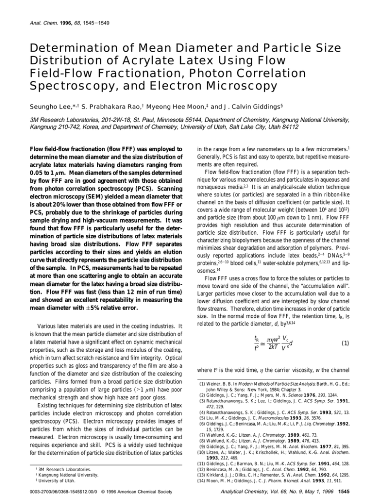
Anal. Chem. 1996, 68, 1545-1549 Determination of Mean Diameter and Particle Size Distribution of Acrylate Latex Using Flow Field-Flow Fractionation, Photon Correlation Spectroscopy, and Electron Microscopy Seungho Lee,*,† S. Prabhakara Rao,† Myeong Hee Moon,‡ and J. Calvin Giddings§ 3M Research Laboratories, 201-2W-18, St. Paul, Minnesota 55144, Department of Chemistry, Kangnung National University, Kangnung 210-742, Korea, and Department of Chemistry, University of Utah, Salt Lake City, Utah 84112 Flow field-flow fractionation (flow FFF) was employed to determine the mean diameter and the size distribution of acrylate latex materials having diameters ranging from 0.05 to 1 µm. Mean diameters of the samples determined by flow FFF are in good agreement with those obtained from photon correlation spectroscopy (PCS). Scanning electron microscopy (SEM) yielded a mean diameter that is about 20% lower than those obtained from flow FFF or PCS, probably due to the shrinkage of particles during sample drying and high-vacuum measurements. It was found that flow FFF is particularly useful for the determination of particle size distributions of latex materials having broad size distributions. Flow FFF separates particles according to their sizes and yields an elution curve that directly represents the particle size distribution of the sample. In PCS, measurements had to be repeated at more than one scattering angle to obtain an accurate mean diameter for the latex having a broad size distribution. Flow FFF was fast (less than 12 min of run time) and showed an excellent repeatability in measuring the mean diameter with (5% relative error. Various latex materials are used in the coating industries. It is known that the mean particle diameter and size distribution of a latex material have a significant effect on dynamic mechanical properties, such as the storage and loss modulus of the coating, which in turn affect scratch resistance and film integrity. Optical properties such as gloss and transparency of the film are also a function of the diameter and size distribution of the coalescing particles. Films formed from a broad particle size distribution comprising a population of large particles (>1 µm) have poor mechanical strength and show high haze and poor gloss. Existing techniques for determining size distribution of latex particles include electron microscopy and photon correlation spectroscopy (PCS). Electron microscopy provides images of particles from which the sizes of individual particles can be measured. Electron microscopy is usually time-consuming and requires experience and skill. PCS is a widely used technique for the determination of particle size distribution of latex particles † 3M Research Laboratories. Kangnung National University. § University of Utah. ‡ 0003-2700/96/0368-1545$12.00/0 © 1996 American Chemical Society in the range from a few nanometers up to a few micrometers.1 Generally, PCS is fast and easy to operate, but repetitive measurements are often required. Flow field-flow fractionation (flow FFF) is a separation technique for various macromolecules and particulates in aqueous and nonaqueous media.2,3 It is an analytical-scale elution technique where solutes (or particles) are separated in a thin ribbon-like channel on the basis of diffusion coefficient (or particle size). It covers a wide range of molecular weight (between 104 and 1012) and particle size (from about 100 µm down to 1 nm). Flow FFF provides high resolution and thus accurate determination of particle size distribution. Flow FFF is particularly useful for characterizing biopolymers because the openness of the channel minimizes shear degradation and adsorption of polymers. Previously reported applications include latex beads,2-4 DNAs,5-9 proteins,2,6-10 blood cells,11 water-soluble polymers,6,12,13 and liposomes.14 Flow FFF uses a cross flow to force the solutes or particles to move toward one side of the channel, the “accumulation wall”. Larger particles move closer to the accumulation wall due to a lower diffusion coefficient and are intercepted by slow channel flow streams. Therefore, elution time increases in order of particle size. In the normal mode of flow FFF, the retention time, tR, is related to the particle diameter, d, by3,6,14 tR t o = πηw2 Vc d 2kT V o (1) where to is the void time, η the carrier viscosity, w the channel (1) Weiner, B. B. In Modern Methods of Particle Size Analysis; Barth, H. G., Ed.; John Wiley & Sons: New York, 1984; Chapter 3. (2) Giddings, J. C.; Yang, F. J.; Myers, M. N. Science 1976, 193, 1244. (3) Ratanathanawongs, S. K.; Lee, I.; Giddings, J. C. ACS Symp. Ser. 1991, 472, 229. (4) Ratanathanawongs, S. K.; Giddings, J. C. ACS Symp. Ser. 1993, 521, 13. (5) Liu, M.-K.; Giddings, J. C. Macromolecules 1993, 26, 3576. (6) Giddings, J. C.; Benincasa, M. A.; Liu, M.-K.; Li, P. J. Liq. Chromatogr. 1992, 15, 1729. (7) Wahlund, K.-G.; Litzen, A. J. Chromatogr. 1989, 461, 73. (8) Wahlund, K.-G.; Litzen, A. J. Chromatogr. 1989, 476, 413. (9) Giddings, J. C.; Yang, F. J.; Myers, M. N. Anal. Biochem. 1977, 81, 395. (10) Litzen, A.; Walter, J. K.; Krischollek, H.; Wahlund, K.-G. Anal. Biochem. 1993, 212, 469. (11) Giddings, J. C.; Barman, B. N.; Liu, M.-K. ACS Symp. Ser. 1991, 464, 128. (12) Benincasa, M. A.; Giddings, J. C. Anal. Chem. 1992, 64, 790. (13) Kirkland, J. J.; Dilks, C. H.; Rementer, S. W. Anal. Chem. 1992, 64, 1295. (14) Moon, M. H.; Giddings, J. C. J. Pharm. Biomed. Anal. 1993, 11, 911. Analytical Chemistry, Vol. 68, No. 9, May 1, 1996 1545 thickness, k Boltzmann’s constant, T the absolute temperature, Vc the cross flow rate, and V o the channel void volume. In eq 1, all parameters except tR and d are constant at a given experimental condition. Thus, the particle diameter can be directly determined from the measured retention time. It is noted that eq 1 is a simplified expression for well-retained samples (tR . to). The full expression of eq 1 is somewhat complicated and will not be discussed here. For all size calculations in this paper, the full expression of eq 1 was used. One of the advantages of flow FFF for particle size determination is that elution time is related to particle size only, and the relationship is straightforward, as shown in eq 1. A flow FFF elution profile can be easily converted to a size distribution of the injected sample. In this paper, flow FFF was used to determine the particle size and its distribution of two different acrylate latex coating materials, one of a narrow and the other of a broad particle size distribution. The results obtained from flow FFF are compared with those obtained from more conventional techniques, such as PCS and electron microscopy. EXPERIMENTAL SECTION Flow Field-Flow Fractionation. A flow FFF system was assembled for this study and is similar to the Model F1000 Universal Fractionator from FFFractionation, LLC (Salt Lake City, UT). The channel has a tip-to-tip length of 27.2 cm and a breadth of 2.0 cm. A Mylar spacer having a thickness of 0.0178 cm was used to construct the channel. The effective channel thickness was determined from the channel void volume, measured by the rapid breakthrough method.15 The measured void volume, V o, was 0.72 mL, and the calculated channel thickness, w, was 0.0143 cm. The channel membrane used for this work is a sheet of YM30 from Amicon (Beverly, MA) made of regenerated cellulose. Carrier fluid is doubly distilled water with 0.05% (w/v) SDS and 0.02% (w/v) NaN3 used as a bactericide. Two Kontron Model 414 HPLC pumps (Kontron Electrolab, London, U.K.) were used for the delivery of carrier liquid to the channel, one for the channel flow, which carries particles down the channel, and another for the cross flow which drives particles toward the accumulation wall. Samples were injected by using a Model 7125 loop injector from Rheodyne, Inc. (Cotati, CA). The injected sample volumes were 5-10 µL of suspended latex particles at the same carrier liquid. Following the entry of the sample, the channel flow was halted for 1 min by diverting the carrier stream to an outside bypass. The stop-flow period of 1 min was slightly longer than the time necessary to drive one channel volume across the channel by the cross flow stream. Upon the completion of the stop-flow procedure, the channel flow was resumed and the run started. Eluted particles were monitored by a 757 absorbance detector from Applied Biosystems (Ramsey, NJ) at a wavelength of 254 nm. The detector signal was collected and processed using the FFF software obtained from FFFractionation, LLC. Photon Correlation Spectroscopy (PCS). The PCS system used in this study is the Model N4MD from Coulter Corp. (Hialeah, FL), which uses a 4 mW He-Ne laser (632.8 nm) as a light source. Experimental parameters were temperature, 25 °C; viscosity, 0.009 P; refractive index, 1.332; sample time, 5.5 µs; prescale, 2; run time, 600 s; 6.96 × 10⁵ counts/s. Electron Microscopy. A JEOL Model 840 high-resolution scanning electron microscope (SEM) and a JEOL Model 100CX (15) Giddings, J. C.; Williams, P. S.; Benincasa, M. A. J. Chromatogr. 1992, 627, 23. 1546 Analytical Chemistry, Vol. 68, No. 9, May 1, 1996 Figure 1. Separation of a mixture of four polystyrene latex standards having nominal diameters of 0.054, 0.135, 0.220, and 0.360 µm, respectively. Channel flow rate, 4.28 mL/min; cross flow rate, 0.99 mL/min; stop-flow, 1 min. Table 1. Comparison of Nominal and Measured Diameters of Polystyrene Latex Standards nominal diam (µm) measd diam (µm) rel error (%) 0.054 0.135 0.220 0.360 0.052 0.130 0.210 0.340 3.7 3.7 4.5 5.6 transmission electron microscope (TEM) were used for electron microscopy of the latex samples. In SEM, about 100 spheres were analyzed for the measurement of the equivalent mean diameter of the particles. In TEM, a small drop of latex suspension was mixed with a 2% solution of phosphotungstic acid (PTA) to improve the contrast of micrographs (“PTA-staining”). Materials. Polystyrene latex standards were obtained from Duke Scientific (Palo Alto, CA). The standards were diluted with the carrier about 100 times and mixed together to prepare a standard mixture. Two acrylate latex materials were prepared at 3M laboratories with slight variations in chemical composition and are arbitrarily labeled as “latex-1” and “latex-2”. RESULTS AND DISCUSSION As discussed earlier, flow FFF separates particles on the basis of size. A flow FFF system was newly assembled and was tested with a mixture of four narrow polystyrene latex standards having nominal diameters of 0.054, 0.135, 0.220, and 0.360 mm. Figure 1 shows a separation of the standards obtained with a channel flow rate (V) of 4.28 mL/min and a cross flow rate (Vc) of 0.99 mL/min. A good separation was obtained with a run time of less than 10 min. The sharp peak shown at the elution time of to (void time) is due to the system transient when the channel flow is resumed after the stop-flow period. Diameters of the standards were calculated from their peak elution times using eq 1, and were compared to their nominal diameters. As shown in Table 1, the calculated diameters agree well with nominal values, with a relative error of <6%. The same flow FFF system was used to determine the mean diameters and size distributions of two acrylate latex materials. Figure 2. Flow FFF elution curves of two acrylate latex materials. Experimental conditions are the same as in Figure 1. Figure 4. Mean diameters determined by unimodal PCS measurements at different scattering angles. Figure 3. Particle size distributions of acrylate latex materials obtained from the elution curves shown in Figure 2. scattering must be removed from the detector response (“light scattering correction”).16 No light scattering correction was performed in this report, as all the necessary optical parameters for the correction were not available for the latex samples used in this study. Thus, the size distributions shown in Figure 3 may not be accurate. The effect of zone broadening must also be removed for accurate determination of size distribution. It has been shown that the effect of zone broadening on the resulting size distribution could be significant for samples having very narrow distributions, but it disappears rather quickly as the distribution becomes broad. The effect of zone broadening was not considered in this report. The same acrylate latex samples were analyzed by photon correlation spectroscopy (PCS). Like flow FFF, PCS is an absolute technique and does not require system calibration.17,18 First, a unimodal analysis method was used, where the mean diameter is determined with an assumption that the size distribution is narrow and has a log-normal distribution profile.19 It has been shown that, as long as the sample distribution is relatively narrow, a unimodal analysis yields a reliable measure of the mean diameter, even if the true distribution is not exactly log-normal. A unimodal analysis was performed at three different scattering angles (30, 62, and 90°), and measurement was repeated at least four times at each scattering angle. The averages of mean diameters obtained at each scattering angle are plotted in Figure 4 as a function of the scattering angle. PCS weights the larger particles more heavily at smaller scattering angles due to the angular dependence of the scattered light intensity.18 This angular dependence decreases as the particle size decreases and becomes negligible for particles whose diameters are much smaller than the wavelength of the incident light. As shown in Figure 4, the measured mean diameter does not vary significantly with the scattering angle for latex-1. This Table 2. Mean Diameter Determined by Flow FFF sample mean diam (µm) latex-1 latex-2 0.124 0.509 Figure 2 shows overlaid elution curves of the latex samples obtained at the same experimental conditions as in Figure 1. Run times are about 5 and 12 min for latex-1 and latex-2, respectively. Elution profiles shown in Figure 2 were converted to the particle size distributions (Figure 3) on the basis of eq 1. The mean diameters were then determined as the first moments of the size distribution curves. Each sample was injected at least four times, and the relative errors in the measured mean diameters were less than (5%. The mean diameters of the samples determined by flow FFF are summarized in Table 2. As can be seen in Figure 3 and Table 2, latex-1 has a narrower size distribution and smaller mean diameter than latex-2. Although the detector used in this study is intended for monitoring UV absorption at 254 nm, light scattering will also contribute to the detector response. To accurately convert a fractogram to a size distribution, the size dependence of light (16) Yang, F.-S.; Caldwell, K. D.; Giddings, J. C. J. Colloid Interface Sci. 1983, 92, 81. (17) Malihi, F. B.; Provder, T.; Koehler, M. E. J. Coatings Technol. 1983, 55, 41. (18) Nicoli, D. F.; McKenzie, D. C.; Wu, J.-S. Am. Lab. 1991, November, 32. (19) Rodreguez, P. Strategies for Routine Sizing Using “Fingerprint” and Multiangle SDP Analysis. Technical Communication, Coulter Corp., Hialeah, FL, 1994. Analytical Chemistry, Vol. 68, No. 9, May 1, 1996 1547 Table 3. Mean Diameter of Latex-2 Measured by PCS Using SDP Analysis Method scattering angle (deg) mean diam (µm) 30 62 90 0.549 0.545 0.502 Figure 6. Size distribution of latex-1 determined by SEM. Figure 5. Size distribution of latex-2 determined by PCS using multiangle SDP analysis method. result indicates that a unimodal PCS measurement made at any single scattering angle will yield an accurate mean diameter for a sample having a relatively small mean diameter and narrow distribution, such as latex-1. The average value of the mean diameters measured at three different scattering angles is 0.119 µm, which is in good agreement (by the difference of 4%) with the result obtained from flow FFF (see Table 2). For latex-2, the measured mean diameter decreases as the scattering angle increases. This result indicates that a unimodal analysis cannot yield an accurate mean diameter nor an accurate size distribution for latex-2. To obtain an accurate mean diameter, latex-2 was further analyzed using a multiangle size distribution processor (SDP) analysis method. Unlike the unimodal analysis, the SDP analysis is carried out without an assumption of the particle size distribution of the sample by way of separating the decay times of the autocorrelation function and mixing these decay times together in a composite autocorrelation function that characterizes all the particles simultaneously.19 The multiangle SDP analysis yields a histogram of the sample size distribution (wt % vs diameter) as well as the mean diameter. Mean diameters measured by SDP analysis at three different angles for latex-2 are summarized in Table 3. A size distribution is regenerated in Figure 5 by combining and normalizing the wt % data obtained at three different scattering angles. As PCS provides wt % data for each size channel (or size range), the size distribution was presented as a wt % histogram rather than a smooth curve, as shown in Figure 3. The mean diameter of the combined distribution shown in Figure 5 is 0.524 µm, which is again in good agreement (by a difference of <3%) with the result obtained by flow FFF (see Table 2). The same latex samples were also analyzed using a highresolution SEM. Figure 6 shows the particle size distribution of latex-1 determined by SEM. The mean diameter was 0.095 µm, with a range from 0.051 to 0.123 µm. The size could not be 1548 Analytical Chemistry, Vol. 68, No. 9, May 1, 1996 Figure 7. Transmission electron micrographs of latex-1 (A) and latex-2 (B). determined for latex-2, as it formed a continuous film on drying with no apparent spheres. This is because the glass transition temperature of latex-2 is about 0 °C, and therefore the particles coalesce to a film even below room temperature. The glass transition temperature of the latex-1 polymer is about 77 °C, and the particles did not coalesce during the drying of the latex. Similar results were obtained from transmission electron microscopy (TEM). Figure 7 shows TEM micrographs of latex-1 and latex-2. Discrete microspheres are seen in micrograph A, while no microsphere is found in micrograph B, due to the formation of film. For polymer particles of low glass transition temperature, where a latex could not be dried without particle aggregation, FFF and PCS become the analytical tools of choice. Table 4. Mean Diameter Determined by Three Different Techniques mean diam (µm) sample flow FFF PCS SEM latex-1 latex-2 0.124 0.509 0.119 0.524 0.095 Table 4 summarizes the mean diameters determined by all three techniques (flow FFF, PCS, and SEM). For both latex-1 and latex-2, the mean diameters determined by flow FFF and PCS are in good agreement (differing by less than 4%). The mean diameter measured by SEM is about 20% lower than the those from flow FFF or PCS, probably due to the shrinkage of particles during SEM measurement (drying and imaging in high vacuum). CONCLUSION Flow FFF is a useful tool for accurately determining size distribution as well as the mean diameter of latex particles. It is fast, simple in principle, and easy to operate. Retention time in flow FFF depends only on the particle size, and thus the elution profile directly represents the particle size distribution of the injected sample (as long as the detector signal is proportional to concentration, and the zone broadening is negligible). If the detector signal is not proportional to concentration (due to light scattering) or the zone broadening is not negligible, appropriate corrections must be made to obtain accurate size distribution. In PCS, a single-angle unimodal analysis yields accurate mean diameters for materials having small diameters and narrow size distributions. For materials having broad size distributions (or large diameters), multiangle measurement is required, as the measured diameter may vary with scattering angle. The FFF results are more unequivocal because the actual size distribution is constructed from the retention volumes. For materials whose size distribution is not known, the PCS method requires unimodal measurement at more than one angle to determine if further analysis is necessary or not. The use of electron microscopy is difficult for latex particles that constitute low glass transition temperature polymers, such as latex-2 in this study, because of the difficulty in maintaining the particles in an uncoalesced state for imaging purposes. Being an elution technique, flow FFF has some additional features that may become useful for some cases. As in chromatography, flow FFF can be coupled with other measurement devices (e.g., ICPMS) for on-line characterization of fractions. If necessary, fractions can be collected for further analysis, such as electron microscopy. Received for review December 5, 1995. February 16, 1996.X Accepted AC9511814 X Abstract published in Advance ACS Abstracts, March 15, 1996. Analytical Chemistry, Vol. 68, No. 9, May 1, 1996 1549
