Technology for Improving the Sensitivity of an X-ray
advertisement

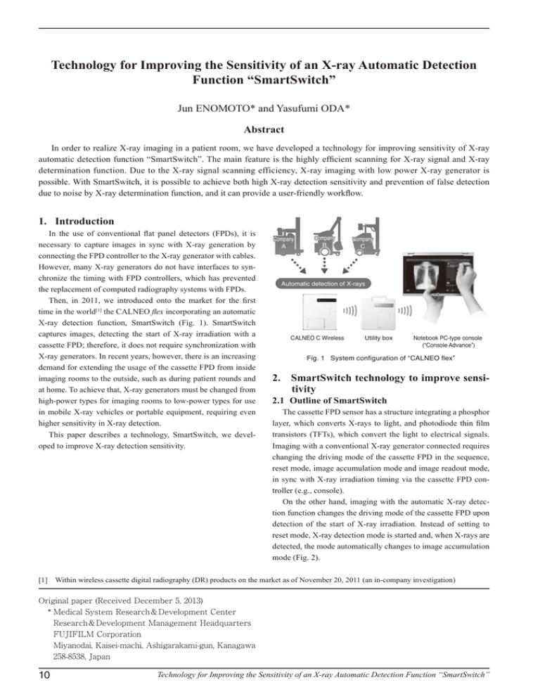
Technology for Improving the Sensitivity of an X-ray Automatic Detection Function “SmartSwitch” Jun ENOMOTO* and Yasufumi ODA* Abstract In order to realize X-ray imaging in a patient room, we have developed a technology for improving sensitivity of X-ray automatic detection function “SmartSwitch”. The main feature is the highly efficient scanning for X-ray signal and X-ray determination function. Due to the X-ray signal scanning efficiency, X-ray imaging with low power X-ray generator is possible. With SmartSwitch, it is possible to achieve both high X-ray detection sensitivity and prevention of false detection due to noise by X-ray determination function, and it can provide a user-friendly workflow. 1. Introduction In the use of conventional flat panel detectors (FPDs), it is necessary to capture images in sync with X-ray generation by connecting the FPD controller to the X-ray generator with cables. However, many X-ray generators do not have interfaces to synchronize the timing with FPD controllers, which has prevented the replacement of computed radiography systems with FPDs. Then, in 2011, we introduced onto the market for the first time in the world[1] the CALNEO flex incorporating an automatic X-ray detection function, SmartSwitch (Fig. 1). SmartSwitch captures images, detecting the start of X-ray irradiation with a cassette FPD; therefore, it does not require synchronization with X-ray generators. In recent years, however, there is an increasing demand for extending the usage of the cassette FPD from inside imaging rooms to the outside, such as during patient rounds and at home. To achieve that, X-ray generators must be changed from high-power types for imaging rooms to low-power types for use in mobile X-ray vehicles or portable equipment, requiring even higher sensitivity in X-ray detection. This paper describes a technology, SmartSwitch, we developed to improve X-ray detection sensitivity. Company A Company B Company C Automatic detection of X-rays CALNEO C Wireless Utility box Notebook PC-type console (“Console Advance”) Fig. 1 System configuration of “CALNEO flex” 2. SmartSwitch technology to improve sensitivity 2.1 Outline of SmartSwitch The cassette FPD sensor has a structure integrating a phosphor layer, which converts X-rays to light, and photodiode thin film transistors (TFTs), which convert the light to electrical signals. Imaging with a conventional X-ray generator connected requires changing the driving mode of the cassette FPD in the sequence, reset mode, image accumulation mode and image readout mode, in sync with X-ray irradiation timing via the cassette FPD controller (e.g., console). On the other hand, imaging with the automatic X-ray detection function changes the driving mode of the cassette FPD upon detection of the start of X-ray irradiation. Instead of setting to reset mode, X-ray detection mode is started and, when X-rays are detected, the mode automatically changes to image accumulation mode (Fig. 2). [1] Within wireless cassette digital radiography (DR) products on the market as of November 20, 2011 (an in-company investigation) Original paper (Received December 5, 2013) * Medical System Research&Development Center Research&Development Management Headquarters FUJIFILM Corporation Miyanodai, Kaisei-machi, Ashigarakami-gun, Kanagawa 258-8538, Japan 10 Technology for Improving the Sensitivity of an X-ray Automatic Detection Function “SmartSwitch” Changes of FPD driving statuses with a conventional X-ray connection method Behavior of the X-ray generator Irradiation switch ON Driving statuses of the imaging section X-ray irradiation X-ray generation signals Connected by cables Output of images (display) Image readout mode X-ray accumulation mode Reset mode Changes of FPD driving statuses with a new automatic X-ray detection function Behavior of the X-ray generator X-ray irradiation Irradiation switch ON Driving statuses of the imaging section X-ray detection mode X-ray accumulation mode Output of images (display) Image readout mode Automatic mode change Fig. 2 Operational state of the X-ray detection system and automatic X-ray connection method To improve X-ray detection sensitivity, we developed two algorithms for (i) the control of scanning to enable high-performance X-ray detection and (ii) the separation of noise from Xrays. The details are as follows. Conventional method In X-ray detection mode, the following cycle is repeated at a high speed: integration and amplification of electrical charges to voltage with a charge amplifier; conversion to digital values with an A/D converter; and X-ray detection with a detection program. To improve the performance of X-ray detection, it is necessary to increase the proportion of the integral time for electrical charges. However, the conventional controller was connected to the cassette FPD as its external unit, requiring time for communication. In addition, the processing speed of the CPU was not sufficient for the parallel processing required. To solve those issues, we have recently developed a dedicated control program and installed it into the processing IC within the cassette FPD. The communication time has thus been minimized. Moreover, we employed a field-programmable gate array (FPGA) for the processing IC, which enables it to perform real-time parallel processing. 2.3 Development of the X-ray determination function In order for the automatic X-ray detection function to sense the start of X-ray irradiation and capture images, a quantity of Xrays not smaller than a threshold level should be irradiated onto the cassette FPD. The quantity of X-rays that reach the cassette FPD depends on the patient’s physique and the output capacity of the X-ray generator. Accordingly, in imaging with a low-power X-ray generator for mobile X-ray vehicles or portable equipment, FUJIFILM RESEARCH & DEVELOPMENT (No.59-2014) ②Positioning ③False detection Threshold X-ray detection Image capture Time SmartSwitch method ①Menu registration X-ray detection signal 2.2 Development of scanning control to enable highperformance X-ray detection X-ray detection signal ①Menu registration Menu registration again after discarding the failed image ②Positioning Threshold X-ray X-ray detection determination ③X-ray imaging ④Completion of imaging Threshold X-ray detection X-ray determination Image capture Time Fig. 3 Control of X-ray determination function it is preferable that the threshold for X-ray signal detection should be set to a low value. However, that often causes false detection when the cassette FPD receives impacts from the patient’s body motion or disturbance noise from peripheral equipment. That means, in general, there is a trade-off between X-ray detection sensitivity and the ability to prevent false detection. To solve that issue, we developed an X-ray determination function to distinguish X-ray signals from noise when the signals exceed the threshold level. The function makes assessments on the signals when the driving status has changed from X-ray detec- 11 General imaging (50 to 80 kW) Imaging during patient rounds (6 to 30 kW) Imaging with portable equipment (2 to 3 kW) Patient room Home Operating room Disaster Incubator Tabletop Conventional method SmartSwitch method Fig. 4 Exposable situation of SmartSwitch tion mode to X-ray accumulation mode with the scanned signals exceeding the threshold. If they are judged to be X-rays, the cassette FPD proceeds to imaging; otherwise, it is set back to X-ray detection mode without imaging the signals (Fig. 3). The function thus prevents false detection from impacts, etc., while keeping high X-ray detection sensitivity even if the quantity of X-rays that reach the cassette FPD is small. Trademarks • “SmartSwitch” and “CALNEO” are registered trademarks of FUJIFILM Corporation. • Any other company names, systems and product names referred to in this paper are generally respective trade names or registered trademarks of other companies. 3. Conclusion Based on the sensitivity improving technology described in this paper, we put into use, for the first time in the world, the high-quality automatic X-ray detection function, SmartSwitch, that can be introduced easily even into existing low-power, analog mobile X-ray vehicles. The function is incorporated into all the CALNEO C-series panels, supporting a variety of imaging situations that require large panels (17 × 17 in.) such as in imaging rooms as well as small panels (24 × 30 cm) such as in incubators (Fig. 4). We will keep seeking to develop technologies to achieve the higher usability of DR imaging systems and to respond to needs at medical sites. 12 Technology for Improving the Sensitivity of an X-ray Automatic Detection Function “SmartSwitch”


