KPL APPLICATION NOTE
SureLINKTM Conjugation Chemistry:
Phosphatase Conjugates and Their Use in Immunodetection
By Danielle Russell, Mwanatumu Mbwana, Mekbib Astatke, Ph.D., Larry Jerome, and Lisa Linske-O’Connell, Ph.D
KPL Research and Development
INTRODUCTION
Immunoassays commonly employ biomolecules conjugated
to a reporter or detection molecule. Enzymes are used as
reporters because of signal amplification via the catalytic
properties of the enzyme, availability of suitable substrates,
and ease of detection of reaction products. Enzyme conjugates are detected by reaction with substrates which yield
either a chromogenic or chemiluminescent reaction product.
Enzymes most frequently used in immunoassays include βgalactosidase (β-gal), horseradish peroxidase (HRP), and
alkaline phosphatase (AP). Each offers unique features that,
under the right conditions, makes it the optimal choice for
immunodetection. Originally isolated from calf intestine, AP
is commonly used in biological assays due to its rapid
turnover, sensitivity, and moderate thermostability.
a second heterobifunctional linker, e.g., SATA (N-Succinimidyl-Sacetylthioacetate), can be used to introduce sulfhydryl groups
through amine-reactive chemistry. Use of such linkers usually
requires deprotection of the sulfhydryl group, adding time and
complexity to the conjugation. Additionally, the EDTA (ethylenediaminetetraacetic acid) used in this protocol chelates the divalent
cations in the active site of AP, thus decreasing enzymatic activity.
SureLINK CONJUGATION CHEMISTRY
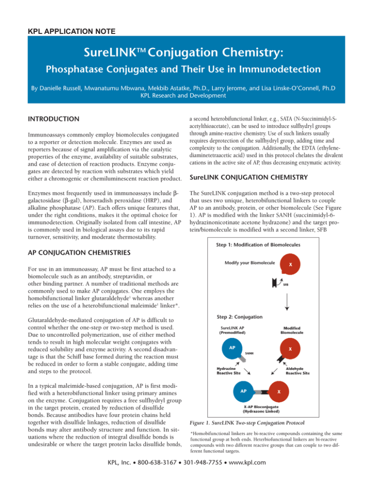
The SureLINK conjugation method is a two-step protocol
that uses two unique, heterobifunctional linkers to couple
AP to an antibody, protein, or other biomolecule (See Figure
1). AP is modified with the linker SANH (succinimidyl-6hydrazinonicotinate acetone hydrazone) and the target protein/biomolecule is modified with a second linker, SFB
Step 1: Modification of Biomolecules
AP CONJUGATION CHEMISTRIES
Modify your Biomolecule
For use in an immunoassay, AP must be first attached to a
biomolecule such as an antibody, streptavidin, or
other binding partner. A number of traditional methods are
commonly used to make AP conjugates. One employs the
homobifunctional linker glutaraldehyde1 whereas another
relies on the use of a heterobifunctional maleimide1 linker*.
Glutaraldehyde-mediated conjugation of AP is difficult to
control whether the one-step or two-step method is used.
Due to uncontrolled polymerization, use of either method
tends to result in high molecular weight conjugates with
reduced solubility and enzyme activity. A second disadvantage is that the Schiff base formed during the reaction must
be reduced in order to form a stable conjugate, adding time
and steps to the protocol.
In a typical maleimide-based conjugation, AP is first modified with a heterobifunctional linker using primary amines
on the enzyme. Conjugation requires a free sulfhydryl group
in the target protein, created by reduction of disulfide
bonds. Because antibodies have four protein chains held
together with disulfide linkages, reduction of disulfide
bonds may alter antibody structure and function. In situations where the reduction of integral disulfide bonds is
undesirable or where the target protein lacks disulfide bonds,
Step 2: Conjugation
SureLINK AP
Figure 1. SureLINK Two-step Conjugation Protocol
*Homobifunctional linkers are bi-reactive compounds containing the same
functional group at both ends. Heterbiofunctional linkers are bi-reactive
compounds with two different reactive groups that can couple to two different functional targets.
KPL, Inc. • 800-638-3167 • 301-948-7755 • www.kpl.com
(succinimidyl 4-formylbenzoate). A stable AP conjugate is
formed when the two modified proteins are subsequently mixed.
Figures 2a and 2b depict the chemical reactions whereby
each biomolecule (AP and the target biomolecule) is separately modified using primary amines (in proteins, the N terminus and R groups of lysine residues supply the primary
amino groups) and one of two different heterobifunctional
modification reagents - SANH or SFB. The primary amine
groups are located on the surface of the protein and are easily accessible for reaction without affecting the stability or
structure of the protein. SANH introduces a reactive
hydrazine group on AP, whereas SFB introduces an active
aldehyde group on the target biomolecule. When the two
modified biomolecules are mixed, the hydrazine and aldehyde groups react in a specific manner to form a stable
hydrazone bond (Figure 3).
Figure 2b. Target Biomolecule Modification: The primary amines of
a protein or biomolecule (X) are reacted with SFB to introduce reactive
aldehyde groups at the sites of primary amines. An N-hydroxysuccinimide leaving group is removed as part of the chemical reaction.
Figure 2a. AP Modification: AP is modified with SANH at the site of
primary amines. In this modification reaction, a N-hydroxysuccinimide
leaving group from the SANH is removed as part of the chemical reaction. The modification introduces a hydrazine, protected as the acetone
hydrazone form.
The SureLINK conjugation chemistry offers several advantages over traditional methods. First, use of this chemistry
assures formation of heteroconjugates, e.g. AP-Ab, with no
formation of homoconjugates, e.g. AP-AP or Ab-Ab.
Nonspecific interactions and polymerization are precluded
by virtue of the reactive moieties on the modified biomolecules. Second, the method provides two points of control,
i.e., the number of modifications per target biomolecule in
the first step and the ratio of the two modified molecules in
the second step. Third, no hazardous reducing agents are
required to stabilize conjugates, thereby eliminating reduced
functionality of biomolecules. Finally, biomolecules modified
with SureLINK heterobifunctional linkers are stable for
extended periods.
Figure 3. Conjugation: At mildly acidic pH, the acetone protecting
group on the hydrazine of SANH is not stable. There is a rapid
exchange with an aromatic aldehyde during conjugation to yield a stable bis-aromatic hydrazone. Acetone is removed as a byproduct. AP is
covalently linked to biomolecule X.
This application note will demonstrate the performance of
SureLINK AP conjugates of a monoclonal antibody, a polyclonal antibody, and streptavidin as immunodetection
reagents in ELISA, Western blot and immunohistology
applications.
KPL, Inc. • 800-638-3167 • 301-948-7755 • www.kpl.com
MATERIALS AND METHODS
Materials: The SureLINK AP Conjugation Kit (Catalog.No. 85-00-01) was
obtained from KPL. The following materials were also obtained from KPL:
polyclonal Goat Anti-Mouse IgG (Catalog No. 01-18-06), Biotin-labeled
Goat Anti-Mouse IgG (Catalog No. 16-18-06), Normal Goat Serum (10%)
(Catalog No. 71-00-27), 10% BSA Diluent/Blocking Solution Concentrate
(Catalog No. 50-61-00), Detector Block (5X) (Catalog No. 71-83-00), Wash
Solution Concentrate (Catalog No. 50-63-00), BCIP/NBT Phosphatase
Substrate (Catalog No. 50-81-07), BluePhos Microwell Phosphatase
Substrate 2-Component (Cat. No. 50-88-02), BluePhos Stop Solution
Concentrate (Catalog No. 50-89-00), PhosphaGLO AP Substrate (Catalog
No. 55-60-03), HistoMark RED Staining System (Catalog No. 55-69-00)
and Contrast GREEN (Catalog No. 71-00-11).
Monoclonal Mouse Anti-Goat IgG was obtained from OEM Concepts
(Catalog No. M2-GG16), Streptavidin from Roche (Catalog No. 115 20679
SQ), Mouse IgG from Stellar Biosystems (Catalog No. MIGG), Goat IgG
from Cappel (Catalog No. 55926), ELISA plates from VWR (Catalog No.
62409-289), formalin-fixed paraffin-embedded (FFPE) human breast tissue
(Catalog No. T1360) and mouse anti-human Epithelial Membrane Antigen
Clone E29 (Cat. No. M0613) from DakoCytomation, Permount from Fisher
(Catalog No. SP15), Xylene Substitute from Shandon (Catalog No.
9990507), and 100% Alcohol Reagent from VWR (Catalog No. VW0470-3).
Precast SDS PAGE (Tris-HCl, 4-20% gradient) gels (Catalog No. 345-0034),
protein sample buffer (Laemmli) (Catalog No. 161-0737), buffers for electrophoresis and transfer (Catalog Nos. 161-0771 and 161-0772), nitrocellulose membrane (0.45 µm pore size) (Catalog No. 162-0235), and the blotting apparatus (PowerPac HC) were obtained from Bio-Rad.
Preparation of conjugates: AP conjugates were prepared according to the
protocol provided in the SureLINK AP Conjugation Kit. Prior to conjugation, monoclonal mouse anti-goat IgG (0.2 mg), polyclonal goat anti-mouse
IgG (0.4 mg), and streptavidin (0.1 mg) were modified with SFB using a 25
molar excess of SFB. AP conjugates of the SFB-modified antibodies were
then prepared using 0.2 mg of SANH-modified AP and 0.1 mg each of SFBmodified antibody to give a final ratio of 2:1 AP to antibody. SFB-modified
streptavidin was conjugated at 0.03 mg to give a final ratio of AP to streptavidin of 2.5:1. Prior to conjugation, Monoclonal Mouse Anti-goat IgG was
dialyzed for 2 hours at 4o C against 0.1M phosphate buffer, pH 7.2, containing 0.15M sodium chloride to remove sodium azide.
Antigen used to test performance of AP conjugates: For demonstration
purposes in ELISA and Western blot, mouse IgG and goat IgG were used as
antigens for AP conjugates of goat anti-mouse IgG and anti-goat IgG,
respectively. Biotinylated goat anti-mouse IgG was used in conjunction with
AP-conjugated streptavidin.
Determination of AP conjugate activity in ELISA: Microplate wells were
coated in duplicate using 2-fold serial dilutions of antigen starting at a dilution of 10 µg/mL. Wells were blocked with 1% BSA Diluent/Blocking
Solution. A 100-µL aliquot of AP conjugate, diluted in 1% BSA
Diluent/Blocking Solution, was added to each well and the plate incubated
for 30 minutes at room temperature. After washing to remove excess conjugate, 100 µL of BluePhos Microwell Substrate (2-Component) was added
to each well and incubated for 15 minutes at room temperature. The reactions were stopped with 100 µL of BluePhos Stop Solution and the optical
density at 630 nm was determined using a 96-well plate reader.
Determination of AP conjugate activity in Western Blotting: Mouse or
goat IgG was prepared in 1X TBS. Each was then diluted 1:3 in Laemmli
buffer without 2-mercaptoethanol to preserve the disulfide bonds in the
antibodies. Samples were heated at 95o C for 3 minutes. Ten microliters of
the sample were then loaded onto wells of precast SDS PAGE (Tris-HCl, 420% gradient) gels. Proteins in the gel were separated by electrophoresis
using the procedure recommended by the manufacturer. Proteins in the gel
were then transferred to nitrocellulose membranes using manufacturer's
suggested settings. Membranes were blocked in 20 mL of Detector Block
solution for 1 hour at room temperature with gentle agitation. Conjugates
were then added directly to the blocking solution and the gels incubated
for 1 hour at room temperature with gentle agitation. Subsequently, membranes were washed in 1X Wash Solution three times for 5 minutes each.
For detection with a chromogenic substrate, the blots were incubated for
15 minutes with BCIP/NBT, washed with distilled water for 10 - 20 seconds, and then dried. For detection with a chemiluminescent substrate, the
blots were incubated for 1 minute with PhosphaGLO, placed in a
hybridization bag, and then exposed to Kodak BioMax film for various time
intervals.
Preparation of slides for immunohistochemistry: Slides of formalin-fixed,
paraffin-embedded (FFPE) human breast tissue were deparaffinized for 5
minutes each in: Xylene Substitute (Shandon), 100% Reagent Alcohol
(VWR), 80% Reagent Alcohol, 40% Reagent Alcohol, 20% Reagent Alcohol,
Distilled water, and then 0.1 M Tris HCl, pH 7.6. The slides were then
incubated in Normal Goat Serum (KPL) for 10 minutes. A positive control
slide was prepared by incubating a slide with mouse anti-human Epithelial
Membrane Antigen Clone E29 diluted 1:50 in 1% Normal Goat Serum for
30 minutes at room temperature. A negative control slide was prepared by
incubating a slide in Tris HCl for 30 minutes at room temperature. Slides to
demonstrate the use of AP conjugates of streptavidin were prepared as follows: slides were incubated in 1 mL of biotinylated goat anti-mouse (2
mg/mL) for 30 minutes, in Tris HCl for 5 minutes, in 1 mL of AP-streptavidin (2 µg/mL in PBS) for 30 minutes, and again in Tris HCl for 5 minutes. Slides to demonstrate the use of AP conjugates of antibodies were prepared as follows: slides were incubated with 1 mL of AP-conjugated polyclonal goat anti-mouse (20 µg/mL in PBS) for 5 minutes followed by incubation in Tris HCl for 5 minutes.
Immunohistochemical staining: Each slide was stained using the
HistoMark RED Staining System. Contrast GREEN was used for counterstaining. For HistoMark RED staining, each slide was incubated for 10 minutes in 1 mL of substrate prepared according to the package insert. Slides
were then washed in distilled water for 2 minutes. One mL of Contrast
GREEN was subsequently added to each slide and each slide was incubated
for 10 minutes. Slides were then washed in distilled water for 2 minutes.
Finally, the slides were incubated in ammonia water solution (2 mL of concentrated ammonium hydroxide in 1L water) for 10 seconds and rinsed in
distilled water for 2 minutes. Slides were briefly rinsed in 80% alcohol,
100% alcohol, and then Xylene Substitute. Slides were then air dried and
mounted with Permount.
Determination of enzyme activity of conjugates in solution: The activity
of AP conjugates in solution was measured at various substrate concentrations. Serial dilutions (1:2) of BluePhos Microwell Substrate Solution A
were prepared in BluePhos Microwell Substrate Solution B. A 100-µL
aliquot of each dilution was added to each of 8 wells of a 96-well plate. The
reaction was initiated by addition of 10 µL (1 ng/well) of conjugate to each
well. After 15 minutes at room temperature, the reactions were stopped
with 100 µL of BluePhos Stop Solution and the optical density of each reaction mixture at 630 nm was then read. Each sample was run in duplicate
and the average of the optical density responses were plotted and analyzed
using Eadie-Hoftsee kinetics. Results were reported as Vmax/Km.
Stability testing of SureLINK AP: Two lots of SureLINK AP (0.2 mg) were
stored lyophilized at 4o C. At various times, samples were conjugated to 0.1
mg of polyclonal goat anti-mouse IgG according to the directions in the kit.
The resulting AP conjugates were subsequently stored at 4o C and enzyme
activity measured as described above.
KPL, Inc. • 800-638-3167 • 301-948-7755 • www.kpl.com
RESULTS AND DISCUSSION
SureLINK Modified AP was used to prepare conjugates of
three frequently used protein binding partners in
immunoassays: a polyclonal antibody, a monoclonal antibody and streptavidin. Results presented below show the
functionality of the conjugates in ELISA, Western blot, and
immunohistology (IHC) applications. For the ELISA and
Western Blot assays, a test system was used with IgG as the
antigen and an anti-IgG secondary antibody as the antibody.
The IHC slides were prepared with an anti-EMA antibody
bound to normal human breast tissue. Also shown are the
results of stability studies of SureLINK AP conjugates.
Use of SureLINK AP Conjugates in ELISA
Experiments were performed to determine the functionality
and sensitivity of SureLINK AP conjugates of goat antimouse IgG (polyclonal), mouse anti-goat IgG
(monoclonal) and streptavidin in an ELISA format. The
antibody conjugates were used to detect antigen that had
been serially diluted and coated onto the surface of microwells. The results shown in figure 4a illustrate that the titration curves for both the AP-labeled polyclonal and APlabeled monoclonal antibodies were similar. The range of
detection was between 8 and 500 ng of the IgG antigen
demonstrating the sensitivity of the antibody conjugates
prepared with SureLINK Modified AP.
SureLINK AP-labeled
polyclonal goat antimouse IgG (0.75
µg/mL)
1000
500
250
125
62
31
16
8
Figure 4b. Detection of mouse IgG (1 µg/well) in an ELISA format
using biotinylated goat anti-mouse IgG (20 to 0.2 ng), SureLINK APlabeled streptavidin (0.3 µg/mL), and BluePhos Microwell
Phosphatase Substrate.
Use of SureLINK AP Conjugates in Western Blotting
The same three SureLINK AP conjugates were used to detect
antigens in Western Blot assays. Antigen preparations were
separated on SDS PAGE gels and then transferred to nitrocellulose membranes. Antigens on the membranes were
then detected with the AP conjugates and either a chromogenic or chemiluminescent substrate. The results in figure 5 a-d show that SureLINK AP conjugates of a polyclonal
or monoclonal antibody or of streptavidin can be used effectively in Western blots. With a chemiluminescent substrate,
significantly lower concentrations of AP conjugates were
used to detect comparable levels of antigen versus that used
with a chromogenic substrate. This is consistent with results
typically obtained using chemiluminescent substrates as compared to chromogenic systems.
500 100 20 4 ng
500 100 20 4 ng
500 100 20 4 ng
250
50 10 2 ng
Figure 5a.
Figure 5b.
Figure 5c.
Figure 5d.
SureLINK AP-labeled
monoclonal goat antimouse IgG (0.5
µg/mL)
Figure 4a. Detection of mouse IgG (1000 to 8 ng) in an ELISA format
using SureLINK AP-labeled polyclonal goat anti-mouse IgG or
SureLINK AP-labeled monoclonal mouse anti-goat IgG and BluePhos
Microwell Phosphatase Substrate.
To test the functionality and sensitivity of SureLINK
AP-labeled streptavidin, a constant amount of mouse
IgG was used to coat the surface of microwells. Serial dilutions of biotinylated goat anti-mouse IgG ranging from 20 to
0.2 ng were then added to the microwells, and the biotinylated goat anti-mouse IgG was detected with the SureLINK
AP-streptavidin conjugate. The results in figure 4b demonstrate detection of biotinylated IgG with the AP-labeled
streptavidin over a broad range of biotinylated antibody. In
summary, SureLINK conjugates of a polyclonal and a monoclonal antibody as well as streptavidin can be used effectively in an ELISA format to detect antigen.
Figure 5a: Western blot prepared using a SureLINK AP conjugate of
polyclonal goat anti-mouse IgG (0.1 µg/mL) and BCIP/NBT
Phosphatase Substrate to detect mouse IgG ranging from 500 to 4 ng
per lane.
Figure 5b: Western blot prepared using a SureLINK AP conjugate of
polyclonal goat anti-mouse IgG (2.5 ng/mL) and PhosphaGLO AP
Substrate to detect mouse IgG ranging from 500 to 4 ng per lane. The
film was exposed to the blot for 10 minutes.
Figure 5c: Western blot prepared using a SureLINK AP conjugate of
monoclonal mouse anti-goat IgG (0.1 µg/mL) and BCIP/NBT
Phosphatase Substrate to detect Goat IgG ranging from 500 to 4 ng per
lane.
Figure 5d: Western blot prepared using a SureLINK AP conjugate of
Streptavidin (0.5 ng/mL), biotinylated goat anti-mouse IgG (25
ng/mL) and PhosphaGLO AP Substrate to detect mouse IgG ranging
from 250 to 2 ng per lane. The film was exposed to the blot for 2 minutes.
KPL, Inc. • 800-638-3167 • 301-948-7755 • www.kpl.com
Vmax/Km
Use of SureLINK AP Conjugates in Immunohistology
Epithelial membrane antigen (EMA) in human breast tissue
was detected using mouse anti-human EMA and either 1)
SureLINK AP conjugated polyclonal goat anti-mouse IgG or
2) biotinylated goat anti-mouse IgG and SureLINK AP-conjugated streptavidin. HistoMark RED was used as substrate
and tissue was counterstained with Contrast GREEN. EMA
is a normal protein in secretory and ductal epithelial cells of
the skin, breast and other glandular organs; it may be a
marker for adenocarcinoma in other organs.2
Lot 1
Lot 2
Months at 4o C
Figure 7. AP activity (Vmax/Km) of two lots of SureLINK APconjugated Goat anti-Mouse IgG after storage at 4o C for the time
intervals indicated.
CONCLUSIONS
A
B
C
D
Figure 6. Detection of EMA in human breast tissue using mouse antiEMA primary antibody and either SureLINK AP-conjugated goat antimouse IgG, 20 µg/mL (A) or biotinylated goat anti-mouse IgG and
AP-conjugated streptavidin, (B). HistoMark RED was used as the AP
substrate and Contrast GREEN was used as the counterstain. No primary antibody was added to the control slides (C and D).
The results shown in figure 6 demonstrate detection of EMA
with both conjugates (figures 6A and 6B); results were similar for both. EMA in the breast tissue was stained brilliant
pink whereas nuclei were counterstained blue-green. EMA
was not detected in the control slides where the primary
antibody was omitted. Only the blue-green color of the
counterstain was visible. As expected, the use of a
biotin/streptavidin system decreased the amount of AP conjugate needed for detection. Based on the results described
above, SureLINK AP conjugates of polyclonal antibodies and
streptavidin can be used to facilitate the detection of tissuespecific markers in IHC applications.
Stability of SureLINK AP-Antibody Conjugates
The results depicted in figure 7 demonstrate the stability of
conjugates made with SureLINK AP Conjugation Kits. Conjugates of goat anti-mouse IgG were prepared with SureLINK
Modified AP and then stored at 4° C. AP activity was determined at intervals during an 8-month period. The results
demonstrate the stability of the conjugates over at least an 8month period SureLINK AP conjugates are kinetically active
even after prolonged storage at 4o C.
SureLINK AP conjugation chemistry enables the facile
preparation of AP conjugates. The resultant SureLINK AP
conjugates are broadly useful in immunoassays. These
include formats such as ELISA, Western blotting, and IHC
applications as described herein but could well be expanded
beyond the realm of such formats to other applications
where AP is a suitable reporter.
As demonstrated in this application note, SureLINK chemistry can be used to prepare conjugates of both monoclonal
and polyclonal antibodies and other proteins such as streptavidin. Although the chemistry has been optimized for use
with proteins, it can readily be used with any biomolecules
containing primary amines or aldehydes as reactive sites.
Furthermore, its use can be expanded to biomolecules lacking either amine or aldehyde reactive groups by derivatizing
such molecules, e.g., amine modifications of nucleic acids.
SureLINK AP conjugation chemistry overcomes many of the
limitations of other conjugation methods. The method is
based on reactions of the heterobifunctional linkers SANH
and SFB with the amino groups of two proteins. Unwanted
cross-linking is virtually eliminated by the formation of
highly specific hydrazone linkages. No reducing agents are
required to stabilize the bond. Disulfide bonds are not
reduced and proteins are not denatured during conjugation.
A forthcoming KPL application note will compare the performance of SureLINK AP conjugates with those produced
using glutaraldehyde and maleimide/thiol chemistry.
The fast, reliable, and reproducible protocol also facilitates
broad application of AP as a reporter enzyme. In 3 hours
with less than 20 minutes of 'hands on' lab time, the reaction is complete and conjugates are ready for immediate use,
as no further purification is required. Moreover, once prepared, SureLINK AP conjugates remain stable, i.e., functionally active, in solution at 4° C for extended periods.
KPL, Inc. • 800-638-3167 • 301-948-7755 • www.kpl.com
REFERENCES
1. Hermanson, Greg T., (1996) Bioconjugate Techniques.
2. ten Berge, R. L., Snijdewint, F. G., von Mensdorff-Pouilly, S.
et al. MUC1 (EMA) is preferentially expressed by ALK positive anaplastic large cell lymphoma, in the normally glycosylated or only partly hypoglycosylated form. J Clin Pathol
2001; 54:933-939.
Related Products
Description
Size
Catalog No.
SureLINK AP Conjugation Kit
3 x 0.5 mg rxn.
85-00-02
SureLINK AP Conjugation Kit
3 x 0.1 mg rxn.
85-00-01
SureLINK HRP Conjugation Kit
6 x 1 mg rxn.
84-00-02
SureLINK HRP Conjugation Kit
6 x 0.1 mg rxn.
84-00-01
SureLINK HRP Conjugation Kit
2 x 0.1 mg rxn.
84-00-03
SureLINK Activated HRP
1.5 mg
84-01-02
SureLINK Activated HRP
0.3 mg
84-01-01
SureLINK Modified AP*
1.0 mg
85-01-02
SureLINK Modified AP*
0.2 mg
85-01-01
SFB Linker
100 mg
80-02-04
SFB Linker
20 mg (4 x 5 mg)
80-02-02
SureLINKTM AP Conjugation Kits
Each kit includes SureLINK Modified AP,
AP Modification Buffer, SFB, AP Conjugation
Buffer and AP Storage Buffer.
SureLINK HRP Conjugation Kits
Each kit includes SureLINK Activated HRP,
HRP Conjugation Buffer, Reducing Reagent
and HRP Storage Buffer.
SureLINK Activated HRP and Modified AP
*SureLINK Modified AP must be used with SFB Linker for successful conjugation of proteins.
Trademarks: SureLINK, Detector and PhosphaGLO are trademarks of KPL.
BluePhos, LumiGLO and HistoMARK are registered trademarks of KPL.
ABTS is a registered trademark of Roche Biochemicals. Permount is a registered trademark of Fisher Scientific. BioMax is a trademark of Eastman
Kodak Company.
SureLINK AP Conjugation Kits and Modified AP are produced with components protected by U.S. Patent Numbers 6,800,728, 5,679,778, 5,420,285,
5,753,520 and 5,206,370.
©2006 KPL, Inc. All rights reserved.
ML316
KPL, Inc. Gaithersburg, Maryland
800.638.3167 301.948.7755
FAX 301.948.0169 www.kpl.com


