Super-resolution microlenses: Attaining the ultimate in optical
advertisement

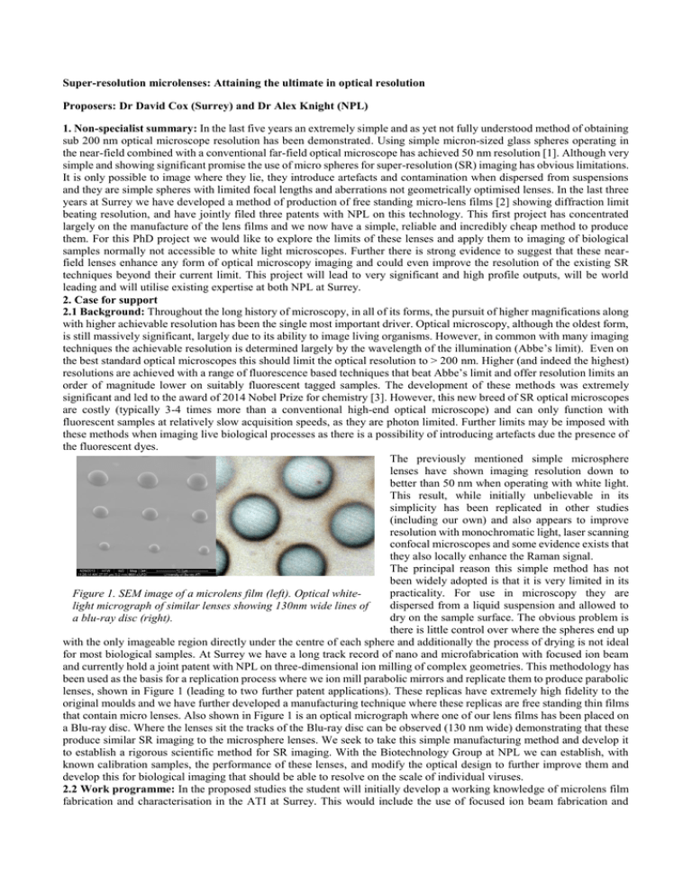
Super-resolution microlenses: Attaining the ultimate in optical resolution Proposers: Dr David Cox (Surrey) and Dr Alex Knight (NPL) 1. Non-specialist summary: In the last five years an extremely simple and as yet not fully understood method of obtaining sub 200 nm optical microscope resolution has been demonstrated. Using simple micron-sized glass spheres operating in the near-field combined with a conventional far-field optical microscope has achieved 50 nm resolution [1]. Although very simple and showing significant promise the use of micro spheres for super-resolution (SR) imaging has obvious limitations. It is only possible to image where they lie, they introduce artefacts and contamination when dispersed from suspensions and they are simple spheres with limited focal lengths and aberrations not geometrically optimised lenses. In the last three years at Surrey we have developed a method of production of free standing micro-lens films [2] showing diffraction limit beating resolution, and have jointly filed three patents with NPL on this technology. This first project has concentrated largely on the manufacture of the lens films and we now have a simple, reliable and incredibly cheap method to produce them. For this PhD project we would like to explore the limits of these lenses and apply them to imaging of biological samples normally not accessible to white light microscopes. Further there is strong evidence to suggest that these nearfield lenses enhance any form of optical microscopy imaging and could even improve the resolution of the existing SR techniques beyond their current limit. This project will lead to very significant and high profile outputs, will be world leading and will utilise existing expertise at both NPL at Surrey. 2. Case for support 2.1 Background: Throughout the long history of microscopy, in all of its forms, the pursuit of higher magnifications along with higher achievable resolution has been the single most important driver. Optical microscopy, although the oldest form, is still massively significant, largely due to its ability to image living organisms. However, in common with many imaging techniques the achievable resolution is determined largely by the wavelength of the illumination (Abbe’s limit). Even on the best standard optical microscopes this should limit the optical resolution to > 200 nm. Higher (and indeed the highest) resolutions are achieved with a range of fluorescence based techniques that beat Abbe’s limit and offer resolution limits an order of magnitude lower on suitably fluorescent tagged samples. The development of these methods was extremely significant and led to the award of 2014 Nobel Prize for chemistry [3]. However, this new breed of SR optical microscopes are costly (typically 3-4 times more than a conventional high-end optical microscope) and can only function with fluorescent samples at relatively slow acquisition speeds, as they are photon limited. Further limits may be imposed with these methods when imaging live biological processes as there is a possibility of introducing artefacts due the presence of the fluorescent dyes. The previously mentioned simple microsphere lenses have shown imaging resolution down to better than 50 nm when operating with white light. This result, while initially unbelievable in its simplicity has been replicated in other studies (including our own) and also appears to improve resolution with monochromatic light, laser scanning confocal microscopes and some evidence exists that they also locally enhance the Raman signal. The principal reason this simple method has not been widely adopted is that it is very limited in its practicality. For use in microscopy they are Figure 1. SEM image of a microlens film (left). Optical whitedispersed from a liquid suspension and allowed to light micrograph of similar lenses showing 130nm wide lines of dry on the sample surface. The obvious problem is a blu-ray disc (right). there is little control over where the spheres end up with the only imageable region directly under the centre of each sphere and additionally the process of drying is not ideal for most biological samples. At Surrey we have a long track record of nano and microfabrication with focused ion beam and currently hold a joint patent with NPL on three-dimensional ion milling of complex geometries. This methodology has been used as the basis for a replication process where we ion mill parabolic mirrors and replicate them to produce parabolic lenses, shown in Figure 1 (leading to two further patent applications). These replicas have extremely high fidelity to the original moulds and we have further developed a manufacturing technique where these replicas are free standing thin films that contain micro lenses. Also shown in Figure 1 is an optical micrograph where one of our lens films has been placed on a Blu-ray disc. Where the lenses sit the tracks of the Blu-ray disc can be observed (130 nm wide) demonstrating that these produce similar SR imaging to the microsphere lenses. We seek to take this simple manufacturing method and develop it to establish a rigorous scientific method for SR imaging. With the Biotechnology Group at NPL we can establish, with known calibration samples, the performance of these lenses, and modify the optical design to further improve them and develop this for biological imaging that should be able to resolve on the scale of individual viruses. 2.2 Work programme: In the proposed studies the student will initially develop a working knowledge of microlens film fabrication and characterisation in the ATI at Surrey. This would include the use of focused ion beam fabrication and replication methods. Most of this methodology is already in place and although the learning curve is quite steep, the process is well understood. One of the aims will also involve use of alternative materials for the lens films to improve mechanical robustness and design of new lens film geometries and lens holders specifically to complement the available optical microscopes at NPL. In parallel, the student will work at NPL with Alex Knight gaining an insight into the state-of-the-art methods and developing an understanding of the limits and strength and weaknesses of the respective techniques. Completion of the above tasks will build on existing understanding and enable the student to further develop new designs and strategies for the development of more advanced lens designs. A key aim of this phase of the proposed research is to establish baseline performance of the respective components of the optical system we will create and to determine the lens performance with conventional optical microscopes with both white and monochromatic light. The SR microscopes are capable of running conventionally and any designs can be transferred to the SR modes. This aspect of the project provides a significant hands-on challenge for the student which if achieved will unlock significant potential to achieve unprecedented imaging resolution beyond the capability of any of the current techniques. Completion of the base-line optical studies will naturally feed into applying these lenses to imaging biological samples and to using the micro-lens system with the current state of the art SR microscope techniques. There is also a plan to operate the lenses on the SR microscope as means of focusing laser light into a fluorescent sample to stimulate increased emission locally, possibly increasing resolution further or increasing acquisition speed of the system. 2.3 Outline project plan: The student will initially focus on developing an understanding of the fundamental principles of microlens design, fabrication and characterisation (work package (WP) 1). This should take approximately 6 months. Once established, the application of the lenses to optical microscopy and base-line performance will be under-taken (WP2) to provide an understanding for subsequent studies. This will be completed by the end of the first year, and provide the material required for the PhD confirmation process. The student will then begin the optimisation of the lens designs along with further fabrication and apply this to obtaining the ultimate resolution possible with the system on both biological and non-biological samples. Additionally, the lenses will be integrated into the existing SR microscopes (WP3). These activities will utilise the remaining time of the project. Specific goals and outputs include: 1) Demonstration and quantification of diffraction-beating resolution with white light on a simple system. 2) Optimisation of the designs and methodologies to maximise resolution in a white light or monochromatic system. 3) Obtaining the ultimate resolution with a hybridised micro-lens SR microscope. 3. Fit to science priority area: The proposed work is directly aligned with major hub research themes of medical physics, and is an excellent example of applying engineering to health. These SR techniques have yielded important biological insights, and NPL have recently shown that they can be used for disease diagnosis [4]. 4. Suitability for PhD study: The project will explore a new field, designing and constructing a super-resolution imaging system which has the potential to set new standards for what is achievable with optical microscopy. Success is likely to be extremely high profile. The project, which is largely experimental, will require the development of fabrication and practical skills along with rigorous scientific method involving complex equipment and cutting edge techniques. The project should not overstretch the capabilities of a student but will provide a serious research challenge, appropriate for a PhD, and is expected to yield high profile publications. 5. Previous collaboration and NPL sponsor details Previous collaboration: David Cox has been collaborating with NPL for more than eleven years. During this period he has worked with almost every division in the laboratory on many projects from internal strategic research programs to very large FP7 projects, spanning quantum technologies, dosimetry, fabrication and materials characterisation. To date this collaboration has led to more than 60 publications during this period. Alex Knight (Biotechnology group NPL) is a Principal Research Scientist and has initiated and led the development of super-resolution microscopies at NPL. Since 2012 this team has published 18 papers on advanced microscopies, and has developed an internationally leading super-resolution microscopy capability. A recent publication [4] describes the use of super-resolution microscopy in the diagnosis of blood clotting disorders. 6. Supporting funding The project is extremely sparing with materials and consumables within Surrey and indeed all of the foreseeable materials are already obtained from previous projects run by DCC. The FIB activity is supported within the ATI as a central facility and also by the existing NPL projects. Access to the microscopes and related consumable at NPL will be provided free of charge (typical non-commercial access charges are £1500 per day). 7. Scientific quality: The proposed work is supported by world-leading activities at both laboratories in FIB fabrication at Surrey and super-resolution microscopy at NPL. The proposed work is interdisciplinary in nature, spanning engineering, physics and biology and will directly benefit both of these laboratories and is in itself of very high scientific quality. It is expected that this work will contribute towards 4* REF outputs. Refs: 1. Optical virtual imaging at 50 nm lateral resolution with a white-light nanoscope. DOI: 10.1038/ncomms1211 2. Manufacture of micro-optical elements for imaging and light guidance. M.T. Langridge, PhD Thesis (2015). 3. https://www.nobelprize.org/nobel_prizes/chemistry/laureates/2014/popular-chemistryprize2014.pdf 4. Super-resolution microscopy as a potential approach to platelet granule disorder diagnosis. Westmoreland et al., (2016) Journal of Thrombosis and Haemostasis, DOI: 10.1111/jth.13269


