Techniques for the Care and Cannulation of AV
advertisement
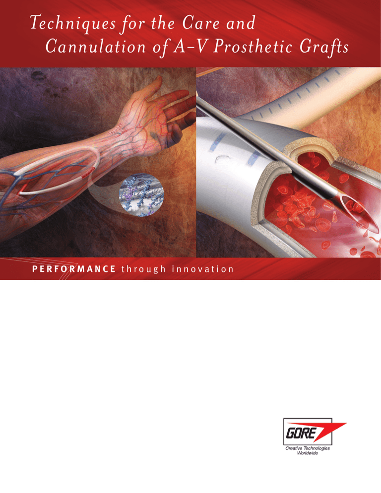
Techniques for the Care and Cannulation of A-V Prosthetic Grafts PERFORMANCE through innovation Techniques for the Care and Cannulation of A-V Prosthetic Grafts The quality of life for hemodialysis patients can be directly related to the life expectancy of their arteriovenous (A-V) grafts. Which is why we recommend the following guidelines for the safe and efficient cannulation of GORE® Vascular Grafts implanted as an A-V hemodialysis access. Taken from the combined experiences of major dialysis centers, we have identified certain techniques that have proven to be of benefit through the years. Through the consistent use of these techniques, you will help cannulation proceed faster and with fewer problems, improving the quality of life for your patients. About Gore Vascular Grafts For more than 35 years meeting the most demanding procedures Recognized by renowned surgeons worldwide for exceptional performance and quality, our vascular grafts are available in a wide range of configurations: • Stretch and non-stretch • Thromboresistant CBAS® Surface a heparin technology with thromboresistant properties • Straight, tapered, and bifurcated • External customizable ring reinforcement or internal radial support These grafts require: • No preclotting • Resist dilatation and the spread of infection • Assure utmost thrombectomy safety Cannulation of GORE® Vascular Grafts After implantation, the physician may require that the patient wait several weeks for the A-V graft to “heal” before cannulation. Typically during this time, there is tissue growth into the outer wall that stabilizes the graft. This is important in the prevention of both infection and hematoma. In some cases, the physician may advise that the patient undergo hemodialysis before adequate healing can take place. Vascular grafts with an early cannulation indication are ideal for patients receiving hemodialysis in the early postoperative period [see SPECIAL CONSIDERATIONS FOR EARLY CANNULATION section]. Early cannulation grafts may be considered in order to avoid a central venous catheter or to permit earlier removal of the catheter. Inside the Microstructure of the CBAS® Surface Heparin molecule Bioactive heparin site Heparin molecules are bonded via end-point linkage mechanism to the surface of the endoprosthesis while retaining heparin’s anticoagulant activity ePTFE fibril The GORE® ACUSEAL Vascular Graft The GORE® ACUSEAL Vascular Graft is a multilayer vascular graft which includes: Tri-Layer Construction of a GORE® ACUSEAL Vascular Graft • An elastomer membrane between the inner and outer layers of expanded polytetrafluoroethylene (ePTFE) Abluminal layer: ePTFE graft • A luminal CBAS® Surface Elastomeric layer • An ePTFE abluminal layer Luminal layer: ePTFE with CBAS® Surface Who benefits from a GORE® ACUSEAL Vascular Graft? • Patients in need of hemodialysis who do not have a central venous catheter 500x magnification 100µ • Patients with a long-term central venous catheter that need a vascular graft • Patients who are prone to develop pseudoaneurysms in their vascular grafts or arteriovenous fistulas • Patients who have experienced seroma formation with other vascular grafts Cannulation of the GORE® ACUSEAL Vascular Graft The graft can be cannulated within 24 hours after implantation. As of this publication, the earliest a GORE® ACUSEAL Vascular Graft has been cannulated for hemodialysis is two hours post-implantation. This patient did not have a central venous catheter. By implanting the graft and permitting early cannulation, the patient was able to avoid a central venous catheter and the subsequent morbidity. Evaluating the A-V Prosthetic Graft 1. DETECTING COMPLICATIONS Initial inspection of the access site should be part of every hemodialysis session, especially after the patient’s graft has healed. Infections, hematomas, and pseudoaneurysms can present problems, making cannulation difficult, even dangerous. Early detection of such problems and prompt referral to the patient’s physician may save the graft and perhaps the patient’s life. The most serious complications are: INFECTION Symptoms: Local swelling, redness, pain, and pus drainage. Should these or other suspicious symptoms be present, notify a physician immediately. Caution: Never insert a needle into the graft through an infected area. To do so is to introduce bacteria directly into the bloodstream [See ASEPTIC PREPARATION section about the importance of rigorous aseptic techniques for reducing chances of infection]. PSEUDOANEURYSM Symptoms: A collection of blood contained by surrounding tissue, also known as false aneurysm. Two factors are primarily responsible: Repeated needle cannulation within the same area compromises the integrity of the prosthetic graft material and the presence of a venous anastomotic or outflow stenosis causes increased intragraft pressure. 1 This pressure may force blood through the needle cannulation holes in the prosthetic graft into the perigraft tissue resulting in a pseudoaneurysm. To halt the growth of pseudoaneurysms, a percutaneous transluminal angioplasty (PTA) of all hemodynamically significant stenoses is performed to reduce the intragraft pressure and decrease the blood flow into the pseudoaneurysm. Other complications of enlarging pseudoaneurysms include breakdown of the overlying skin, spontaneous bleeding, and acute rupture of the pseudoaneurysm capsule. Chance of prosthetic graft infection is also greater. 2 The result of repeated needle cannulation at one site, “one-site-itis” or “cannulation site laceration (lesion)” is pseudoaneurysm. The importance of cannulating along the entire length of the prosthetic graft cannot be overstated. HEMATOMA Symptoms: Unchecked bleeding from a graft puncture site. Blood spreads between the tissue and the graft wall, resulting in swelling and discoloration. Do not attempt to insert a needle through a hematoma. The needle will often clot, making it necessary to puncture the graft at a new site. This reduces the available sites and complicates cannulation since pressure on the non-usable puncture must be maintained to prevent enlargement of the hematoma. Prompt referral to a physician for removal and correction of the cause of the hematoma may be indicated depending on the severity of the hematoma. Careful technique during and after cannulation will greatly reduce the number of hematomas [see CANNULATION TECHNIQUE section]. Evaluating the A-V Prosthetic Graft 2. CHECKING THE FLOW IN THE GRAFT It is important to check for blood flow in the graft because reduced blood flow not only makes hemodialysis more difficult, but it also increases the chance for a graft occlusion. How to check for adequate flow: • Palpate the entire length of the graft for a strong thrill. A thrill feels like a consistent vibration under the skin, different from the feel of a pulse. A bounding pulse indicates a stenosis somewhere in the circuit. A thrill indicates sufficient blood flow. Right Arm Left Arm • If unable to palpate a thrill, listen with a stethoscope for the sound, or bruit, made by the blood rushing through the graft. • Changes in either the strength or nature of these two diagnostic procedures should be noted on the patient’s chart. Do not cannulate in the absence of either a thrill or a bruit. Dialysis access case planning form Knowing the direction of the blood flow in the dialysis graft is also important. The venous needle should be placed with the direction of blood flow, assisting in the normal pattern of venous return. Ideally, the surgeon provides a diagram indicating the location of the graft and direction of the blood flow. If a diagram is not available, you can determine flow directions with this simple technique; How to check for flow direction: • Apply momentary pressure to the mid-point of the graft with your finger. Note the thrill in the graft. • The side with the strongest pulsation is the direction from which blood enters the graft, the arterial side. 3. NEEDLE SELECTION As a general guideline, always select the smallest gauge and shortest length needle that will achieve the required flow rate for the dialysis machine. In most cases, a one-inch needle is adequate and helps reduce the chance of damaging the back wall of the graft. • A needle with an ultrathin wall and a back eye can be useful in this regard. • The length of the needle chosen may vary with the depth of the graft in the tissue. Evaluating the A-V Prosthetic Graft 4. Assess puncture site It is important to know what graft sites have been used during the course of hemodialysis in order to avoid “one-site-itis.” To track this history, keep a chart to map the position and date of puncture. Use the following to evaluate the efficacy of a puncture site: HOW TO EVALUATE THE EFFICACY OF A PUNCTURE SITE: • Puncture sites should be approximately one centimeter apart along the straight portion of the graft. • Let two to three weeks elapse before puncturing closer than one centimeter from a previous site. Graft section with “one-site-itis” Avoid the following areas for puncturing: • Sites within three centimeters from where the graft is sewn to the artery or the vein. • Along the tightly curved portion of a loop graft because it is difficult to properly position the needle. • Along the portion of the loop graft where external reinforcing rings help prevent kinking. Cannulation should be considered a surgical procedure with risks of contamination and infection. For this reason, it is important that you perform aseptic preparation of the skin. Aseptic Preparation protection and precaution: • You may choose to wear sterile gloves, depending on procedures followed in your dialysis center. Avoid touching a disinfected puncture site with unprotected hands, non-sterile gloves or instruments, or dialysis equipment. • Surgical masks are warranted since a high proportion of hemodialysis patients (32–81%) carry higher than normal levels of Staphylococcus aureus in their nares.3 preparation: • Upon entering the dialysis unit, patients should wash their access site using an antibacterial soap and water. • Applying isopropyl alcohol prior to the antiseptic may aid in removing dirt and oils from the skin. • Inspect the graft for possible needle cannulation sites and identify appropriate sites. • Cleanse the area with a solution of the following: –2% chlorhexidine gluconate / 70% isopropyl alcohol. Using a back and forth friction, scrub for 30 seconds. This antiseptic has a rapid (30 second) and persistent (up to 48 hours) antimicrobial activity on the skin. –70% alcohol and / or 10% povidone iodine.4 Alcohol has a short bacteriostatic action time and should be applied in a rubbing motion for one minute immediately prior to needle cannulation.4 Povidone iodine needs to be applied for two to three minutes and must be allowed to dry prior to needle cannulation.4 • Allow the area to dry. Do not blot the solution.4 Cannulation Technique Needle Direction and Blood Flow: • For dialysis using two needles, the arterial needle may be positioned either with, or against, the blood flow. However, less turbulence will result if the needle points in the direction of the blood flow. The venous (or return needle must always be positioned in the direction of blood flow. • In single-needle dialysis, the needle must always point in the direction of blood flow. Step 1: Pull the skin over the cannulation site taut in the opposite direction of needle insertion. Excessive pressure to the cannulation site may cause the graft to flatten making cannulation difficult. Step 1 Step 2: Usually the bevel of the needle faces upward and is introduced into the skin at an angle determined by graft configuration, location, and depth. Step 3: Gently insert the needle through the graft wall while maintaining this angle. Step 2 Step 3 Holding the graft in place may aid in accurately piercing the graft wall. Watch for blood flashback into the cannula. If the blood flashback does not appear or seems sluggish, verify the needle position by attempting to irrigate the needle and tubing with a syringe. A decrease or lack of blood flashback may occur because the: • bevel of the needle is pressed against the graft wall • needle is only partly in the graft lumen • needle has passed through the back wall of the graft • patient has low blood pressure • graft has low blood flow due to obstruction After confirming an adequate blood flashback, continue to insert the needle for no more than one-eighth of an inch to ensure the needle tip is positioned well inside the graft. Step 4: Continue to introduce the needle until it has been inserted up to the hub. Step 4 Step 5 Step 5: Moving the shaft close to, and nearly parallel with, the skin surface may minimize the chance of puncturing the back wall of the graft during full insertion. After Dialysis is Completed Please note that during all phases of needle insertion, care must be taken not to contaminate the disinfected area around the puncture site. Investigate unusual resistance or pain occurring during cannulation. Once the needle is fully inserted and the wings taped, the patient should not experience discomfort. Persistent pain may indicate needle puncture of the back wall of the graft. In this condition, flow will often be sluggish and erratic upon aspiration. Correct such problems before continuing dialysis. Step 6: Upon completion of dialysis, carefully withdraw the needle and apply digital compression to the exit site to halt bleeding. Mild compression is more effective when applied to the area where the needle entered the graft, rather than where it entered the skin. Maintain light pressure with a cotton ball or folded gauze dressing over the site of graft puncture, until the bleeding stops. Typically, 10–15 minutes of compression is needed to reach hemostasis. Step 6 Inspect the puncture site for any external sign of abnormal bleeding. There is a fine balance between enough pressure to prevent needle hole bleeding and excessive compression, which may result in graft thrombosis. The decision to use adjustable arm clamps to control bleeding should be made on a patient-bypatient basis. Indicate and date the needle puncture site on the patient’s chart. GORE® ACUSEAL Vascular Graft with cannulation needle through graft wall Special Consideration for Early Cannulation of the GORE® ACUSEAL Vascular Graft In selected cases, a physician may decide that a patient must undergo hemodialysis shortly after the vascular access graft has been implanted. Extra precautions must be taken with these patients because the danger of venous outflow damage, hematoma formation, and infection is great. Postoperative swelling may make it difficult to locate the graft and place the needle. A misplaced needle may damage the graft or puncture the back wall. Gentle digital pressure can be used to temporarily displace the swelling. This makes it easier to locate the graft by touch or by listening for the bruit with a stethoscope. A sketch by the surgeon can be extremely helpful in these cases. GORE® ACUSEAL Vascular Graft: flexibility without kinking Absolute adherence to aseptic technique is critical in early cannulation. It is advisable to wear sterile gloves and face masks since surgical incisions have not had sufficient time to heal adequately. After dialysis is complete, pressure should be applied to the graft puncture site until the bleeding stops. This typically takes 10–15 minutes. Certain dialysis units successfully employ the following practices for cannulation within two weeks of implantation: • Local infiltration of Lidocaine • Graft movement prevented during cannulation • Swift, clean puncture • Small (17 gauge) needles • 200–250 ml / min blood flow for the entire dialysis session • Low dose heparin A List of Reminders • Inspect the access site for any complications • Assess blood flow in the graft and determine its direction • Select the smallest, shortest needle possible • Disinfect the chosen puncture site and do not touch again • Rotate the puncture sites every session • Insert the needle through the graft at an appropriate angle • Minimize the chance of puncturing the back wall of the graft during insertion • Evaluate the adequacy of the blood flow into and out of the needles • Upon needle removal, non-occlusive pressure on the graft puncture site is needed until bleeding stops References and Bibliography References 1. Vesely TM. Use of stent grafts to repair hemodialysis graft–related pseudoaneurysms. Journal of Vascular & Interventional Radiology 2005;16(10):1301-1307 2. National Kidney Foundation DOQI Guidelines. Clinical Practice Guidelines and Clinical Practice Recommendations. Guideline 6. Treatment of arteriovenous graft complications. Graft Degeneration and Pseudoaneurysm Formation (CPG 6.2, CPG 6.3). http://www.kidney.org/professionals/kdoqi/guideline_ upHD_PD_VA/va_guide6.htm Published 2006. Accessed August 18, 2011. 3. Boelaert JR, Van Landuyt HW, Gordts BZ, De Baere YA, Messer SA, Herwaldt LA. Nasal and cutaneous carriage of staphylococcus aureus in hemodialysis patients: the effect of nasal mupirocin. Infection Control & Hospital Epidemiology 1996;17(12):809-811. 4. National Kidney Foundation DOQI Guidelines. Clinical Practice Guidelines and Clinical Practice Recommendations. Guideline 3. Cannulation of fistulae and grafts and accession of hemodialysis catheters and port catheter systems. http://www.kidney.org/professionals/kdoqi/guideline_upHD_PD_VA/va_guide3.htm Published 2006. Accessed August 18, 2011. BIBLIOGRAPHY Thermann F, Wollert U. Proximalization of the arterial inflow: new treatment of choice in patients with advanced dialysis shunt-associated steal syndrome? Annals of Vascular Surgery 2009;23(4):485-490. Peden EK. Pro/Con - 1997 DOQI access guidelines. Five things it got wrong. Presented at the Controversies in Dialysis Access Meeting; December 8-10, 2005; Scottsdale, AZ. Journal of Vascular Access 2005;6(3):149-150. Dember LM, Beck GJ, Allon M, et al; Dialysis Access Consortium Study Group. Effect of Clopidogrel on early failure of arteriovenous fistulas for hemodialysis. A randomized controlled trial. Journal of the American Medical Association 2008;299(18):2164-2171. Chemla ES, Korrakuti L, Makanjuola D, Chang RW. Vascular access in hemodialysis patients with central venous obstruction or stenosis: one center’s experience. Annals of Vascular Surgery 2005;19(5):692-698. Morsy MA, Khan A, Chemla ES. Prosthetic axillary-axillary arteriovenous straight access (necklace graft) for difficult hemodialysis patients: a prospective single-center experience. Journal of Vascular Surgery 2008;48(5):1251-1254. Thibodeaux LC, Reyes AA. Initial experience with an intrawall, radially supported expanded polytetrafluoroethylene graft for vascular access. In: Henry ML, ed. Vascular Access for Hemodialysis –IX. Los Angeles, CA.: W.L. Gore & Associates, Inc. & Bonus Books, Inc;2005:19:195-201. MacReady N. The D & T Report. The fistula fight. Dialysis & Transplantation 2007;36(8):434-435. Goldin I, Shemesh D, Zaghal I, Berelowitz D, Olsha O. Evaluation of a 6 mm heparin-bonded vascular graft versus standard graft in prosthetic arteriovenous access- first clinical results. Abstract presented at the 5th International Congress of the Vascular Access Society (VAS); June 11-13, 2007; Nice, France. Abstract P-034. Goldin I, Shemesh D, Zaghal I, Berelowitz D, Olsha O. Evaluation of 6mm heparin-bonded Propaten® graft versus standard graft in prosthetic arteriovenous access–first clinical results. Poster presented at the 5th International Congress of the Vascular Access Society (VAS); June 11-13, 2007; Nice, France. Kopriva D, Moustapha A. Axillary artery to right atrium shunt for hemodialysis access in a patient with advanced central vein thrombosis: a case report. Annals of Vascular Surgery 2006;20(3):418-421. Besarab A. K/DOQI update. Abstract presented at the 10th Biannual Symposium on Dialysis Access. Vascular Access for Hemodialysis X Symposium; May 18-19, 2006; Phoenix, AZ. Pages 34-43. Chemla ES. Exotic surgery. Abstract presented at the 10th Biannual Symposium on Dialysis Access. Vascular Access for Hemodialysis X Symposium; May 18-19, 2006; Phoenix, AZ. Page 85. Lurie DK. Disease management teams for enhancement of graft survival in patients undergoing hemodialysis. Abstract presented at the 10th Biannual Symposium on Dialysis Access. Vascular Access for Hemodialysis X Symposium; May 18-19, 2006; Phoenix, AZ. Page 94-96. Chemla ES, Morsy M, Anderson L, Makinjuola D. Complex bypasses and fistulas for difficult hemodialysis access: a prospective, single-center experience. Seminars in Dialysis 2006;19(3):246-250. Davidson IJA, Bartsch CC, Bravo K, Gable D, Munschauer C. Stretch expanded polytetrafluoroethylene graft with intrawall radial support system: an advance in vascular access graft design. Journal of Vascular Access 2004;5(3):93-98. Papalois VE, Haritopoulos KN, Labruzzo C, Farrington K, Hakim NS. Reversal of steal syndrome following creation of arteriovenous fistula by banding with a Gore-Tex cuff: a new technique. International Surgery 2001;86(4):210-212. Dolmatch B. Pro/Con - The magic bullet? The best solution for AV graft restenosis so far. Presented at the Controversies in Dialysis Access Meeting; December 8-10, 2005; Scottsdale, AZ. Journal of Vascular Access 2005;6(3):124-126. Saad TF. Pro/Con - The magic bullet? Too early to tell. Presented at the Controversies in Dialysis Access Meeting; December 8-10, 2005; Scottsdale, AZ. Journal of Vascular Access 2005;6(3):126-127. Schild AF, Baltodano NM, Alfieri K, Livingstone J, Raines JK. Pro/Con - Fistula first. Better AV grafts now: improved surgical techniques and graft conduits that all surgeons should know about. Presented at the Controversies in Dialysis Access Meeting; December 8-10, 2005; Scottsdale, AZ. Journal of Vascular Access 2005;6(3):143-147. Davidson IJA, Brava K, Munschauer C, Nichols D, Frawley WH. Conversion of failed native vein fistulas to PTFE grafts yields 90% access function at one year. In: Henry ML, ed. Vascular Access for Hemodialysis –IX. Los Angeles, CA.: W.L. Gore & Associates, Inc. & Bonus Books; 2005:20:203-206. Rooijens PPGM, Burgmans JPJ, Yo TI, et al. Autogenous radial-cephalic or prosthetic brachial-antecubital forearm loop AVF in patients with compromised vessels? A randomized, multicenter study on the patency of primary hemodialysis access. Journal of Vascular Surgery 2005;42(3):481-487. Bünger CM, Kröger J, Kock L, Henning A, Klar E, Schareck W. Axillary-axillary interarterial chest loop conduit as an alternative for chronic hemodialysis access. Journal of Vascular Surgery 2005;42(2):290-295. Thibodeaux LC, Reyes AA. Initial experience with an intrawall, radially supported ePTFE graft for vascular access. Abstract presented at the 9th Biennial Symposium on Dialysis Access. Vascular Access for Hemodialysis IX Symposium; May 6-7, 2004; Lake Buena Vista, FL. Page 74. Davidson IJ, Brava K, Munschauer C, Nichols D. Conversion of failed native vein fistulas to PTFE grafts yields 90% access function at one year. Abstract presented at the 9th Biennial Symposium on Dialysis Access. Vascular Access for Hemodialysis IX Symposium; May 6-7, 2004; Lake Buena Vista, FL. Page 75. Rooijens PPGM, Burgmans JPJ, Tordoir JHM, et al. Autogenous radial-cephalic or prosthetic brachial-antecubital forearm loop AVF? - A randomized prospective multicenter study of the patency of primary hemodialysis vascular access. Abstract presented at the 9th Biennial Symposium on Dialysis Access. Vascular Access for Hemodialysis IX Symposium; May 6-7, 2004; Lake Buena Vista, FL. Page 33. Gallkowski U, Treiber W, Sures T. Preliminary results of a heparin bonded ePTFE graft for AV-access surgery. Abstract presented at the 9th Biennial Symposium on Dialysis Access. Vascular Access for Hemodialysis IX Symposium; May 6-7, 2004; Lake Buena Vista, FL. Page 96. Davidson IJA, Munschauer CE. Preliminary experience with a new intrawall radially supported expanded PTFE graft. In: Henry ML, ed. Vascular Access for Hemodialysis -VIII. Arlington Heights, IL: W.L. Gore & Associates, Inc. & Access Medical Press; 2002:7:95-101. Davidson IJA, Nichols D, Munschauer CE. PTFE graft survival following conversion from failed wrist native arteriovenous fistula. In: Henry ML, ed. Vascular Access for Hemodialysis -VIII. Arlington Heights, IL: W.L. Gore & Associates, Inc. & Access Medical Press; 2002:8:103-106. Davidson IJA. PTFE bridge grafts. In: Davidson IJA, ed. Access for Dialysis: Surgical & Radiologic Procedures. 2nd Ed. Georgetown, TX: Landes Bioscience; 2002:4:45-81. Bünger CM, Kröger J, Kock L, Henning A, Klar E, Schareck W. Axillary-axillary interarterial chest loop conduit as an alternative for chronic hemodialysis access. Journal of Vascular Surgery 2005;42(2):290-295. Davidson I, Munschauer C. Preliminary experience with a new intrawall radially supported ePTFE graft. Abstract presented at the 8th Biannual Symposium on Dialysis Access. Vascular Access for Hemodialysis VIII; May 9-10, 2002; Rancho Mirage, CA. Page 23. Benedetto B, Lipkowitz G, Madden R, et al. Use of cryopreserved cadaveric vein allograft for hemodialysis access precludes kidney transplantation because of allosensitization. Journal of Vascular Surgery 2001;34(1):139-142. Lumsden AB. Prospective, randomized multicenter trial of stepped Venaflo e-polytetrafluoroethylene grafts compared with Impra® stepped e-polytetrafluoroethylene grafts in hemodialysis. Abstract presented at the 7th Biannual Symposium in Dialysis Access. Vascular Access for Hemodialysis VII; May 4-5, 2000; San Antonio, TX. Page 11. Gagne PJ, Martinez J, DeMassi R, et al. The effect of a venous anastomosis Tyrell vein collar on the primary patency of arteriovenous grafts in patients undergoing hemodialysis. Journal of Vascular Surgery 2000;32(6):1149-1154. Hakaim AG, Scott TE. Durability of early prosthetic dialysis graft cannulation: results of a prospective, nonrandomized clinical trial. Journal of Vascular Surgery 1997;25(6):1002-1006. Veldenz HC, Lenz BJ, Dennis JW, Khansarinia S, Atteberry LR. Superiority of standardthickness PTFE grafts in hemodialysis access. In: Henry ML, Ferguson RM, eds. Vascular Access for Hemodialysis-V. Chicago, IL: W.L. Gore & Associates, Inc. & Precept Press; 1997:316-322. Hudson PC. Early cannulation of vascular access sites for dialysis. Dialysis & Transplantation 1996;25(8):523-526. Bartlett ST, Schweitzer EJ, Roberts JE, et al. Early experience with a new ePTFE vascular prosthesis for hemodialysis. American Journal of Surgery 1995;170(2):118-122. Brouwer DJ. Cannulation camp: basic needle cannulation training for dialysis staff. Dialysis & Transplantation 1995;24(11):606-612. Brouwer DJ. Hemodialysis: a nursing perspective. In: Henry ML, Ferguson RM, eds. Vascular Access for Hemodialysis IV. Chicago, IL: W.L. Gore & Associates, Inc. and Precept Press; 1995:131-151. Colonna JO II, Swanson J, Shaver TR. Successful early use of the stretch PTFE graft. In: Henry ML, Ferguson RM, eds. Vascular Access for Hemodialysis IV. Chicago, IL: W.L. Gore & Associates, Inc. and Precept Press; 1995:273-276. Dawidson IJA, Ar’Rajab A, Melone LD, Poole T, Griffen D, Risser R. Early use of the GORETEX stretch graft. In: Henry ML, Ferguson RM, eds. Vascular Access for Hemodialysis IV. Chicago, IL: W.L. Gore & Associates, Inc. & Precept Press; 1995:109-117. Derenoncourt FJ. PTFE for A-V access: six years of experience with 310 reinforced and stretch grafts. In: Henry ML, Ferguson RM, eds. Vascular Access for Hemodialysis IV. Chicago, IL: W.L. Gore & Associates, Inc. & Precept Press; 1995:286-291. Tordoir JHM, Hofstra L, Leunissen KML, Kitslaar PJEHM. Early experience with stretch polytetrafluoroethylene grafts for haemodialysis access surgery: results of a prospective randomised study. European Journal of Vascular & Endovascular Surgery 1995;9(3):305-309. Tordoir JHM, Hofstra L, Bergmans DCJJ, Hoeks APG, Leunissen KML, Kitslaar JEHM. Stretch versus standard expanded PTFE grafts for hemodialysis access. In: Henry ML, Ferguson RM, eds. Vascular Access for Hemodialysis-IV. Chicago, IL: W.L. Gore & Associates, Inc. & Precept Press; 1995:277-285. Sands JJ, Miranda CL. Prolongation of hemodialysis access survival with elective revision. In: Henry ML, Ferguson RM, eds. Vascular Access for Hemodialysis IV. Chicago, IL: W.L. Gore & Associates, Inc. and Precept Press; 1995:197-202. Hartigan MF. Vascular access and nephrology nursing practice: existing views and rationales for change. Advances in Renal Replacement Therapy 1994;1(2):155-162. Dawidson I, Melone D. Preliminary experience with a new PTFE graft for vascular access. In: Henry ML, Ferguson RM, eds. Vascular Access for Hemodialysis-III. Chicago, IL: W.L. Gore & Associates, Inc. & Precept Press; 1993:133-136. Simoni EJ, Jain KM, Munn JS. Use of 6 mm stretch PTFE grafts for early use in hemodialysis. In: Henry ML, Ferguson RM, eds. Vascular Access for Hemodialysis-III. Chicago, IL: W.L. Gore & Associates, Inc. & Precept Press; 1993:142-145. Taylor B, Sigley RD, May KJ. Fate of infected and eroded hemodialysis grafts and autogenous fistulas. American Journal of Surgery 1993;165(5):632-636. Hill SL, Donato AT. Complications of dialysis access: a six year study. American Journal of Surgery 1991;162(3):265-267. Fillinger MF, Reinitz ER, Schwartz RA, et al. Graft geometry and venous intimal-medial hyperplasia in arteriovenous loop grafts. Journal of Vascular Surgery 1990;11(4):556-566. Joffe B, Mordechay I. Conservative treatment of polytetrafluoroethylene graft infection. Vascular Surgery 1989;23(6):464-469. Rizzuti RP, Hale JC, Burkart TE. Extended patency of expanded polytetrafluoroethylene grafts for vascular access using optimal configuration and revisions. Surgery, Gynecology & Obstetrics 1988;166(1):23-27. Bell DD, Rosental JJ. Arteriovenous graft life in chronic hemodialysis: a need for prolongation. Archives of Surgery 1988;123(9):1169-1172. Zibari GB, Rohr MS, Landreneau MD, et al. Complications from permanent hemodialysis vascular access. Surgery 1988;104(4):681-686. McKenna PJ, Leadbetter MG. Salvage of chronically exposed Gore-Tex vascular access grafts in the hemodialysis patient. Plastic & Reconstructive Surgery 1988;82(6):1046-1049. Raju S. PTFE grafts for hemodialysis access: techniques for insertion and management of complications. Annals of Surgery 1987;206(5):666-673. Rittgers SE, Garcia-Valdez C, McCormick JT, Posner MP. Noninvasive blood flow measurement in expanded polytetrafluoroethylene grafts for hemodialysis access. Journal of Vascular Surgery 1986;3(4):635-642. Fivush BA, Bock GH, Guzzetta PC, Salcedo JR, Ruley EJ. Vancomycin prevents polytetrafluoroethylene graft infections in pediatric patients receiving chronic hemodialysis. American Journal of Kidney Diseases 1985;5(2):120-123. Tanchajja S, Mohaideen AH, Avram MM, Eisenberg MM. Management of infection associated with prosthetic graft exposure in angioaccess. Vascular Surgery 1985;19(2):117-121. Mandel SR, McDougal EG. Popliteal artery to saphenous vein vascular access for hemodialysis. Surgery, Gynecology & Obstetrics 1985;160(4):358-359. Moosa HH, Peitzman AB, Thompson BR, Webster MW, Steed DL. Salvage of exposed arteriovenous hemodialysis fistulas. Journal of Vascular Surgery 1985;2(4):610-612. Taucher LA. Immediate safe hemodialysis into arteriovenous fistulas created with a new tunneler: an 11 year experience. American Journal of Surgery 1985;150(2):212-215. Hinsdale JG, Lipkowitz GS, Hoover EL. Vascular access for hemodialysis in the elderly: results and perspectives in a geriatric population. Dialysis & Transplantation 1985;14(10):560-565. Gifford RRM. Management of tunnel infections of dialysis polytetrafluoroethylene grafts. Journal of Vascular Surgery 1985;2(6):854-858. Majeski JA. Forearm arterial venous shunt for hemodialysis. American Surgeon 1985;51(11):630-631. Collier PE, Saracco GM, Diamond DL. Streptokinase and transluminal angioplasty in reclamation of AV fistula. Contemporary Surgery 1984;24:69-71. Connolly JE, Brownell DA, Levine EF, McCart PM. Complications of renal dialysis access procedures. Archives of Surgery 1984;119(11):1325-1328. Anderson CB, Sicard GA, Etheredge EE. Primary and secondary operations for vascular access. In: Bergan JJ. ed Evaluation and Treatment of Upper and Lower Extremity Circulatory Disorders, Pennsylvania, PA: Grune & Stratton; 1983:279-306. Etheredge EE, Haid SD, Maeser MN, Sicard GA, Anderson CB. Salvage operations for malfunctioning polytetrafluoroethylene hemodialysis access grafts. Surgery 1983;94(3):464-470. Slooff MJH, Smits PJH, Lichtendahl DHE, Van Der Hem GK. Non-thrombotic complications of PTFE grafts for hemodialysis. Proceedings of the European Dialysis & Transplant Association 1982;19:234-237. Hodgkinson DJ, Shepard GH. Coverage of exposed GORE-TEX dialysis access graft with local sublimis myocutaneous flap. Plastic & Reconstructive Surgery 1982;69(6):1010-1012. Sabanayagam P, Sorkin HL, Soricelli RR, Clark JE. A prospective random study comparing GORE-TEX graft and bovine graft A.V. fistulae for maintenance hemodialysis. Journal of the European Society for Artificial Organs 1982:8-14. Bolton W, Cannon JA. Seroma formation associated with PTFE vascular grafts used as arteriovenous fistulae. Dialysis & Transplantation 1981;10(1):60-63. Mukherjee D. Rescue of failed forearm arteriovenous access grafts using an externally supported polytetrafluoroethylene graft. American Journal of Surgery 1993;166(3):306-307. Humphries Jr. AL, Nesbit Jr. RR, Caruana RJ, Hutchins RS, Heimburger RA, Wray CH. Thirty-six recommendations for vascular access operations: lessons learned from our first thousand operations. American Surgeon 1981;47(4):145-151. Mayer DA, Zingale RG, Tsapogas MJ. Duplex scanning of expanded polytetrafluoroethylene dialysis shunts: impact on patient management and graft survival. Vascular Surgery 1993;27(9):647-658. Rapaport A, Noon GP, McCollum CH. Polytetrafluoroethylene (PTFE) grafts for haemodialysis in chronic renal failure: assessment of durability and function at three years. Australian & New Zealand Journal of Surgery 1981;51(6):562-567. Ballard JL, Bunt TJ, Malone JM. Major complications of angioaccess surgery. American Journal of Surgery 1992;164(3):229-232. Anderson CB, Etheredge EE, Sicard GA. One hundred polytetrafluoroethylene vascular access grafts. Dialysis & Transplantation 1980;9(3):237-238. Halstuk K, Berns A. Aneurysmal disease and puncture site complications in vascular access. In: Sommer BG, Henry ML, eds. Vascular Access for Hemodialysis -II. Chicago, IL: W.L. Gore & Associates, Inc. & Precept Press; 1991:75-84. Anderson CB, Etheredge EE. Subcutaneous tunneler for vascular access grafts for hemodialysis. Surgery, Gynecology & Obstetrics 1980;150(4):569-570. Mehta S. Statistical summary of clinical results of vascular access procedures for hemodialysis. In: Sommer BG, Henry ML, eds. Vascular Access for Hemodialysis-II. Chicago, IL: W.L. Gore & Associates, Inc. & Precept Press; 1991:145-157. Pontari MA, McMillen MA. The straight radial-antecubital PTFE angio-access graft in an era of high-flux dialysis. American Journal of Surgery 1991;161(4):450-453. Schulak JA, Lukens ML, Mayes JT. Salvage of thrombosed forearm polytetrafluoroethylene vascular access grafts by reversal of flow direction and venous bypass grafting. American Journal of Surgery 1991;161(4):485-487. Rosental JJ, Bell DD, Gaspar MR, Movius HJ, Lemire GG. Prevention of high flow problems of arteriovenous grafts. American Journal of Surgery 1980;140(2):231-233. Anderson CB, Sicard GA, Etheredge EE. Bovine carotid artery and expanded polytetrafluoroethylene grafts for hemodialysis vascular access. Journal of Surgical Research 1980;29(2):184-188. Sabanayagam P, Schwartz AB, Soricelli RR, Lyons P, Chinitz J. A comparative study of 402 bovine heterografts and 225 reinforced expanded PTFE grafts as AVF in the ESRD patient. Transactions - American Society for Artificial Internal Organs 1980;1(26):88-91. W. L. Gore & Associates, Inc. Flagstaff, AZ 86004 +65.67332882 (Asia Pacific) 00800.6334.4673 (Europe) 800.437.8181 (United States) 928.779.2771 (United States) goremedical.com Products listed may not be available in all markets. GORE®, GORE-TEX®, ACUSEAL, PERFORMANCE THROUGH INNOVATION, and designs are trademarks of W. L. Gore & Associates. CARMEDA® and CBAS® are trademarks of Carmeda AB, a wholly owned subsidiary of W. L. Gore & Associates, Inc. © 2011 – 2013 W. L. Gore & Associates, Inc. AP0064-EN2 MAY 2013
