Colourful butterfly wings - Wageningen UR E
advertisement

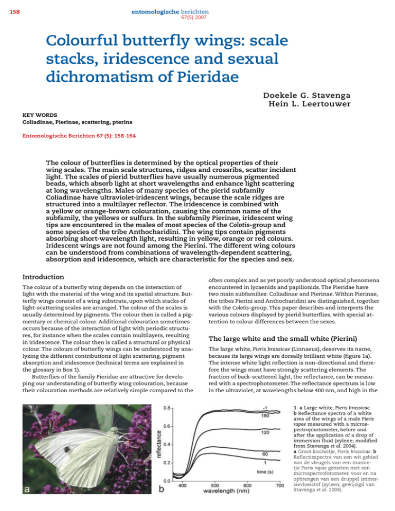
158 entomologische berichten 67(5) 2007 Colourful butterfly wings: scale stacks, iridescence and sexual dichromatism of Pieridae Doekele G. Stavenga Hein L. Leertouwer KEY WORDS Coliadinae, Pierinae, scattering, pterins Entomologische Berichten 67 (5): 158-164 The colour of butterflies is determined by the optical properties of their wing scales. The main scale structures, ridges and crossribs, scatter incident light. The scales of pierid butterflies have usually numerous pigmented beads, which absorb light at short wavelengths and enhance light scattering at long wavelengths. Males of many species of the pierid subfamily Coliadinae have ultraviolet-iridescent wings, because the scale ridges are structured into a multilayer reflector. The iridescence is combined with a yellow or orange-brown colouration, causing the common name of the subfamily, the yellows or sulfurs. In the subfamily Pierinae, iridescent wing tips are encountered in the males of most species of the Colotis-group and some species of the tribe Anthocharidini. The wing tips contain pigments absorbing short-wavelength light, resulting in yellow, orange or red colours. Iridescent wings are not found among the Pierini. The different wing colours can be understood from combinations of wavelength-dependent scattering, absorption and iridescence, which are characteristic for the species and sex. Introduction The colour of a butterfly wing depends on the interaction of light with the material of the wing and its spatial structure. Butterfly wings consist of a wing substrate, upon which stacks of light-scattering scales are arranged. The colour of the scales is usually determined by pigments. The colour then is called a pigmentary or chemical colour. Additional colouration sometimes occurs because of the interaction of light with periodic structures, for instance when the scales contain multilayers, resulting in iridescence. The colour then is called a structural or physical colour. The colours of butterfly wings can be understood by analyzing the different contributions of light scattering, pigment absorption and iridescence (technical terms are explained in the glossary in Box 1). Butterflies of the family Pieridae are attractive for developing our understanding of butterfly wing colouration, because their colouration methods are relatively simple compared to the often complex and as yet poorly understood optical phenomena encountered in lycaenids and papilionids. The Pieridae have two main subfamilies: Coliadinae and Pierinae. Within Pierinae, the tribes Pierini and Anthocharidini are distinguished, together with the Colotis-group. This paper describes and interprets the various colours displayed by pierid butterflies, with special attention to colour differences between the sexes. The large white and the small white (Pierini) The large white, Pieris brassicae (Linnaeus), deserves its name, because its large wings are dorsally brilliant white (figure 1a). The intense white light reflection is non-directional and therefore the wings must have strongly scattering elements. The fraction of back-scattered light, the reflectance, can be measured with a spectrophotometer. The reflectance spectrum is low in the ultraviolet, at wavelengths below 400 nm, and high in the 1. a Large white, Pieris brassicae. b Reflectance spectra of a white area of the wings of a male Pieris rapae measured with a microspectrophotometer, before and after the application of a drop of immersion fluid (xylene; modified from Stavenga et al. 2004). a Groot koolwitje, Pieris brassicae. b Reflectiespectra van een wit gebied van de vleugels van een mannetje Pieris rapae gemeten met een microspectrofotometer, voor en na opbrengen van een druppel immersievloeistof (xyleen; gewijzigd van Stavenga et al. 2004). entomologische berichten 67(5) 2007 2. Electron microscopic photographs of scales of a male small white, Pieris rapae. a Cover scales partly overlap ground scales (scanning electron microscopy, SEM). The upper lamina of the scales features longitudinal ridges. b Part of a white scale (SEM). The ridges are connected by crossribs, which are adorned by numerous oval-shaped beads. c Transmission electron microscopic (TEM) section of a white scale, showing the beads at the upper lamina of the scale. The lower lamina of the scale is more or less flat. d A scale from one of the black spots (SEM), showing that in this case the crossribs do not have beads (a, bar: 25 μm; b-d, bar: 2 μm; modified from Stavenga et al. 2004). Electronenmicroscopische foto's van schubben van een mannetje klein koolwitje Pieris rapae. a Dekschubben overlappen deels de onderschubben (scanning-electronenmicroscopie, SEM). b Deel van een witte schub (SEM). De richels zijn verbonden door dwarsribben, waaraan talrijke ovale kralen hangen. c Transmissie-electronenmicroscopische (TEM) foto van een witte schub, waarin de kralen aan de bovenlaag van de schub te zien zijn. De onderlaag van de schub is min of meer vlak. d Een schub van een van de zwarte vlekken (SEM), waarbij te zien is dat de kralen aan de dwarsribben ontbreken (a, maatstreep: 25 μm; b-d, maatstreep: 2 μm; gewijzigd van Stavenga et al. 2004). and the lower lamina is virtually smooth (figure 2c). The upper lamina rests via pillars, so-called trabeculae, on the lower lamina. The scales at the dorsal side of the wings of P. rapae are virtually all white, but some scales are black. The colour difference is related to a structural difference: the crossribs of the white scales carry oval-shaped beads, which contain pigment that absorbs exclusively ultraviolet light (figure 2b, c), whereas the black scales do not have beads (figure 2d). The ridges and crossribs of the black scales contain melanin pigment, which absorbs ultraviolet as well as visible light (Stavenga et al. 2004). Structures with a refractive index different from their environment scatter incident light. The material of the ridges and crossribs of butterfly wing scales is chitin, which has a refractive index of about 1.57, distinctly higher than the refractive index value 1 of air. The beads probably have a still higher refractive index, but that is as yet uncertain. It is clear that the scales act as light scatterers, because the scattering by the ridges, crossribs as well as the beads of the white scales is substantially reduced when an immersion fluid like xylene, which has a refractive index 1.49, replaces the air, so that the difference in refractive difference distinctly drops. In other words, the immersion fluid causes a reduced reflectance (figure 1b). The beads of the scales of P. rapae contain a pigment that absorbs in the ultraviolet (UV) and this causes a low UV-reflectan- visible wavelength range, at wavelengths above 450 nm, as shown in the closely related small white P. rapae (Linnaeus) (figure 1b). Insight into the physical basis of the reflectance can be obtained by applying immersion fluid. The reflectance spectrum (figure 1b, time t = -1 s) changes abruptly when a drop of immersion fluid, for instance xylene, is applied to the wing (at time t = 0 s). It causes an immediate decrease in the long-wavelength reflectance, but the reflectance gradually recovers when the immersion fluid evaporates (figure 1b, times t = 60, 120, 180 s). This result can be readily understood from the structure of the wings (Ghiradella 1984, Nijhout 1991). The wings of pierids are, like those of all butterflies (and moths), studded with scales, partly overlapping as tiles on a roof (figure 2a). The scales are flat and measure about 50 x 200 μm2. The upper lamina features characteristic longitudinal ridges, connected by crossribs (figure 2b, d), 3. Reflectance spectra of various parts of the wings of the male black jezebel, Delias nigrina (inset: up – dorsal side, and down – ventral side), measured with a fiber optic spectrometer (modified from Stavenga et al. 2006). Reflectiespectra van verschillende delen van de vleugels van een mannetje Delias nigrina (inzet: boven – dorsale zijde, onder – ventrale zijde), gemeten met een fiber-optische spectrometer (gewijzigd van Stavenga et al. 2006). 159 160 entomologische berichten 67(5) 2007 4. Reflectance spectra from the dorsal forewing of the male orange tip, Anthocharis cardamines. Inset: up - normal RGB photograph; down - UV photograph. Scale bar: 1 cm. Reflectiespectra van de dorsale voorvleugel van een mannetje oranjetip, Anthocharis cardamines. Inzet: boven - normale kleurenfoto, onder – UV foto. Maatstreep: 1 cm. ce (figure 1b). The human eye does not see into the ultraviolet wavelength range and therefore the high reflectance throughout the visible wavelength range of the wings of the whites causes the observed white colour. Butterflies, as all insects, see well into the UV and will therefore detect the low UV reflectance of the Pieris wings. Consequently, they will perceive the wings of P. brassicae and P. rapae as strongly coloured. Other pierids, for instance members of the subfamily Coliadinae, have pigments that absorb not only UV light, but also in the blue, green and yellow wavelength ranges, depending on the type of pigment. As a consequence, the colour of the scales is then yellow, orange or red, respectively (Rutowski et al. 2005). The black jezebel (Pierini) A beautiful example of a pierid with different types of pigmented scales is the black jezebel, Delias nigrina Fabricius (figure 3). The dorsal side of the wings of the male is white with black wing tips, as in P. brassicae and P. rapae, but ventrally the wings are brown-black with yellow and red bands. Reflectance spectra measured from various wing areas (indicated by numbers in figure 3) show a low reflectance at short wavelengths and a high reflectance in the long wavelength range, except in location 6, a brown-black area, where the reflectance is low at all wavelengths. The reflectance spectra 1 and 3 in figure 3 were taken at lo- cations 1 and 3 at the dorsal side of the wing. These locations were opposed at the ventral side by red bands, whereas location 2 of the dorsal side of the wing had brown-black scales at the ventral side. Reflectance spectrum 2 (of location 2) is more or less flat at all wavelengths above 450 nm, but spectra 1 and 3 are only flat between 450 and 550 nm. The latter spectra rise above 550 nm, to flatten again at wavelengths above 700 nm. The reflectance rise happens precisely in the wavelength range where the reflectance of the red band at the ventral wing side rapidly increases. The explanation of the rise in the reflectance spectra 1 and 3 above 550 nm is therefore that the dorsal scales are slightly transmittant. Long-wavelength incident light is obviously strongly reflected, but a fraction is transmitted, and this light is back-scattered by the red scales at the ventral side of the wings and subsequently partly passes the dorsal scales. This transmitted light slightly but significantly contributes to the reflectance measured at the dorsal side (Stavenga et al. 2006). The orange tip (Anthocharidini) Strikingly orange coloured scales cover the outer parts of the dorsal forewings of the male orange tip, Anthocharis cardamines (Linnaeus) (figure 4). The remaining parts of the dorsal side of the wings are covered by white scales. Reflectance spectra measured from the two wing areas can be immediately understood from the presence of two different pigments in the orange and white scales. The pigments appreciably absorb short-wavelength light up to about 450 and 550 nm respectively. The great orange tip (Colotis-group) The dorsal side of the wings of males of the great orange tip, Hebomoia glaucippe (Linnaeus) (Colotis-group; figure 5), resemble those of A. cardamines (figure 4), at least for a human observer. The reflectance spectrum of the main, white area of the dorsal side of the wing (numbered 2 in figure 5) is indeed virtually identical to reflectance spectra of the white wings of P. brassicae (figure 1b), D. nigrina (figure 3) and A. cardamines (figure 4). However, different from A. cardamines, the wing tips have a high reflectance in the ultraviolet (figure 5, spectrum 1), as is also seen in UV photographs. Scanning electron microscopy of the wing scales shows the origin of the UV reflectance (figure 6a). The scales can be distinguished in cover scales and ground scales, where the cover scales largely overlap the ground scales. The ridges of the cover scales of male Hebomoia appear to have a special construction. 5. The male great orange tip, Hebomoia glaucippe, has dorsal forewing tips that reflect substantially in the long-wavelength range (1, inset up). The scales in the remaining part of the dorsal side of the wings strongly reflect visible light, also very similar as in the orange tip, Anthocharis cardamines. The dorsal tips reflect however in the ultraviolet (inset down, scale bar: 2 cm), which is very different from the orange tip (see figure 4). Het mannetje van de grote oranjetip, Hebomoia glaucippe, heeft dorsale voorvleugelpunten die aanzienlijk reflecteren in het lange-golflengtegebied (1, inzet boven). De schubben in het overige dorsale deel van de vleugels reflecteren zichtbaar licht, net als in het oranjetipje, Anthocharis cardamines. De dorsale vleugelpunten reflecteren echter ook ultraviolet (inzet beneden; maatstreep: 2 cm), wat niet het geval is bij het oranjetipje (zie figuur 4). entomologische berichten 67(5) 2007 6. a Part of a cover scale, overlapping a ground scale, from the orange tip of a male Hebomoia glaucippe. Both cover and ground scales have crossribs with beads. The ridges of the cover scale (black arrow) are more densely spaced than the ridges of the ground scale (white arrow) and are strongly folded into lamellae (scale bar: 2 μm). b The lamellae form a multilayer stack, so that incident light arriving at a surface of a lamella is partly reflected and transmitted. The lamellae act together as an interference reflector for short-wavelength, ultraviolet light. a Deel van een dekschub die een onderschub overlapt in de oranje vleugelpunt van een mannetje Hebomoia glaucippe. Zowel de dekschub als de onderschub hebben dwarsribben met kralen. De richels van de dekschub (zwarte pijl) zitten dichter op elkaar dan die van de onderschub (witte pijl) en zijn geplooid in lamellen (maatstreep: 2 μm). b De lamellen vormen een multilaag, waarbij invallend licht door de afzonderlijke lagen deels gespiegeld en deels doorgelaten wordt. De multilaag blijkt te werken als een interferentiespiegel voor kortgolvig ultraviolet licht. They consist of a stack of narrow plates (lamellae), with distances of about 0.1 μm. The plates partly reflect and transmit incident light and the transmitted and reflected light interferes because of the wave nature of light and the small plate distance (figure 6b). The interference is constructive at ultraviolet wavelengths, thus causing the substantial reflectance of the cover scales in the ultraviolet. Both cover- and ground scales at the dorsal side of the wing tip of male Hebomoia have a large amount of pigmented beads, which are primarily responsible for the orange coloured dorsal side of the wing tips. Sexual dichromatism of the autumn leaf vagrant (Colotis-group) Many pierid butterfly species display sexual dichromatism: males and females have differently coloured wings. A clear example is the autumn leaf vagrant, Eronia leda Boisduval (figure 7). The bright orange dorsal side of the wing tips of the male strongly reflect UV light (Ghiradella et al. 1972, Ghiradella 1991, Stavenga et al. 2006). The spectral characteristics of the wing tips are very similar to those of the male great orange tip (figure 5). The dorsal side of the wings of the female lack the UV iridescent tips. They have an overall whitish-yellow colour. The ventral sides of both males and females are more indistinctly coloured (figure 7). This shows that the sexual dichromatism of E. leda is not restricted to the short-wavelength iridescence of the male wing tips, but that also the pigment density differs. This is 7. Sexual dichromatism of the autumn leaf vagrant, Eronia leda (D: dorsal; V: ventral; columns 1 and 3 are UV photographs and columns 2 and 4 RGB photographs). The wings reflect yellow light, because ultraviolet as well as blue absorbing pigment suppresses light scattering by the scale structures in the short-wavelength range. The wing tips of the male reflect ultraviolet as well as orange light, in a similar way as in the great orange tip, Hebomoia glaucippe (figure 5); scale bars: 1 cm. Sexueel dichromatisme van Eronia leda (D: dorsaal; V: ventraal; de foto's van de kolommen 1 en 3 zijn opgenomen met ultraviolet licht en die van de kolommen 2 en 4 zijn normale kleurenfoto's). De vleugels reflecteren geel licht, omdat de lichtverstrooiing door de schubstructuren in het kortgolvig gebied onderdrukt wordt door zowel ultraviolet- als blauwabsorberend pigment. De vleugelpunten van het mannetje reflecteren UV en oranje licht, net als bij de grote oranjetip, Hebomoia glaucippe (figure 5); maatstrepen: 1 cm. 161 162 entomologische berichten 67(5) 2007 8. Phylogeny (left, simplified from Braby et al. 2006) and sexual dichromatism of Pieridae (the photographs in columns 1 and 3 were taken with ultraviolet illumination, those in columns 2 and 4 are normal RGB photographs). a Colias electo (subfamily Coliadinae), b Red tip, Colotis danae (Colotis-group). c Orange tip, Anthocharis cardamines (Anthocharidini). d Japanese small white, Pieris rapae crucivora (Pierini). Fylogenie (links, naar Braby et al. 2006) en sexueel dichromatisme van Pieridae (foto's in kolom 1 en 3 zijn UV foto's, die in kolom 2 en 4 kleurenfoto's). a Colias electo (subfamilie Coliadinae), b roodtip, Colotis danae (Colotis-groep). c oranjetip, Anthocharis cardamines (Anthocharidini). d Japanse klein koolwitje, Pieris rapae crucivora (Pierini). done by varying the number of pigmented beads (Giraldo & Stavenga 2007). Different colouration methods in male and female Pieridae The colouration of pierid butterflies can be compared with their phylogeny (Braby et al. 2006). Quite extreme examples of differently coloured males and females are encountered among the pierid subfamily Coliadinae, or sulfurs. The males of many Colias-species have wings that dorsally iridesce in the UV over virtually the complete wing surface, whereas the females lack such ornamentation (figure 8). The overall colour of Coliadinae is sometimes brown-orange, but often bright yellow colours are seen, for instance in the brimstone, Gonepteryx rhamni (Linnaeus) and in Eurema-species. Among the subfamily Pierinae, the Pierini are mostly white, but the various Anthocharidini take an intermediate position. Their tip is orange (as in the European orange tip, A. cardamines) or yellow (as in the Japanese yellow tip, Anthocharis scolymus Butler) and the remaining part of the dorsal side of the wing can be yellow or white. Comparing the different tribes of the Pierinae, we thus can speculate that the colouration has shifted from orange to yellow to white, with a coloured tip as an intermediate case. The male members of the Colotis-group have usually ultraviolet or violet/blue iridescent wing tips (figure 8). There are of course many variations and gradations. Males of the Japanese small white, Pieris rapae crucivora Boisduval, are more brightly white than the females, and the male wings reflect less than the female wings in the ultraviolet (Obara 1970), but the two sexes of the European small white Pieris r. rapae (Lin- Box 1. Glossary 9. Structural formulae of the three main pterins occurring in pierid wings and their absorption spectra. Structuurformules van de drie belangrijkste pterines in vleugels van Pieridae en hun absorptiespectra. reflectance - the fraction of incident light that is reflected by a material transmittance - the fraction of incident light that is transmitted wavelengths and (complementary) colours - the wavelength range of ultraviolet light is 300-400 nm, and that of visible light (for humans) 400-700 nm. For most insects, the wavelength range of visible light is 300-600 nm, but some butterflies extend the range up to 650, or even 700 nm. When a substance is illuminated with ultraviolet as well as white light, and the substance contains a pigment that absorbs exclusively ultraviolet light, the remaining light then is white. When the pigment also absorbs violet and blue light (wavelength range 400-500 nm), yellow light remains. When the pigment absorbs light of the wavelength range 300-550 nm, the remaining light is seen as orange, and when the pigment absorbs in the wavelength range 300-600 nm, only red light remains. entomologische berichten 67(5) 2007 10. Summary diagram of the wing tip reflectance of the male great orange tip, Hebomoia glaucippe. The lamellae of the cover scale ridges reflect ultraviolet, but not visible light. Blue light is absorbed by pigment in the beads at the crossribs. The pigment does not absorb in the orange-red wavelength range, but instead the beads strongly scatter light, which therefore results in a high reflectance. The dashed lines represent the minimum and maximum reflectance. The reflectance is minimal where multilayer reflectance is negligible and pigment absorption is maximal. The reflectance is maximal where the multilayer reflectance is high, and where pigment absorption is minimal and light scattering by the beads is maximal. Schema van de verschillende optische mechanismen die de kleuren in de vleugelpunt veroorzaken bij het mannetje grote oranjetip, Hebomoia glaucippe. De lamellen van de dekschubben reflecteren ultraviolet, maar geen (voor mensen) zichtbaar licht. Blauw licht wordt geabsorbeerd door het pigment in de kralen aan de dwarsribben. Het pigment absorbeert niet in het oranje en rode golflengtegebied, maar de kralen verstrooien dat licht sterk, hetgeen resulteert in een hoge reflectie. De onderbroken lijnen geven de minimum en maximum reflectie weer. De reflectie is minimaal als de multilaagreflectie verwaarloosbaar is en de absorptie van het pigment maximaal. De reflectie is maximaal bij golflengten waar de multilaagreflectie hoog, de absorptie van het pigment minimaal en de lichtverstrooiing door de kralen maximaal is. naeus) differ only slightly in colouration (Obara & Majerus 2000, Giraldo & Stavenga 2007). Similarly, the males of some species of the Coliadinae subfamily do not have ultraviolet reflecting wings (Kemp et al. 2005). In conclusion, although sexual dichromatism seems to be a very general phenomenon among pierid butterflies, this appears to be not universal. Pterins, the pigments of pierid wings Previous work has demonstrated that the pigments of pierid wings belong to the class of pterins (Kayser 1985, figure 9). The various pterins have different absorption spectra, which can be restricted to the ultraviolet or can extend into the yellow or green wavelength range. Different pterins can be simultaneously expressed in one and the same scale (Giraldo & Stavenga 2007), apparently providing the butterflies with a flexible colouration palette. The pterins can be extracted from the wing scales by applying ammonium hydroxide (ammonia) solutions. The main pterins found in pierid wings are leucopterin, xanthopterin and erythropterin, which absorb maximally in the ultraviolet, violet, and blue wavelength range, respectively (figure 9). the latter case thus is that the reflectance is high in the orange/ red as well as in the UV, and the reflectance is low at medium wavelengths, in the blue. A further important determinant of the wing colour is the way the multilayers and absorbing pigments are arranged on the wing. For instance, only cover scales will have the UV-reflecting multilayers (figure 6). These structures must be situated above the pigmented beads, because the absorbing pigments should otherwise already have reduced incident ultraviolet light before it would reach the multilayers. Furthermore, by piling several scattering scales on top of one another very high reflectances can be realized at wavelengths where there is no absorption by pigments. The wings of P. brassicae, for example, are very densely packed with white scales, which together create the very white colour of the large white. The mechanisms of pierid wing colouration are by now well understood. The next step is to better understand the evolutionary forces that determine the sexual dichromatism, or for that matter, that determine animal colouration. Acknowledgements The butterflies described in this paper were studied in the butterfly col- How to colour the wings of Pieridae lections of the National Museum of Natural History Naturalis (Leiden, We can conclude that there are several optical mechanisms that together colour the wings of pierids, three of which are summarized in figure 10. The principal colouring methods are illustrated with the reflectance spectrum of the orange tip of a male H. glaucippe (figure 5). In the long-wavelength range, light scattering by beads and other scale structures dominates. When the scales contain pigments, either concentrated in granular beads or diffusely in the ridges and crossribs, these will reduce the scattered light in the wavelength range of their absorption spectrum, resulting in a low reflectance. When the scales possess a multilayer or another periodical arrangement, they are structurally coloured. As we have seen above, the latter happens in the dorsal side of the wing tips of male Hebomoia. The total effect in ments at the manuscript) and the Royal Museum for Central Africa curator Dr. R. de Jong, who also provided valuable constructive com(Tervuren, Belgium, curator Dr. U. Dall'Asta), which was supported by the EU via SYNTHESYS. Colias erate specimens were obtained from Prof. K. Arikawa, Sokendai, Hayama, Japan. The EOARD provided financial support (grant 063027). 163 164 entomologische berichten References Braby MF, Vila R & Pierce NE 2006. Molecular phylogeny and systematics of the Pieridae (Lepidoptera : Papilionoidea): higher classification and biogeography. Zoological Journal of the Linnean Society 147: 238-275. Ghiradella H 1984. Structure of iridescent lepidopteran scales: variations on several themes. Annals of the Entomological Society of America 77: 637-645. Ghiradella H 1991. Light and color on the wing: structural colors in butterflies and moths. Applied Optics 30: 3492-3500. Ghiradella H, Aneshansley D, Eisner T, Silberglied R & Hinton HE 1972. Ultraviolet reflection of a male butterfly: Interference color caused by thin-layer elaboration of wing scales. Science 178: 1214-1217. Giraldo MA & Stavenga DG 2007. Sexual dichroism and pigment localization in the wing scales of Pieris rapae butterflies. Pro- 67(5) 2007 ceedings of the Royal Society B 274: 97-102. Kayser H 1985. Pigments. In: Kerkut GA & Gilbert LI (eds) Comprehensive insect physiology, biochemistry and pharmacology, Vol. 10: 367-415. Pergamon. Kemp DJ, Rutowski RL & Mendoza M 2005. Color pattern evolution in butterflies: a phylogenetic analysis of structural ultraviolet and melanic markings in North American sulphurs. Evolutionary Ecology Research 7: 133-141. Nijhout HF 1991. The development and evolution of butterfly wing patterns. Smithsonian Institution Press. Obara Y 1970. Studies on the mating behavior of the white cabbage butterfly, Pieris rapae crucivora Boisduval. III. Near-ultraviolet reflection as the signal of intraspecific communication. Zeitschrift für vergleichende Physiologie 69: 99-116. Obara Y & Majerus MEN 2000. Initial mate re- cognition in the British cabbage butterfly, Pieris rapae rapae. Zoological Science 17: 725-730. Rutowski RL, Macedonia JM, Morehouse N & Taylor-Taft L (2005). Pterin pigments amplify iridescent ultraviolet signal in males of the orange sulphur butterfly, Colias eurytheme. Proceedings of the Royal Society B 272: 2329-2335. Stavenga DG, Giraldo MA & Hoenders BJ 2006. Reflectance and transmittance of light scattering scales stacked on the wings of pierid butterflies. Optics Express 14: 4880-4890. Stavenga DG, Stowe S, Siebke K, Zeil J & Arikawa K 2004. Butterfly wing colors: scale beads make white pierid wings brighter. Proceedings of the Royal Society B 271: 1577-1584. Ingekomen 5 december 2006, geaccepteerd 27 juni 2007. Samenvatting Kleurrijke vlindervleugels: reflecterende schubben en sekse-afhankelijke kleuren van Pieridae De kleur van vlinders wordt bepaald door de optische eigenschappen van de vleugelschubben. De schubstructuren, richels en dwarsribben verstrooien invallend licht. De schubben van vlinders van de familie Pieridae bevatten meestal een grote hoeveelheid korrels, die licht van korte golflengten absorberen en licht van langere golflengten sterk verstrooien. Mannetjes van de subfamilie Coliadinae hebben vleugels met een weerschijn in het ultraviolet, omdat de richels van de schubben een multilaagstructuur hebben. Deze weerschijn wordt gecombineerd met een gele of oranje-bruine kleur. Bij de subfamilie Pierinae komen vleugels met weerschijn ook voor, maar dan uitsluitend op de bovenkant van de voorvleugelpunt van mannetjes van de meeste soorten in de Colotis-groep en minder bij soorten van de tribus Anthocharidini. De vleugelpunten bevatten ook pigmenten die kortgolvig licht absorberen, waardoor ze een gele, oranje of rode kleur hebben. De weerschijn komt niet voor bij vlindersoorten van het tribus Pierini. De kleuren van vlindervleugels worden bepaald door combinaties van golflengte-afhankelijke lichtverstrooiing, absorptie en iridescentie, waarbij de combinaties karakteristiek zijn voor de soort en het geslacht. Doekele G. Stavenga & Hein L. Leertouwer Department of Neurobiophysics University of Groningen Nijenborgh 4 9747 AG Groningen D.G.Stavenga@rug.nl
