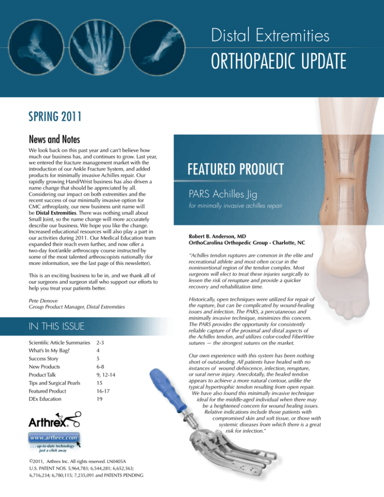
Distal Extremities
orthopaedic update
Spring 2011
News and Notes
We look back on this past year and can’t believe how
much our business has, and continues to grow. Last year,
we entered the fracture management market with the
introduction of our Ankle Fracture System, and added
products for minimally invasive Achilles repair. Our
rapidly growing Hand/Wrist business has also driven a
name change that should be appreciated by all.
Considering our impact on both extremities and the
recent success of our minimally invasive option for
CMC arthroplasty, our new business unit name will
be Distal Extremities. There was nothing small about
Small Joint, so the name change will more accurately
describe our business. We hope you like the change.
Increased educational resources will also play a part in
our activities during 2011. Our Medical Education team
expanded their reach even further, and now offer a
two-day foot/ankle arthroscopy course instructed by
some of the most talented arthroscopists nationally (for
more information, see the last page of this newsletter).
This is an exciting business to be in, and we thank all of
our surgeons and surgeon staff who support our efforts to
help you treat your patients better.
Pete Denove
Group Product Manager, Distal Extremities
in this issue
Scientific Article Summaries 2-3
What’s In My Bag?
4
Success Story
5
New Products
6-8
Product Talk
9, 12-14
Tips and Surgical Pearls
15
Featured Product
16-17
DEx Education
19
©2011, Arthrex Inc. All rights reserved. LN0405A
U.S. PATENT NOS. 5,964,783; 6,544,281; 6,652,563;
6,716,234; 6,780,115; 7,235,091 and PATENTS PENDING
Featured Product
PARS Achilles Jig
for minimally invasive achilles repair
Robert B. Anderson, MD
OrthoCarolina Orthopedic Group - Charlotte, NC
“Achilles tendon ruptures are common in the elite and
recreational athlete and most often occur in the
noninsertional region of the tendon complex. Most
surgeons will elect to treat these injuries surgically to
lessen the risk of rerupture and provide a quicker
recovery and rehabilitation time.
Historically, open techniques were utilized for repair of
the rupture, but can be complicated by wound-healing
issues and infection. The PARS, a percutaneous and
minimally invasive technique, minimizes this concern.
The PARS provides the opportunity for consistently
reliable capture of the proximal and distal aspects of
the Achilles tendon, and utilizes color-coded FiberWire
sutures ­— the strongest sutures on the market.
Our own experience with this system has been nothing
short of outstanding. All patients have healed with no
instances of wound dehiscence, infection, rerupture,
or sural nerve injury. Anecdotally, the healed tendon
appears to achieve a more natural contour, unlike the
typical hypertrophic tendon resulting from open repair.
We have also found this minimally invasive technique
ideal for the middle-aged individual when there may
be a heightened concern for wound healing issues.
Relative indications include those patients with
compromised skin and soft tissue, or those with
systemic diseases from which there is a great
risk for infection.”
Scientific Article Summaries
TightRope Syndesmosis Fixation System
More Physiologic Means of Achieving Syndesmosis Fixation
Robert Klitzman, MD; Heng Zhao, PhD; Li-Qun Zhang, PhD; Greg Strohmeyer, BS; Anand Vora,
MD, “Suture-Button Versus Screw Fixation of the Syndesmosis: A Biomechanical Analysis,”
Foot and Ankle International, January 2010: 69-75.
• Rigid fixation of the syndesmosis with screw fixation may be problematic in allowing
physiologic motion of the syndesmosis.
• TightRope fixation allowed movement in the sagittal plane, which more closely mimicked
the movement of the native syndesmosis than did the screw construct.
CONCLUSION:
The results of this study suggest that suture-button fixation is a viable alternative to screws in the
fixation of syndesmotic injuries. We believe it is a more physiologic type of fixation, which has the
ability to maintain a reduction of the syndesmosis and negate a second surgery for removal of
fixation.
CMC Mini TightRope
Mini TightRope CMC Arthroplasty Provides Faster Recovery and
Improves Patient Quality of Life
Christopher A. Cox, MD; Dan A. Zlotolow, MD; Jeffrey Yao, MD, “Suture-Button Suspensionplasty After Arthoscopic Hemitrapeziectomy for Treatment of Thumb Carpometacarpal Arthritis,”
Arthroscopy: The Journal of Arthroscopic and Related Surgery, February 2010: 1395-1403.
• Offers an advantage over currently described techniques in that the period of postoperative
immobilization is decreased and range-of-motion exercises are initiated earlier.
• Enhances patient rehabilitation rate and the elimination of external implants greatly
improves quality of life.
CONCLUSION:
The results of the study suggest that suture-button fixation provides an alternative to traditional
surgical repair methods for basal joint arthritis. This described technique of using suture-button
fixation for suspension of the thumb ray after hemi-trapezial or complete trapezial excision offers
an advantage over currently described techniques in that the period of postoperative immobilization is decreased and range-of-motion exercises are initiated earlier. This enhances the rate at
which outpatients rehabilitate. We believe that this earlier recovery, along with the elimination of
external implants, greatly improves their quality of life.
2
Mini Pushlock Anchors
Knotless Anchors Improve Soft Tissue Irritation in the Modified Brostrom Procedure
James A. Bynum, MD; John M. Crates, MD; Jorge Aziz-Jacobo, MD; F. Alan Barber, MD,
“Modified Brostrom Technique Using Knotless Suture Anchors,” Techniques in Foot & Ankle Surgery,
March 2010.
• Brostrom reconstructions utilizing suture anchors have good results and low complication rates.
• Prevents problems associated with subcutaneous knot irritation.
CONCLUSION:
Brostrom reconstructions utilizing suture anchors have good results and low complication rates.
The technique presented here improves upon the suture anchor technique by addressing concerns
about subcutaneous knot irritation, which have been a source of postsurgical patient dissatisfaction.
Micro SutureLassos and Small Joint Bio-SutureTaks
Early Results of Arthroscopic Lateral Ankle Ligament Reconstruction Promising
Peter G. Mangone, MD, “Early Results of Arthroscopic Lateral Ankle Ligament Reconstruction
Promising,” Orthopedics Today, September 2010.
• Arthroscopic Brostrom utilizing specifically engineered micro suturelassos aid in calculated
suture passage through the ATFL.
• Smaller incisions and less overall morbidity will support quicker postoperative rehabilitation.
CONCLUSION:
We are in the dawn of a new era. Arthroscopic Brostrom techniques are providing solid fixation,
allowing patients to return to functional activity. Anterior drawer tests were negative in most
patients after arthroscopic Brostrom-Gould-type technique.
3
What’s in My Bag?
Featuring: James McWilliam, MD
James McWilliam, MD
New York Foot & Ankle
Harrison, NY
Arthrex Ankle Fracture System
In this interview, Dr. James McWilliam (A) shares his surgical
technique tips, current successes with the Arthrex Ankle Fracture
System, and thoughts on using this product in the future. Before
getting in to the interview, here are some basics on the system:
Plates: This set includes a variety of fracture-specific anatomically
contoured plates, engineered to address even the most difficult
fracture patterns. These consist of: Third Tubular Plates, 3.5 mm
Reconstruction Plates, Lateral and Medial Hook Plates and a
Complex Fibular Fracture Plate. All plates designed for the fibula
have modifications that allow for use of the TightRope or
syndesmotic screws.
Q: Has the TightRope changed the way you treat syndesmotic
injuries in relation to ankle fractures? Any tips for reduction?
A: The TightRope has made me think a lot more about subtle
syndesmotic injury and its effect on late outcomes. Previously, I
assessed syndesmotic instability fluoroscopically with external
rotation and the Cotton test. More recently, I began visually
inspecting the syndesmosis in Weber B and C fractures and
have been surprised by the frequency of what I might call
“subtle” instability. Due to the more physiologic nature of the
TightRope, I feel much more sanguine about fixing these subtly
unstable ankles.
Screws: This set includes the following: 2.7 mm locking screws,
3.0 mm cancellous screws, 3.5 mm locking and nonlocking screws,
4.0 mm cancellous screws, 4.0 mm short and long thread solid
screws, and 4.0 mm short and long thread cannulated screws.
Q: There are both medial and lateral hook plates included in the
comprehensive ankle fracture set. What indications would
you use these plates for? The set also includes 4.0 mm
cannulated screws – how do you decide what type of fixation
you should use?
Instrumentation: Designed to be the most comprehensive set
available, it includes improved basic small fragment instrumentation
for the treatment of the majority of fracture patterns.
A: The lateral hook plates are outstanding for any transverse fibula
fracture or fibular osteotomy. A 4.0 mm screw placed in an
intramedullary fashion through the hook of the plate provides
excellent axial compression.
“The comprehensive set of plates and screws,
as well as the outstanding retractors and
clamps make this set definitive in its scope for
the treatment of ankle fractures.”
I use the medial hook plate for comminuted and osteoporotic
medial malleolar fractures. In elderly patients, the medial
malleolus will frequently fragment with the placement of
malleolar screws. This can be avoided by placing the plate
extraperiosteally with the hook, providing excellent stable soft
tissue fixation via the deltoid ligament. The plate also acts as a
washer allowing for compression across the fracture via a
traditionally placed “malleolar” screw, obviating fear of
malleolar fragmentation.
The medial hook plate also provides excellent fixation in
patients requiring malleolar osteotomy for osteochondral lesions.
Q: What is the most important distinguishing feature about this
set against the competition’s system?
A: The comprehensive set of plates and screws, as well as the
outstanding retractors and clamps, make this set definitive in its
scope for the treatment of ankle fractures.
Q: How do you prepare for the reduction and fixation of the
ankle? Do you scope your ankle fractures?
Q: If you had to provide one pearl of wisdom to surgeons who will
use this system in the future, what would you say?
A: Respect the posterior malleolus and its effect on syndesmosis
when fractured.
Pre-op
Post-op
A: As always, I obtain three views of the ankle. I have been using
gravity-stress views on nondisplaced fractures for the past few
years and have been surprised by how many of these have
demonstrated significant instability and are subsequently treated
with surgery. I have not routinely scoped my otherwise
uncomplicated fractures, but my increasing roster of late scopes
after ankle fractures has made me begin to rethink this stance.
4
X-rays courtesy of Dr. Joe Koscielniak, Hobart, IN
Success Story
Use of TightRope with Ankle
Syndesmosis Injuries in the NFL
I
initially started using TightRope because of the problems
I was having with screw fixation of syndesmosis injuries.
In higher-level athletes, those problems became bigger problems. Broken screws in elite athletes can create so much stress in
the area they can erode the cortex of the fibula. I’ve seen it lead
to stress fractures in three different professional football players.
Because of this, I would get nervous about allowing them to play
early in their recovery. When the TightRope came out, I found the
avoidance of screw breakage very appealing.
I have especially had problems with using screws in NFL
athletes. One in particular, an offensive lineman, had a pretty
routine syndesmosis injury fixed with two 4.5 mm screws. Both
screws broke, however, and when we went to take them out the
screw fragments had to be left in. The remaining screw ended
up slightly eroding the medial cortex of his fibula and created a
stress fracture. We then had to take the screw fragment out from
the medial cortex of the tibia using a trephine. He subsequently
developed a stress fracture of his tibia three months later. Bottom
line is, the injury and subsequent complications wound up
essentially putting him out of the NFL. This increased my
awareness of the reality of the complication of screw breakage
and what a problem it can be, especially in terms of time lost or
further surgery. That alone made me more willing to use a less
rigid fixation for syndesmosis injuries. My early and successful
use of the TightRope in high school and college players made me
willing to use it in NFL players.
My experience with the TightRope: it doesn’t break and it hasn’t
led to the complications I was seeing with the other types of
fixation. I also see fewer problems and complications from
allowing the athlete to participate in their sport once the injury is
healed.
My technique using the TightRope might be a little different than
most. I don’t rely on just the TightRope; I believe in fixing the
syndesmosis anatomically and in repairing the ligaments so that
once the ligaments are healed it takes the stress off the fixation. At
that point, I am willing to allow athletes to return to their sport or
advance their activity towards that.
My post-op protocol after using the TightRope is a little bit variable
depending on the patient, but at 6 weeks if I repair the ligament
then I am willing to let them start running and start cutting. I have
had players re-enter the sport as early at 6 - 6.5 weeks post-surgery
in the NFL without any adverse outcomes or complications.
Daniel Cooper, MD
The Carrell Clinic
Dallas, TX
“My experience with TightRope: It hasn’t led
to the complications I was seeing with other
types of fixation.”
an elite or professional athlete patient population. If their body
feels normal to them, they are going to push it because it is their
mentality. So, an inherent problem with screw fixation is that
you may not be able to control what they are doing at an early
stage. They may be doing high impact loading and running, and
you aren’t always aware. Therein lies another advantage of a
somewhat flexible construct – even if the patient is somewhat
noncompliant, they won’t have the complications and problems
I discussed earlier.
I would recommend everyone to at least strongly consider using
the TightRope because I think that by using it, you avoid the risk
of screw breakage. Not everyone that breaks a screw is going to
have stress fractures or problems, but that potential is there and
we are always nervous about letting patients play or start activity
with the hardware in. Conversely, we also get nervous about
taking the hardware out and letting them play immediately. So I
repair the ligament anatomically at the time I do the surgery and
let the patient play with the fixation in.
When there is a push to get the player back without them going
on “Injured Reserve,” it is appealing to the player to have the
option of having the TightRope put in. It’s obviously appealing
to them to know they’re unlikely to develop complications from
hardware.
TightRope has made me look at ankle sprains and syndesmotic
sprains a little bit differently. I used to know I could fix these
injuries and get good results, but I would worry about the
athlete’s compliance post-op. It was also really problematic to
hold them out of play for 12 weeks, which is what the norm was
with screw fixation. I think TightRope has given me the piece of
mind that I don’t have to worry as much about their compliance
with the protocol, I don’t need to try to talk them into waiting
three or four months before they push it when they have been
feeling great early on, and I don’t worry about screws breaking.
Patient compliance is always an issue after fixing syndesmosis.
Over my career I have seen the patient feels good postoperatively
once the syndesmosis is secure, even with screw fixation. Even to
the degree that many times they are noncompliant, particularly in
5
New Products
New Arthrex Small Joint
Arthroscopy Instrument Set
This set of instruments was designed for the foot and ankle surgeon
to eliminate the need to borrow instruments from larger joint sets
and offer a comprehensive solution for small joint arthroscopy.
This complete set of instruments includes: ring-handled graspers
and punches, as well as curettes, osteotomes, elevators and
Chondro Picks for the daily work of the small joint arthroscopist.
In addition to the standard instrumentation, this unique set is
available with the optional Arthrex Ankle GPS System for pinpoint
pin and screw placement. Specialty instruments for OCD carving
and elevation are also available.
Appropriate Sizing for Small Joints: Ring-handled instruments
have 2.75 mm diameters. The other instruments are sized and
designed specifically for foot and ankle applications.
Innovative Design: One-of-a-kind designs, like the optional
Arthrex Ankle GPS System and specialized OCD instruments
provide a complete and unique offering to the small joint
arthroscopist.
AR-8655S
Complete Set for the OR: The tray holds all of the commonly used
instruments, so there is no need to pull multiple sets.
Quality Construction: Ring-handled instruments use friction-free
Teflon® bearings and come with a lifetime warranty against
manufacturing defects.
Arthrex Ankle GPS System
Designed specifically for the ankle,
the Arthrex Ankle GPS System offers
precision positioning and placement
of pins and screws. The guide is designed
to easily allow placement of 1.1 mm and 1.6 mm
K-wires in one, two, and four-hole patterns for simple
and effective retrograde OCD drilling. A 2.4 mm guide pin
sleeve is also included to allow precise cannulated screw
placements for bony fusions. This set is available separately
or as part of the Ankle Arthroscopy Instrument Set.
6
AR-8655GS
Ankle Arthroscopy
New Arthrex Noninvasive Ankle Distractor
A simple straight-forward design that allows quick
distraction with a turn of the tensioning wheel.
• One-piece construction for ease of setup
and operation
AR-1712
• Connects to the bed with a Clark Rail Adapter
• Combined with the Arthrex Ankle Distraction Strap, this is the complete set up for your next
ankle arthroscopy case
AR-1713
The Noninvasive Ankle Distraction Strap
• Made of strong nylon strapping material with
soft nonslip foam pads for patient comfort
and secure hold.
• This easy to use, one-size-fits-all device
offers effective traction and grip, which gives
the surgeon a distinct advantage over current
distraction devices.
Nonslip Pads of AR-1712
The Small Joint Limb Holder
• Has an adjustable post for Clark Rail
attachment
• A small limb tourniquet or optimal foam
insert may be used for limb fixation
• Also ideal for elbow and pediatric knee
arthroscopy
AR-1506
7
New Products
Cannulated Screw System Instrument Set
A complete screw system for fixation of the forefoot and midfoot, this
comprehensive set stands alone. The small screw system is a cannulated,
partially-threaded titanium alloy screw system that is indicated for use in
bone reconstruction, osteotomy, arthrodesis, joint fusion, fracture repair,
and fracture fixation of bones appropriate for the size of the device. With
self-drilling and self-tapping headed and headless compression screws
and diameters ranging from 2.0 mm to 4.0 mm, the small cannulated
screw system provides extensive versatility for surgical procedures of the
foot all-in-one system.
AR-8737S
• All small screws are manufactured from titanium alloy to provide
consistent strength.
• Screws are Type II anodized, a superior material on the market.
• Pilot drills, Countersinks, and drivers have corresponding
color-coded banding to match screw diameter, simplifying the
pairing of instrumentation with screw selection.
• While the screws are self-drilling, Cannulated Drill Bits and Triple
Threat Devices are included for use in hard cortical bone.
• Triple Threats (all-in-one near cortex drill, countersink, and depth
gauge) were designed to have a unique instrument in your set.
This device may speed up OR time and aid in implantation.
AR- 1318FT
AR-1319FT
AR-8737-12, -13, -14
Micro, Mini, and 3.5 mm Corkscrew FT
The Micro, Mini, and 3.5 mm Corkscrew FT Suture Anchors are designed
with a fully-threaded length to create maximum cortical purchase in smaller
bones. Using an internal drive mechanism and suture eyelet, these titanium
anchors enable surgeons to secure threads in the best bone – the cortex.
Fully Threaded: For maximum cortical purchase
Convenient: Predrill the cortex with included K-wire and insert the anchor
AR-1915FT
Preloaded with 2-0 FiberWire: For superior strength and handling ease
Preloaded with Smaller Tapered Needles: To save surgical time
8
Product Talk
LPS Screw System
AR-8946S
LPS 4.5 mm Titanium Screws
The workhorse of the foot and
ankle, the LPS 4.5 mm Cannulated
Lag Screw is ideal for fractures and
fusions of the lower extremity. With
a lower profile head and deeper
threads than a traditional AO screw,
the LPS 4.5 mm screw purchases
bone better and keeps a lower profile. This is a benefit in the
foot and ankle, where weight-bearing loads are significant and
soft tissue coverage may be minimal.
“Titanium Screws: Easy to use, quick, incredible
fixation with improved thread design, and cost
effective...the perfect combination.”
– Dr. Anand Vora, Illinois Bone and Joint Institute
Low Profile Head: Almost 1 mm shorter than a traditional AO
4.5 mm screw, while still using a 3.5 mm hex
Better Pull-out: 25% better than a standard AO 4.5 mm screw
Deeper Threads: Using a 2.4 mm Guide Pin allows the threads
to be deeper than a standard AO screw
Self-Drilling/Tapping: Speeds up the insertion process
5.5 mm Jones Fracture Screw
The 5.5 mm low profile Jones Fracture Screw is designed to
provide excellent stability for the stresses found at the base of
the 5th metatarsal. Whether used for acute fractures or chronic
nonunions, this screw is designed to provide stout IM fixation
for healing this difficult sports injury.
LPS 6.7 mm
Cannulated Lag Screws
Working closely with a team of top
foot and ankle surgeons, Arthrex
lowered the head profile by 1 mm, increased thread purchase by
lengthening and deepening the threads to increase pull-out by
30%, in comparison to a standard AO screw. This makes the screw
ideal for the high-demand, low-coverage applications in the foot.
The 6.7 mm screws are available with 4.5 mm and 5.5 mm screws
in a comprehensive set that will include a subtalar/ankle targeting
guide to improve accuracy and speed in the OR. A limited set of
MCO appropriate lengths (40 - 60 mm) of 6.7 mm LPS Screws are
available in a tandem tray with the Tenodesis system as a complete
solution for flatfoot reconstructions.
Increased Shaft Diameter: For greater resistance to the
micro-motion that may lead to the nonunions common for this
pathology
Low Profile Head: 1 mm shorter than a traditional AO 6.5 mm
screw, while still using a 3.5 mm hex
Solid Titanium Design: For greater strength against bending
loads
Deeper Threads: Using a 2.4 mm Guide Pin that allows the
threads to be deeper than a standard AO screw
Cortical Thread Design: For excellent purchase in the cortical
bone
Longer Threads: 18 mm thread length is designed specifically
for the foot
Bullet Nose Tip: For guidance down the IM canal of the 5th
metatarsal
Self-Drilling/Tapping: Speeds up the insertion process
Low Profile Head: To minimize soft tissue irritation in this area
of minimal coverage and high shoe pressures
Improved Instruments: For guidance down the IM canal of the
5th metatarsal
Better Pull-out: 30% better than a standard AO 6.5 mm screw
Assisted Targeting: Parallel and C-Ring Guide Pins enable quick
and accurate placement
Type II Anodized Titanium: The best material on the market
Complete Set: Housed with the 4.5 and 6.7 mm screws for a
complete solution
9
Trifecta!
The Winning Combination for
Arthroscopic Ankle Fusions
1
Preparation is Paramount
Arthrex Bone Cutter
• Designed for aggressive soft tissue and bone resection
• Multiple options: 3.8 mm, 4.0 mm, 5.0 mm, 5.5 mm
Arthrex BLURRTM
• A unique device designed for maximum bone
and tissue resection. Available in 5 mm size
2
Bone Cutter
BLURR
Facilitate a Biologic Response
Autologous Conditioned Plasma (ACP)
•Double syringe system allows for rapid and efficient
preparation of platelet rich plasma
•Concentration of platelets and growth factors within
a plasma layer, while removing the degradative
white blood cells
StimuBlastTM Demineralized Bone Matrix (DBM)
JRF
•Moldable bone void filler with osteoinductive and osteoconductive properties
•Unique Reverse Phase Medium (RPM) carrier thickens
when reaching body temperature
Optimize the bone healing
environment with natural growth
factors from ACP and JRFStimuBlast
3
Fixation You Can Count On
Arthrex 6.7 mm Screw
•Designed specifically for the foot & ankle surgeon
•Better pull-out – 30% better than standard 6.5 mm AO Screw
•Deeper threads – Using a 2.4 mm Guide Pin allows threads to be deeper than standard AO Screw
•18 mm and 28 mm thread length options designed for foot & ankle •Self-drilling with reverse cutting flutes
•Best material on the market – Type II Anodized Titanium
Comparison between
Arthrex 6.7 mm Screw
and the AO 6.5 mm Screw
Arthrex 6.7 mm Screw
1 mm shorter head height
than AO Screw
Arthrex 6.7 mm
2.3 mm
core-to-thread
differential
Arthroscopic ankle fusion utilizing
Arthrex 6.7 mm Screws in conjunction
with ACP and JRFStimuBlast
AO 6.5 mm
1.7 mm
core-to-thread
differential
For more information go to:
www.footscrews.arthrex.com
© 2010, Arthrex Inc. All rights reserved.
Product Talk
Mini TightRope System
2.7 mm Mini TightRope
(AR-8911DS)
Mini TightRope Disposables Kit
The Mini TightRope provides an alternative to both
pin and screw fixation. The advantages include:
• An absence of protruding hardware
• A second procedure is not required for removal
of a screw
• Far less joint disruption than that caused by a
3.5, 4, 4.5, 6.5, or 7.3 mm screw
For more complex fractures, this technique can
easily be combined with other fixation techniques.
The Mini TightRope provides a new approach to
treatment of Lisfranc ligament disruptions.
Oblong Button placed lateral
to 2nd metatarsal
3.5 mm Metal Mini TightRope FT
The new 3.5 mm Metal Mini TightRope FT is now available.
• Minimally invasive dorsal medial single incision
• Anchor construct stabilizes the metatarsal cuneiform
joint and acts as a ‘backstop’ to help prevent recurrence
of the deformity
• IM angle correction with or without an osteotomy
• Can be used with a distal osteotomy in cases of larger
IM angles or semi-rigid deformities
1
* Not cleared in the United States
Metal Mini TightRope FT
(AR-8917DS)
The lateral capsular structures
are released, followed by the
manual reduction of the 1st
intermetatarsal space.
2
Insert a Guidewire, starting on
the medial cortex of the 1st
metatarsal, at least 1.5 - 2.5 cm
distal to the 1st M-C joint aiming
toward the base of the 2nd
metatarsal.
Note: Surgeon should utilize an
x-ray or C-arm to ensure proper
placement of the tip of the pin.
12
Product Talk
Trim-It Pins
for Hammertoe Repair (PIP Fusions)
Trim-It Spin Pin Instrument Set
(AR-4156S)
Trim-It Pins
Trim-It Spin Pin (AR-4151DS)
Use Trim-It Pins for:
• Flexed toe fusions
2
mm Pin with
Metal Tip (AR-4152DS)
• Faster bathing
• Faster into footwear
1.5 mm Pin (AR-4156DS)
“I have never, ever had a patient choose the metal external pins
after I advised them that I could do their toe surgery with external
pins or an all-inside absorbable approach. They instinctively
understand the advantages. With all the technological advances
in the world, they simply wish for the same in foot surgery.“
– Dr. Luke Cicchinelli, East Valley Foot and Ankle Specialists, Mesa, AZ
Standard Fusion
3
Pass the step drill over the
Guidewire until the pin tip of
the drill penetrates the medial
cortex of the 2nd metatarsal.
Confirm proper alignment with
fluoroscopy. Remove the drill bit
and the K-wire.
Note: Do not penetrate the
medial cortex of the 2nd
metatarsal farther than 3 mm
(length of the step drill).
Flexed Toe Fusion
4
Pass the cutting punch/tap through
the 1st metatarsal and the 2nd
metatarsal, making sure not to
advance the instrument beyond the
lateral wall of the 2nd metatarsal
base. Confirm on fluoroscopy.
5
6
Advance the Mini TightRope FT
on the driver through the 1st
metatarsal and thread the anchor
into the 2nd metatarsal. Confirm
on fluoroscopy.
Manually reduce the intermetatarsal angle and tighten the trailing
medial button over the 1st metatarsal. Use at least three half-hitches
to tie off suture and lock button in
place medially. Cut the suture ends
long enough to allow the knot and
suture to lay down, reducing knot
prominence.
Note: You can visualize the anchor
only by observing the metal tip.
The bioabsorbable anchor is 6 mm
past the metal driver tip.
13
Product Talk
The Plaple
for Akin Osteotomy
AR-8714S
Introducing the Plaple
The Plaple provides efficient, low-profile fixation for a number of meta-diaphyseal indications
such as:
• Akin or Moberg phalangeal osteotomies
• Metatarsal or malleolar osteotomies
• Interphalangeal arthrodesis
The system is made of stainless steel implants and is ideally suited for fixation metaphyseal to
diaphyseal bone. The barbed traditional staple leg of the Plaple is inserted into the metaphyseal
portion of the bone, whereas the stainless steel 2.3 mm low profile screw provides either
unicortical or bicortical fixation in the diaphyseal bone.
Plaple sizes are 12, 15, and 20 mm bridge lengths and have screws that range in length from
8 mm to 24 mm. The ergonomically designed impactor makes the insertion of the implant
quick, as the surgeon is able to securely grab the Plaple and impact it into the bone with one
piece of instrumentation.
The Plaple can also be used
for Biplanar Chevron.
Pictured: Biplanar Chevron
using two Plaples and a
Mini TightRope FT
Pre-Op
14
Post-Op
Tips and Surgical Pearls
George Quill Jr., MD
Louisville Orthopaedic Clinic
Louisville, KY
Q: Why did you design the Plaple?
A: I designed the Plaple as a natural extension to the LPS system for small and medium-sized bone fixation that is typically meta-diaphyseal. Previously, there were no fixation options that could provide stable, bicortical diaphyseal fixation and easy, quick metaphyseal fixation, especially when the fixation is juxta-articular.
Q: What benefits does it have over other staple devices?
A: Other truly stapling devices do not penetrate diaphyseal/cortical bone as readily as they do metaphyseal/cancellous bone, no matter how sharp the staple legs may be. As such, one leg penetrates the bone faster than the other and the staple toggles back and forth as
it is inserted. On a closer level, this means that the staple legs are really loose in their respective bone channels which become much bigger than the staple leg and, therefore, fixation and pull-out are poor at best.
Q: Is this technique difficult to perform for Akin osteotomies?
A: The Akin osteotomy should be a quick, straight-forward procedure as it is rarely performed as the sole forefoot procedure and must be done as an “add-on” to other, often complex, foot procedures (think double osteotomy for hallux valgus, salvage or revision foot
surgery, etc.). Traditional methods of fixation for this osteotomy are cumbersome and
unreliable, especially if the lateral cortex is violated by the osteotomy or the bone is of poor quality. There is very little soft tissue for coverage here as well. The Plaple provides
reproducible, simple, quick fixation by simply pushing or tapping the staple leg into the metaphyseal side and passing a screw distally.
Q: What pearls can you offer sales reps about minimizing complications to pass along to their surgeons for their first few cases?
A: Leave enough metaphyseal bone proximal to the osteotomy so that the staple leg will be out of the 1st MTP joint. The Plaple will not seat all the way to the bone if the staple leg
hits hard subchondral bone on its way in (this is actually a good thing as it naturally prevents the surgeon from accidentally penetrating the joint with the staple leg). It wouldn’t hurt to use a mini C-arm for the first few cases.
Most Akins only require the 12 mm Plaple. The bigger ones are for other indications,
such as TMT fixation/fusion, malleolar osteotomies, and Hand & Wrist applications.
15
Featured Product
PARS Achilles Jig
for minimally invasive Achilles repair
Percutaneous Achilles Repair System
for Acute Midsubstance Achilles Ruptures
• Minimal incision reduces risk of wound complications
• Provides the ability to create a locking stitch to provide a stronger repair
• Specialized suture kits with eight pieces of different colored FiberWire
Side view of Achilles Jig
2
The proximal portion of
the tendon is grasped
with an Alice Clamp
or some other grasping
device.
1
Incision planning. The incision is placed approximately
1 cm proximal to the palpable rupture in the Achilles
tendon.
16
3
The Jig is advanced
proximally. The muscle
belly will usually stop
the Jig at an adequate
level.
4
Pass the Guide Pin with
Nitiol loop through the
numbered holes using
the different colored
FiberWire suture.
5
6
7
Pull the Jig slowly out of
the operative site.
Continue to pull the Jig
slowly down until all
the suture is out of the
wound.
Illustration showing all
of the sutures once they
have been pulled out of
the wound.
Robert B. Anderson, MD
OrthoCarolina Orthopaedic Group
Charlotte, NC
AR-8860S
Log In to Arthrex.com to watch a surgical technique video by
Dr. Robert B. Anderson, OrthoCarolina Orthopedic Group, and to
view the entire surgical technique guide (LT0464).
8
9
Pull the #2 suture through the Achilles
tendon to the other side by pulling on
the nonlooped side of the blue looped
suture.
Pull on the #2 suture to lock the stitch
in place. You are now left with two
transverse sutures (#1 and #5) and one
locked suture. Tag these sutures with
a hemostat to get them out of the way,
while you prepare the distal side of the
tendon.
10
Place the Jig in the distal
part of the incision and
perform the exact same
steps on the distal
portion of the tendon.
11
You are left with three
sutures proximally and
three distally, ready for
reapproximation of the
tendon.
12
With the foot in maximum plantarflexion, tie
all like-colored sutures
together with four to
eight surgeon’s knots.
13
Final repair. The wound can be closed with the suture of
the surgeon’s choice. Postoperative routine is left to the
surgeon’s preference.
17
Bone-like
BioComposite Tenodesis Screws
The Arthrex Bio-Tenodesis System facilitates combined
interference screw and suture anchor fixation to maximize
intraosseous tendon, ligament or graft fixation strength.
Advancements in Tenodesis
Technology continues to evolve with our novel Bio-Tenodesis System.
Within the last year we have added a full line of implants in BioComposite and PEEK vented implants.
The BioComposite Tenodesis Screws are composed of 85% PLLA and 15% ß-TCP. The superior
performance of our implants has not changed, and clinical reports suggest ß-TCP is safe and has
excellent potential for orthopaedic applications.
Arthrex Tenodesis System
•
•
•
•
Comprehensive screw selection in BioComposite, PLLA and PEEK
Unique patented blind tunnel tensioning system with flexibility to perform pull-through technique
Complete disposable system for simplified OR stocking
Proven clinical track record with over 20 years of experience in ligament reconstruction
links missing
Lateral Ankle
Reconstruction
FDL Tendon Transfer
FHL Tendon Transfer
Flexor to Extensor
Transfer
FDL/EDL Transfer Using
the Plantar Approach
Distal extremities Education
The Year in Review, and Distal Extremities
Medical Education for 2011
2010 was a very exciting year for Distal Extremities Medical
Education. We had the opportunity to expand our educational
horizons to supplementary locations; besides our main
headquarters in Naples, and the ones in Scottsdale and
Los Angeles, the wet labs at the Surgery Center of Manhattan
(SOM) and the University of California at Irvine (UCI) were
added to our list of training facilities.
We held our first Faculty Symposium with participation of
32 surgeons from our educational force exchanging their
expertise on our surgical techniques. We had our first
Foot & Ankle and Orthobiologics course. We continued to
train our fast-growing Distal Extremities sales force, and we
put into effect new formats for surgeon education. Surgeons’
demands to participate in our educational events have been
reflected in the 70% expansion of courses in 2010.
The educational plan for 2011 looks even brighter. In addition
to our standard courses, some disorder-specific and more
arthroscopy courses have been added to the academic
calendar. The number of courses is going to significantly
increase, as we have more facilities and the manpower
to support them; this will drastically boost the amount of
opportunities for surgeons to participate in our labs.
We also have a game plan in the works to support a good
number of local and regional training events. We are going
to have two Foot & Ankle Fellows courses and our first
Hand & Wrist Fellows Course.
We will continue to work hard to train foot & ankle, hand
& wrist, and trauma surgeons in the safe use of our cutting
edge techniques to help them treat their patients better.
Felix Riano, MD
Senior Clinical Specialist, Distal Extremities Medical Education
Distal Extremities Product Development Team
Toll-free: 800-933-7001
Pete Denove
Karen Gallen
Chris Powell
RJ Choinski
Lindsey Dorill
Michael Karnes
Michelle Morar
Jerome Gulvas
Jesse Moore
Stephanie Crabtree
Jamie Wenman
Group Product Manager
Engineering Manager
Product Manager
Product Manager
Associate Product Manager
Associate Product Manager
Senior Project Engineer
Senior Designer
Project Engineer
Associate Project Engineer
Administrative Assistant
2011 Roadshow Locations:
•
•
•
•
•
•
Las Vegas, NV (Foot & Ankle)
Boston, MA (Hand & Wrist)
Chicago, IL (Foot & Ankle)
Tracy, CA (Hand & Wrist), San Antonio, TX (Hand & Wrist)
Tracy, CA (Foot & Ankle)
3/26/11
6/18/11
8/6/11
10/15/11
11/5/11
12/3/11
2011 Specialty Course Locations:
•
•
•
•
•
•
Naples, FL (Foot & Ankle Fellows Course)
4/8 – 4/9/11
Naples, FL (Hand & Wrist Fellows Course)
4/29 – 4/30/11
Irvine, CA UCI (Foot & Ankle Arthroscopy Course)
5/13 – 5/14/11
Naples, FL (Advanced Ankle Instability & Sports Course) 6/3 – 6/4/11
Naples, FL (Foot & Sports Injuries Course) 10/8/11
Naples, FL (Foot & Ankle Symposium)
12/9 – 12/10/11
2011 Foot & Ankle Cadaver Lab Locations:
•
•
•
•
•
Los Angeles, CA
Naples, FL
Naples, FL
Los Angeles, CA
Naples, FL
For more information, contact
your Arthrex sales representative
Need to find your sales representative?
Call Arthrex Customer Service at 1-800-934-4404
4/2/11
5/7/11
8/20/11
9/24/11
10/15/11
Arthrex Ankle Fracture
Management System
Combines TightRope® and a comprehensive
plate and screw system specifically designed
for ankle fractures
• Complete ankle fracture management system
combines anatomically contoured, low profile locking
and nonlocking plates
• Medial and Lateral Hook Plates for distal fractures
• TightRope button nesting holes for the treatment of
concomitant syndesmotic injury
Comprehensive Ankle Fracture
Management System
Anatomically designed
fracture-specific plates
For more information go to:
http://ankleFXsystem.arthrex.com
©2011, Arthrex Inc. All rights reserved.

