RESEARCH NEWS & VIEWS
fmax (GHz)
1,000
InP HEMT
GaAs mHEMT
Si MOSFET
GaAs pHEMT
Graphene FET
Graphene FET Wu et al.
100
10
1
1
10
100
1,000
fT (GHz)
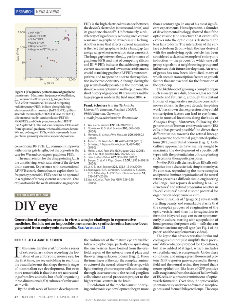
Figure 1 | Frequency performance of graphene
transistors. Maximum frequency of oscillation,
fmax, versus cut-off frequency, fT, for graphene
field-effect transistors (FETs) and competing
radiofrequency FETs: indium phosphide high
electron mobility transistor (InP HEMT), gallium
arsenide metamorphic HEMT (GaAs mHEMT),
silicon metal-oxide-semiconductor FET (Si
MOSFET), and GaAs pseudomorphic HEMT
(GaAs pHEMT). The red stars designate FETs made
from ‘epitaxial’ graphene, whereas blue stars denote
Wu and colleagues’1 FETs, which were made from
graphene grown by chemical vapour deposition.
conventional RF FETs, fmax commonly improves
with shorter gate lengths, but the opposite is the
case for Wu and colleagues’ graphene FETs.
The main reason for the disappointing fmax is
the unsatisfying, weak saturation of the device’s
drain current. Experience with conventional
RF FETs clearly shows that, to exploit their full
frequency potential, FETs need to be operated
in a regime of strong current saturation. One
explanation for the weak saturation in graphene
FETs is the high electrical resistance between
the device’s electrodes (source and drain) and
its graphene channel12. Unfortunately, a reliable way of significantly reducing such contact
resistance in graphene devices is still lacking.
Another issue that affects current saturation
is the fact that graphene lacks a bandgap (an
energy range where no electron states can exist).
The huge gap between the fmax performance of
graphene FETs and that of competing silicon
and III-V FETs indicates that achieving strong
current saturation and low contact resistance is
crucial to making graphene RF FETs more competitive, and to open the door to their application in electronic circuitry. Although closing the
gap seems hardly possible at the moment, we
should remain optimistic and keep in mind the
short history of graphene RF transistors and the
huge progress made in the field since 2008. ■
Frank Schwierz is at the Technische
Universität Ilmenau, Postfach 100565,
98684 Ilmenau, Germany.
e-mail: frank.schwierz@tu-ilmenau.de
1. Wu, Y. et al. Nature 472, 74–78 (2011).
2. Novoselov, K. S. et al. Science 306, 666–669
(2004).
3. Morozov, S. V. et al. Phys. Rev. Lett. 100, 016602
(2008).
4. Avouris, Ph. Nano Lett. 10, 4285–4294 (2010).
5. Schwierz, F. Nature Nanotechnol. 5, 487–496
(2010).
6. Meric, I. et al. Tech. Dig. IEDM, paper 21.2 (2008).
7. Lin, Y.-M. et al. Science 327, 662 (2010).
8. Liao, L. et al. Nature 467, 305–308 (2010).
9. Berger, C. et al. J. Phys. Chem. B 108, 19912–19916
(2004).
10.Li, X. S. et al. Science 324, 1312–1314 (2009).
11.Ekanayake, S. R., Lehmann, T., Dzurak, A. S., Clark,
R. G. & Brawley, A. IEEE Trans. Electron Devices 57,
539–547 (2010).
12.Wu, Y. Q. et al. Tech. Dig. IEDM 226–228
(2010).
REG E NERATIVE MED ICINE
DIY eye
Generation of complex organs in vitro is a major challenge in regenerative
medicine. But it is not an impossible one: an entire synthetic retina has now been
generated from embryonic stem cells. See Article p.51
ROBIN R. ALI & JANE C. SOWDEN
I
n this issue, Eiraku et al.1 provide a series
of extraordinary videos recording the formation of an embryonic mouse eye: for
the first time, we see unfolding in real time
the beautiful events that shape the early stages
of mammalian eye development. But even
more remarkable is that these are not recordings from live animals, but of self-organizing
three-dimensional (3D) cultures of embryonic
stem cells.
By the sixth week of human development,
4 2 | NAT U R E | VO L 4 7 2 | 7 A P R I L 2 0 1 1
the rudiments of the mature eye are visible:
bilayered optic cups, partially encapsulating
the lens vesicles, have formed from the eyefield region of the anterior neural plate and
the overlying surface ectoderm (Fig. 1). From
the inner layer of the cup, the complex laminar
structure of the neural retina will develop, with
light-sensing photoreceptor cells connecting
through interneurons to the retinal ganglion
cells whose axonal processes project to the
higher visual centres in the brain.
Elucidation of the mechanisms underlying embryonic eye development began more
© 2011 Macmillan Publishers Limited. All rights reserved
than a century ago. In one of his most significant experiments, Hans Spemann, a founder
of developmental biology, showed that if the
optic vesicle (the structure that eventually
evolves into the optic cup) is destroyed, the
lens fails to form. The interaction of the surface ectoderm (from which the lens derives)
with the underlying optic vesicle has been
considered a classical example of embryonic
induction — the process by which one cell
group signals to a neighbouring group and
influences their future development. An array
of genes has now been identified, many of
which encode transcription factors or growth
factors that are essential for the formation of
the optic cup.
The likelihood of growing a complex organ
such as an eye in a dish, however, has seemed
remote and futuristic, although this distant
frontier of regenerative medicine constantly
moves closer. In the past decade, inspiring
work2 has shown that expression of eye-field
transcription factors can lead to eye formation in unusual locations along the body of
Xenopus frogs. Moreover, following the
generation of human embryonic stem (ES)
cells, it has proved possible3,4 to direct their
differentiation towards the retinal lineage
and generate both retinal pigmented epithelium (RPE) and retinal neurons (Fig. 1). Cellculture approaches have mainly sought to
maximize the development of specific cell
types with the potential aim of transplanting
such cells for therapeutic purposes.
In vitro, RPE cells derived from ES cells selforganize into a characteristic simple monolayer.
By contrast, reproducing the more complex
and precise laminar organization of the neural
retina presents a difficult tissue-engineering
challenge. But reports describing lens-like
structures5 and retinal progenitor rosettes in
ES-cell cultures6 hinted at some potential for
organization of eye tissue in vitro.
Now, Eiraku et al.1 (page 51) reveal with
startling beauty and remarkable clarity that
the complex process of evagination of the
optic vesicle, and then its invagination to
form the bilayered cup, can occur spontaneously in culture, starting with a population of
homogeneous pluripotent cells — cells that can
differentiate into any cell type (see Fig. 1 of the
paper1 and the supplementary videos).
The key to this advance was that Eiraku and
colleagues did not just simplify their previous7 differentiation protocol for ES cultures,
but also added Matrigel, which includes
extracellular-matrix components. Under these
conditions, and using a green fluorescent protein (GFP) reporter gene expressed in the eye
field and the neural retina, they found that a
neuro-epithelium-like layer of GFP-positive
cells evaginated from the sides of hollow balls
of ES cells, in a process reminiscent of opticvesicle formation. Over time, the optic vesicles
spontaneously underwent dynamic morphogenesis and formed bilayered cups. The cups
NEWS & VIEWS RESEARCH
a
b
Surface ectoderm
Mesenchyme
Neuroepithelium
c
Lens
vesicle
Lens
Neural
retina RPE
Optic vesicle
Lens
placode
Bilayered
optic cup
Photoreceptor cells
Interneurons
Ganglion cells
Figure 1 | Eye development. a, At early stages of eye development, the surface ectoderm thickens and
invaginates together with the underlying neuroepithelium of the optic vesicle. b, The inner layer of
the bilayered optic cup gives rise to neural retina and the outer layer gives rise to the retinal pigmented
epithelium (RPE) (c). The mature neural retina (c) comprises three cellular layers: photoreceptors,
interneurons (horizontal, amacrine and bipolar cells), and retinal ganglion cells. Eiraku et al.1 ­generated
optical cups in vitro from embryonic stem cells.
appropriately expressed the distinctive molecular markers of both the neural retina and the
RPE, confirming their identity; another indicator was visible as RPE pigmentation.
An even more striking proof that these are
genuine retinas is that, in culture, the synthetic optic cups undergo cell differentiation.
Indeed, retinal progenitor cells — the multipotential cells of the neural retina — divided
and differentiated into all the main retinal
neuronal cell types, including photoreceptors.
These events seem to follow the normal temporal sequence of retinal tissue formation, and
the resulting cells were correctly organized in
the appropriate cellular layer.
But even though optic cups can now be
grown in culture from ES cells, we still don’t
fully understand the principles underlying
their development. For instance, it is surprising that optic cups can form independently
of any interaction of the neuroepithelial cells
with surface ectoderm or mesenchymal tissue that would normally surround them in a
developing embryo (Fig. 1). Eiraku et al.1 propose that the ES-cell-derived retinal cells have
a latent intrinsic order, and that collections of
cells can self-pattern and undergo dynamic
morphogenesis by obeying a sequential combination of local rules and internal forces
within the epithelium.
However, Eiraku and colleagues’ powerful in vitro system has great potential as it
can be manipulated to define the molecular
interactions that are essential for eye development. Moreover, if functional outer rod
segments — where the protein complexes
responsible for phototransduction are located
— can be produced in longer-term cultures,
this 3D system will be invaluable for functional studies examining the response of the
retina to light.
What’s more, development of an equivalent human 3D system could offer the
prospect of disease modelling and drug
testing using induced pluripotent stem cells
generated from patients’ tissues. Most forms
of untreatable blindness result from the loss
of photoreceptor cells, leaving other retinal
neurons intact. In mice, transplantation of
photoreceptor precursor cells isolated from
the developing mouse retina can repair adult
retinas8. A major challenge is to obtain sufficient numbers of photoreceptor precursors
at the appropriate stage of development from
a renewable cell source. This 3D system for
culturing ES cells1 may solve that problem
by providing synthetic retinas at defined
stages of development from which precursors can be isolated more readily for use in
transplantation. ■
Robin R. Ali is in the Department of Genetics,
Institute of Ophthalmology, University College
London, London EC1V 9EL, UK.
Jane C. Sowden is in the Developmental
Biology Unit, Institute of Child Health,
University College London, London WC1N
1EH, UK.
e-mails: r.ali@ucl.ac.uk;
j.sowden@ich.ucl.ac.uk
1. Eiraku, M. et al. Nature 472, 51–56 (2011).
2. Zuber, M. E., Gestri, G., Viczian, A. S., Barsacchi, G. &
Harris, W. A. Development 130, 5155–5167
(2003).
3. Lamba, D. A., Karl, M. O., Ware, C. B. & Reh, T. A.
Proc. Natl Acad. Sci. USA 103, 12769–12774
(2006).
4. Osakada, F. et al. Nature Biotechnol. 26, 215–224
(2008).
5. Yang, C. et al. FASEB J. 24, 3274-3283 (2010).
6. Meyer, J. S. et al. Proc. Natl Acad. Sci. USA 106,
16698–16703 (2009).
7. Ikeda, H. et al. Proc. Natl Acad. Sci. USA 102,
11331–11336 (2005).
8. MacLaren, R. E. et al. Nature 444, 203–207
(2006).
O C EA N O GR AP HY
When glacial giants
roll over
The energy released by capsizing icebergs can be equal to that of small
earthquakes — enough to create ocean waves of considerable magnitude. Should
such ‘glacial tsunamis’ be added to the list of future global-warming hazards?
ANDERS LEVERMANN
A
bout half of Greenland’s annual ice loss
occurs through solid-ice discharge;
in Antarctica such calving processes
account for almost all ice loss. The resulting
icebergs come in various sizes and shapes,
some several hundred metres high. Immediately after they break off, when their height
exceeds their horizontal extent, these floating giants can be unstable and capsize. In a
paper in the Annals of Glaciology, Mac­Ayeal
and colleagues1 have estimated the energy that
is released when icebergs roll over. They find
that this can be as large as that of an earthquake of magnitude 5–6 on the Gutenberg–
Richter scale, depending on the iceberg’s
dimensions.
As one of several possibilities, a proportion
© 2011 Macmillan Publishers Limited. All rights reserved
of this energy can generate a surface gravity
wave — a tsunami. MacAyeal et al. provide a
theoretical analysis of the potential of iceberg
capsize to generate tsunamis. Assuming a
simplified, but not completely unrealistic, rectangular geometry, they calculate the potential
energy before and after capsizing. The difference is the energy released from the roll-over.
Thin icebergs do not carry a lot of potential
energy, whereas ice-cube-shaped icebergs,
which are as thick as they are high, have the
same potential energy before and after turning over. Thus the most energy is released by
icebergs that are half as thick as they are high
— and can be equivalent to the explosion of
several thousand tonnes of TNT.
According to MacAyeal and colleagues1,
energy release increases with the fourth power
of an iceberg’s height (Box 1). But not all of
7 A P R I L 2 0 1 1 | VO L 4 7 2 | NAT U R E | 4 3


