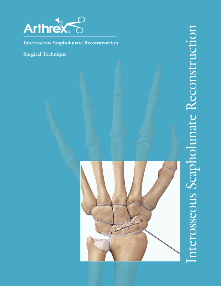
Surgical Technique
Interosseous Scapholunate Reconstruction
Interosseous Scapholunate Reconstruction
Interosseous Scapholunate Reconstruction
The treatment of scapholunate ligament tears remains
difficult and controversial. The pathology represents a
spectrum of injury that ranges from a sprain of the ligament
to a partial tear to the volar, central, or more commonly
dorsal component of the S-L ligament. Progression leads
to complete tears of all three components, disruption
of secondary stabilizers and subsequent DISI deformity.
Ultimately, arthritic changes ensue. Direct repair of
all three components of the S-L ligament cannot be
performed reliably.
The 3.5 mm SwiveLock SL reconstruction is achieved
by utilizing a strong anchor construct that incorporates
a combination of a biologic tendon graft reconstruction
into bony tunnels with static SutureTape™ InternalBrace™
reinforcement. Together, this construct supports the
connection between the bones so that graft incorporation
can take place. This reconstruction is best suited for tears
of all three components of the scapholunate interosseous
ligament. Partial tears of the dorsal and central portions
of the S-L ligament can be reconstructed using the dorsal
reconstruction technique.
1
Make a 4-inch longitudinal incision starting between the bases
of the 2nd and 3rd metacarpals and extending proximally
between the 3rd and 4th compartments. PIN and/or AIN
neurectomy is optional. Expose the scapholunate interval via
a dorsal approach with an inverted T-capsulotomy. The transverse
portion of the capsulotomy is taken down directly off the distal
radius. Incise enough capsule to adequately visualize the entire
dorsal surface of the lunate and scaphoid bones. Try to preserve
the lateral/dorsal blood supply to the scaphoid.
Interosseous reconstruction of the scapholunate ligament
with the 3.5 mm DX SwiveLock® SL is indicated for
complete tears of the scapholunate ligament where there
is inadequate ligament to repair, the carpal cartilage is
preserved (no arthrosis) and the carpus is reducible. The
goal of the reconstruction is to address the scapholunate
diastasis as well as the scaphoid flexion and lunate extension.
Contraindications for SwiveLock SL technique include
inflammatory arthritis, previous and/or current infection,
carpal arthrosis (SLAC wrist), irreducible carpus, pediatric
patients, preexisting hardware in the carpal bones, large
cystic changes in the carpal bones and patients with
unusually small anatomy.
2
Harvest a 2 mm wide slip of the extensor carpi radialis
brevis (ECRB) tendon at its insertion on the base of the
3rd metacarpal. Alternatively, a slip of ECRL can be used.
It is crucial to keep the graft width at 2 mm. Using a larger
graft will complicate insertion into both the eyelet of the
anchor and the drill hole. A tendon stripper is often useful in
retrieving the required 10 cm or longer graft, and can be used
in an intratendinous fashion.
3
Whipstitch both ends of the graft with 2-0 FiberLoop®. It is
important to stretch out the tendon to avoid tendon creep
during the healing process.
5
Using the joysticks, the scaphoid and lunate are “open-booked”
to access the radial side of the scaphoid and ulnar aspect of the
lunate. The central point of the scaphoid is determined and
a 0.054” Guidewire is placed into this central axis. Check the
position on fluoroscopy to confirm that the drill will be within
the confines of the bone.
4
Place one K-wire into the waist of the scaphoid, and one into
the proximal/ulnar side of the lunate to act as joysticks. Place
the joysticks in a way that avoids future bone tunnels, and
when clamped together helps to reduce the diastasis and
rotational deformity. Avoid over-reduction of the scaphoid
flexion/lunate extension. If there are any remaining soft
tissues joining the scaphoid to the lunate, they may be incised
in order to allow complete diastasis between the bones.
6
Drill using the 3.5 mm Cannulated Drill and drill guide to
1 cm depth stop.
Surgical Technique
7
The central point of the lunate matching the central point
of the scaphoid bone is located and a 0.054” Guidewire is
placed into the central axis of the lunate. Drill using the
3.2 mm Cannulated Drill to 1 cm depth stop, creating an
inside-out tunnel in the lunate. The 1 cm tunnel is within the
confines of the lunate and does not exit out of the far cortex.
9
Place the final 0.054” K-wire into the distal pole of the
scaphoid and confirm via fluoroscopy prior to overdrilling.
Use the 3.5 mm Cannulated Drill to the 1 cm depth stop.
Clear any soft tissue and remnants of bone surrounding, and
in the drill holes, to facilitate insertion of the graft.
8
A separate outside-in tunnel in the lunate is made starting
from the dorsal-ulnar corner of the lunate and connecting
to the central, inside-out tunnel. The Guidewire is aimed from
the dorsal-ulnar corner of the lunate near the lunotriquetral
ligament attachment and aimed at a 45° angle toward the
central tunnel. Overdrill using the 3.2 mm Cannulated Drill to
create a connected tunnel through the lunate.
10
Place the forked eyelet of the SwiveLock® SL onto the tendon
graft about 3 mm from the end of the graft and secure both
limbs of the FiberLoop® into the notch on the SwiveLock
tab. Additionally, place a 1.3 mm SutureTape™ over the graft
and forked eyelet and secure onto the SwiveLock tab. The
SutureTape acts as the InternalBrace™ reinforcement.
Interosseous Scapholunate Reconstruction
11
Insert the SwiveLock® SL into the scaphoid until the leading
end of the threaded anchor body is flush to the bone. Hold
the square tab steady while turning the knob clockwise with
forward pressure until the laser line is just below the level
of the bone. Do not countersink the anchor. Remove the
handle. If it does not disengage easily, turn the square tab
counterclockwise to disengage the handle from the anchor.
12
Shuttle the tendon graft and one limb of the 1.3 mm SutureTape™
through the lunate tunnel. (The extra limb of SutureTape can be
cut flush with the anchor body). Reduce the scaphoid and lunate
and clamp the joystick K-wires together.
A 3 mm x 8 mm Tenodesis Screw™ is used to secure the tendon
graft and SutureTape in the lunate hole.
Optional: If the length of the tunnel is less than 8 mm, the anchor body
from a 3.5 mm DX SwiveLock SL can be used. The tunnel should be
overdrilled with the 3.5 mm drill if this fixation method is preferred.
13
An additional 3.5 DEX SwiveLock SL is used to capture the
tendon graft and SutureTape near the distal pole of the scaphoid
drill hole. Twisting the graft and SutureTape together can
facilitate placement into the forked eyelet. Insert into the
scaphoid. Apply counter pressure to the volar scaphoid tubercle
while advancing the anchor. If resistance is met, slow and
constant pressure downward will allow the SwiveLock SL
to insert and auto-tension the construct.
14
Cut off any excess tendon graft and FiberLoop®. If you haven’t
previously placed a scapho-capitate K-wire, do so now prior
to removing the joystick K-wires. Close the capsule and dorsal
incision.
Post-Op: A forearm-based thumb spica splint or cast is worn
for 6-8 weeks. Any supplemental K-wires can be removed and
hand therapy is started at this time. The splint is worn for an
additional six weeks.
DX SwiveLock SL
AR-8978P
Ordering Information
DX SwiveLock SL, 3.5 mm x 8.5 mm, w/forked eyelet
AR-8978P
DX SwiveLock® SL Disposable Kit (AR-8978DS) includes:
Drill Guide
Tap (for use with 3.2 mm Cannulated Drill Bit)
3.2 mm Cannulated Drill Bit (for all suture constructs)
3.5 mm Cannulated Drill Bit (for constructs with graft incorporation)
Guidewires, 1.35 mm
2-0 FiberLoop®
SutureTape™ Options:
1.5 mm LabralTape™ 1.3 mm SutureTape w/needles
AR-7276 (white)
AR-7276T (white/black)
AR-7500
This description of technique is provided as an educational tool and clinical aid to assist properly licensed medical professionals
in the usage of specific Arthrex products. As part of this professional usage, the medical professional must use
their professional judgment in making any final determinations in product usage and technique.
In doing so, the medical professional should rely on their own training and experience and should conduct
a thorough review of pertinent medical literature and the product’s Directions For Use.
2016, Arthrex Inc. All rights reserved. LT1-00047-EN_B
©


