Adrenal Glands
advertisement
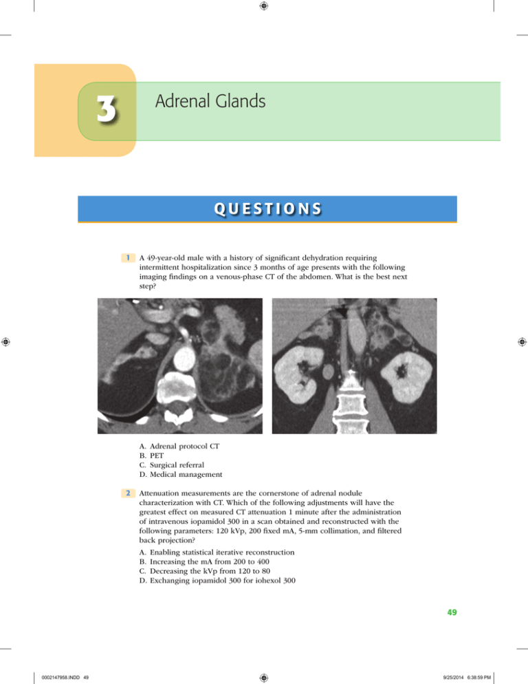
3 Adrenal Glands QUESTIONS 1 A 49-year-old male with a history of significant dehydration requiring intermittent hospitalization since 3 months of age presents with the following imaging findings on a venous-phase CT of the abdomen. What is the best next step? A. Adrenal protocol CT B. PET C. Surgical referral D.Medical management 2 Attenuation measurements are the cornerstone of adrenal nodule characterization with CT. Which of the following adjustments will have the greatest effect on measured CT attenuation 1 minute after the administration of intravenous iopamidol 300 in a scan obtained and reconstructed with the following parameters: 120 kVp, 200 fixed mA, 5-mm collimation, and filtered back projection? A. Enabling statistical iterative reconstruction B. Increasing the mA from 200 to 400 C. Decreasing the kVp from 120 to 80 D.Exchanging iopamidol 300 for iohexol 300 49 0002147958.INDD 49 9/25/2014 6:38:59 PM 50 Genitourinary Imaging 3 A 50-year-old male with history of clear cell renal cell carcinoma status ­ post–left nephrectomy presents for characterization of a right adrenal nodule detected on follow-up imaging. Images represent dual-echo gradient-recalled echo T1-weighted imaging with the following echo times at 1.5 Tesla: 2.2 msec (image on LEFT) and 4.4 msec (image on RIGHT). Signal intensity measurements within the adrenal mass are as follows: 144 (image on LEFT) and 175 (image on RIGHT) (18% signal loss). If there are no comparison studies, what is the best next step? A. Tissue sampling B. Follow-up in 12 months C. Ignore (benign finding) D.Adrenal protocol CT 4 A 63-year-old female with a history of gastric cancer presents for characterization of a 3.3-cm adrenal mass. There are no comparison images. Regions of interest drawn in the center of the mass on triphasic adrenal protocol CT are as follows: (a) unenhanced: 16 Hounsfield units; (b) 1-minute delay: 55 Hounsfield units; and (c) 15-minute delay: 28 Hounsfield units. What is the calculated absolute percent washout? A. 29% B. 49% C. 69% D.89% 0002147958.INDD 50 9/25/2014 6:38:59 PM Adrenal Glands51 5 A 54-year-old female with node-positive invasive lobular breast cancer has a 2.1-cm homogeneous right adrenal mass. Adrenal protocol CT is performed utilizing 1-minute and 15-minute delays. Relative washout is calculated to be 50%. Which of the following is the most likely diagnosis? A. Pheochromocytoma B. Metastasis C. Adenoma D.Adrenocortical carcinoma 6 A 60-year-old male with hypertension and right lower quadrant pain undergoes a CT of the abdomen and pelvis to rule out appendicitis. An incidental 1.5-cm homogeneous right adrenal mass is identified. He has no other relevant medical history. What is the estimated risk that this adrenal nodule is malignant? A. 1 in 5 B. 1 in 50 C. 1 in 500 D.<1 in 1,000 7 A 65-year-old male ICU patient with a complicated medical history presents with new findings in the adrenal glands. The unenhanced CT image on the left was obtained 2 weeks after the unenhanced CT image on the right. What is the best next step? A. Adrenal protocol CT B. Percutaneous biopsy C. Medical management D.Follow-up in 6 to 12 months 8 A 40-year-old female with postprandial pain and no other past medical history undergoes a right upper quadrant ultrasound. The gallbladder and biliary tree are normal, but an incidental 2.0-cm mass is identified in the right suprarenal fossa. An abdominal MRI follows confirming that the mass arises from the right adrenal gland. India ink artifact is observed within the mass encircling a 1.2-cm internal nodule. What is the most likely diagnosis? A. Pheochromocytoma B. Myelolipoma C. Collision tumor D.Adrenocortical carcinoma 0002147958.INDD 51 9/25/2014 6:39:00 PM 52 Genitourinary Imaging 9 A 23-year-old female with MEN-IIb syndrome and hypertension presents for an I123-MIBG scan. In which of the following organs is uptake on an I123-MIBG scan always abnormal? A. Myocardium B. Salivary glands C. Adrenal glands D.Bones E. Colon 10 A 25-year-old male with a hereditary paraganglioma–pheochromocytoma syndrome undergoes an I123-MIBG study. What is the best diagnosis? A. Myocarditis B. Hepatic metastatic disease C. Normal study D.Nodal metastatic disease (neck) 11 T1-weighted dual-echo gradient-recalled echo imaging is used for the detection of intracellular lipid and macroscopic fat. With this sequence, when fat and water protons are out of phase, fat protons are shifted a certain distance along the frequency-encoding axis depending on the receiver bandwidth and field strength. What is the approximate calculated fat/water chemical shift at 1.5 Tesla? Use the following formula: Shift = (fat / water resonant frequency separation [in ppm]) × (field strength[T]) × 42 MHz / T A. 85 Hz B. 220 Hz C. 440 Hz D.550 Hz 0002147958.INDD 52 9/25/2014 6:39:00 PM Adrenal Glands53 12 A 56-year-old male with no history of malignancy and suspected primary hyperaldosteronism undergoes an adrenal protocol CT demonstrating a 1.5cm left adrenal nodule with 70% absolute washout. The right adrenal gland is normal. What is the best next step? A. Adrenal protocol MRI B. Left adrenalectomy C. Percutaneous biopsy D.Adrenal vein sampling 13 A 59-year-old female with suspected primary hyperaldosteronism and a 1.3cm unilateral left adrenal nodule undergoes adrenal vein sampling. Which description best characterizes the adrenal venous anatomy? A. Right: A single vein drains into the IVC; Left: A single vein drains into the left renal vein. B. Right: Three veins drain into the IVC; Left: Three veins drain into the left renal vein. C. Right: A single vein drains into the right renal vein; Left: A single vein drains into the IVC. D.Right: Three veins drain into the right renal vein; Left: Three veins drain into the IVC. 0002147958.INDD 53 9/25/2014 6:39:01 PM 54 Genitourinary Imaging 14 A 55-year-old male with non–small cell lung cancer and a 1.5-cm homogeneous right adrenal nodule presents for triphasic adrenal protocol CT. Which of the following image–time combinations has been shown to be most effective for adrenal nodule characterization? A. Unenhanced, 1 minute postcontrast, 20 minutes postcontrast B. Unenhanced, 1 minute postcontrast, 15 minutes postcontrast C. Unenhanced, 1 minute postcontrast, 10 minutes postcontrast D.Unenhanced, 1 minute postcontrast, 5 minutes postcontrast 15 A 62-year-old female with invasive ductal carcinoma of the breast and an indeterminate 1.5-cm left adrenal nodule undergoes a staging PET study. Which of the following adrenal nodule characteristics has the highest negative predictive value for metastasis? A. Standardized uptake value (SUV) maximum >5 B. Standardized uptake value (SUV) maximum <5 C. Qualitative uptake greater than background liver D.Qualitative uptake less than background liver 16 A 38-year-old female with early-onset diabetes mellitus presents with a dark rash covering her neck and armpits. CT imaging is performed and demonstrates a 4.9-cm left adrenal mass. An image representative of the entire mass is shown below. The patient has no history of malignancy. What is the best next step? A. Adrenal protocol CT B. Adrenal protocol MRI C. Percutaneous biopsy D.Open surgical resection 17 One of the fundamental sequences of adrenal protocol MR is T1-weighted dual-echo gradient-recalled echo imaging for the detection of intracellular lipid and macroscopic fat. Which of the following correctly describes a difference between gradient echo imaging and spin echo imaging? A. Gradient echo imaging usually has a longer TR. B. Gradient echo imaging usually has a larger flip angle. C. Gradient echo imaging has a refocusing RF pulse, and spin echo imaging does not. D.Gradient echo imaging is more affected by residual transverse magnetization. 0002147958.INDD 54 9/25/2014 6:39:01 PM Adrenal Glands55 18 A 70-year-old female with lung cancer and an indeterminate 2.2-cm left adrenal mass presents for percutaneous biopsy. Which of the following positional maneuvers will most effectively reduce the risk of pneumothorax during the biopsy while minimizing risk to other structures? A. Decubitus, ipsilateral side down B. Decubitus, contralateral side down C. Prone D.Supine 19 A 32-year-old male (body mass index: 22 kg/m2) with intermittent diaphoresis, palpitations, and early-onset hypertension (190/110) presents for adrenal protocol CT. What is the serious adverse event risk of administering intravenous low-osmolality iodinated contrast material to a patient with pheochromocytoma who is not receiving alpha- and beta-blockade? A. 80% chance of a serious adverse event B. 30% chance of a serious adverse event C. 5% chance of a serious adverse event D.<1% chance of a serious adverse event 20 A 62-year-old male with clinical-stage T1cN0Mx Gleason 5 + 4 = 9 prostate cancer and no signs of adrenal hyperactivity presents for MR characterization of a 2.4-cm right adrenal nodule. Images represent dual-echo gradient-recalled echo T1-weighted imaging with the following echo times at 1.5 Tesla: 2.2 msec (image on left) and 4.4 msec (image on right). What is the best next step? A. Ignore (benign finding) B. Adrenal protocol CT C. Percutaneous biopsy D.Right adrenalectomy 21 Homogeneous loss of signal intensity within an adrenal nodule <4 cm on opposed-phase T1-weighted dual-echo gradient-recalled echo imaging is compatible with an adrenal adenoma in most cases. What organ is best used as an internal reference standard to determine the degree of signal loss within an adrenal nodule? A. Liver B. Pancreas C. Kidney D.Spleen 0002147958.INDD 55 9/25/2014 6:39:01 PM 56 Genitourinary Imaging 22 A 60-year-old female with right lower quadrant abdominal pain undergoes a CT of the abdomen and pelvis demonstrating perforated appendicitis and an incidental finding in the left adrenal gland. The following image is representative of the entire gland. Which of the following is most strongly associated with this abnormality? A. Hemangioblastoma B. Renal agenesis C. Pheochromocytoma D.Polysplenia 23 A 58-year-old male with mild hypertension (140/90) managed with one medication and no history of malignancy undergoes an abdominal MRI that shows a 3.2-cm enhancing mass in the right adrenal gland. No intracellular lipid or macroscopic fat is identified. The left adrenal gland is normal, and the patient is asymptomatic. What is the best next step? A. Right adrenalectomy B. Alpha- and beta-adrenergic blockade C. Percutaneous biopsy D.Laboratory evaluation 0002147958.INDD 56 9/25/2014 6:39:02 PM Adrenal Glands57 24 A 45-year-old male with a right adrenal myelolipoma undergoes MR imaging. The following T2-weighted images were acquired without (left) and with (right) a conventional inversion recovery technique. Along what vector do the protons align in conventional inversion recovery imaging immediately following the preparation radiofrequency pulse (applied before the excitation pulse)? A. 0 degrees (with main magnetic field) B. 90 degrees (perpendicular to main magnetic field) C. 180 degrees (opposed to main magnetic field) D.270 degrees (perpendicular to main magnetic field) 25 A 51-year-old male with no comparison imaging and a recently diagnosed obstructing sigmoid colon cancer presents for staging CT of the chest, abdomen, and pelvis. Imaging demonstrates the known colon mass, enlarged regional lymph nodes, and a 2.2-cm left suprarenal abnormality. What is the best next step? A. Adrenal protocol CT B. Biopsy C. Aspiration D.Ignore (benign finding) 0002147958.INDD 57 9/25/2014 6:39:03 PM 58 Genitourinary Imaging 26 A 28-year-old male with suspected pheochromocytoma is scheduled to undergo an I131-MIBG scan to confirm the diagnosis and evaluate for distant disease. Which organ is at greatest risk of radiation-induced carcinogenesis if no precautionary measures are taken? A. Adrenal glands B. Spleen C. Myocardium D.Thyroid 27 A 4-year-old girl with recurrent abdominal pain and unexplained fevers presents with a right suprarenal mass. What is the most likely diagnosis? A. Sarcoma B. Neuroblastoma C. Adrenocortical carcinoma D.Pheochromocytoma 0002147958.INDD 58 9/25/2014 6:39:03 PM Adrenal Glands59 28 Where is the organ of Zuckerkandl? A. Near the carotid bifurcation B. Near the aortic bifurcation C. Within the middle ear D.Along the urinary bladder wall 29 Pheochromocytoma is associated with several tumor-forming genetic syndromes. Which of the following is an example of that? A. Multiple endocrine neoplasia (MEN) type I B. Beckwith-Wiedemann syndrome C. Neurofibromatosis type I D.Hereditary leiomyomatosis renal cell cancer (HLRCC) syndrome 30 A 75-year-old male with altered mental status undergoes an abdominopelvic CT examination that demonstrates an abnormal left adrenal gland. The right adrenal gland is normal. Which of the following best explains this imaging finding? A. Pheochromocytoma B. Metastasis C. Remote hemorrhage D.Adrenocortical carcinoma 0002147958.INDD 59 9/25/2014 6:39:04 PM 60 Genitourinary Imaging A N S W E R S A N D E X P L A N AT I O N S 1 Answer D. Large, bulky, bilateral macroscopic fat-containing adrenal masses is indicative of adrenal myelolipomas in the setting of congenital adrenal hyperplasia (CAH). These patients require medical management for their endocrine abnormalities. The most common cause of CAH is 21-hydroxylase deficiency, which results in salt wasting, female virilization, and hyperandrogenism. Most patients with 21-hydroxylase deficiency are detected during newborn screening. Adrenal protocol CT is not indicated for adrenal masses that contain macroscopic fat because it will add no additional information. PET and/or operative referral are not indicated because the masses are not malignant. References: German-Mena E, Zibari GB, Levine SN. Adrenal myelolipomas in patients with congenital adrenal hyperplasia: review of the literature and a case report. Endocr Pract 2011;17:441–447. Nermoen I, Rorvik J, Holmedal SH, et al. High frequency of adrenal myelolipomas and testicular adrenal rest tumours in adult Norwegian patients with classical congenital adrenal hyperplasia because of 21-hyroxylase deficiency. Clin Endocrinol 2011;75:753–759. 2 Answer C. Changing kVp has a direct effect on CT number measurements. Higher kVp imaging, particularly with multidetector CT, is susceptible to artificial increases in measured attenuation due to “pseudoenhancement” effects. Lower kVp imaging is closer to the k-edge of iodine, resulting in increased attenuation at lower kVp within iodine-containing (i.e., enhanced) structures. This fact is often exploited for high-contrast examinations like CT angiography. Lower kVp imaging is associated with greater image noise, less radiation dose, and greater measured iodine attenuation than higher kVp imaging. Changing mA, enabling statistical iterative reconstruction, and/or switching between contrast agents with similar iodine concentrations will have a negligible effect on CT number measurements. References: Kaza RK, Platt JF, Goodsitt MM, et al. Emerging techniques for dose optimization in abdominal CT. Radiographics 2014;34:4–17. Wang ZJ, Coakley FV, Fu Y, et al. Renal cyst pseudoenhancement at multidetector CT: what are the effects of number of detectors and peak tube voltage? Radiology 2008;248:910–916. 3 Answer A. The calculated signal intensity index ([IP − OP]/IP) of the right adrenal mass is 18% ([175 − 144]/175). Although the signal intensity index threshold varies with field strength (1.5 vs. 3.0 Tesla), sequence type (2D vs. 3D), and other parameters, homogeneous masses with a signal intensity index ≥16.5% are often thought to meet criteria for a lipid-rich adenoma. However, there are some exceptions (as in this case). Metastases known to contain “intracellular lipid” such as clear cell renal cell carcinoma and hepatocellular carcinoma can mimic adenomas on chemical shift imaging. Therefore, tissue sampling is the next best step (Answer A). This mass, though homogeneous and meeting the signal intensity index threshold, was proven to be a clear cell renal cell carcinoma metastasis. Another caveat to the use of the signal intensity index is for large masses (e.g., ≥4 cm) because adrenocortical carcinoma can also demonstrate signal loss on opposed-phase imaging. Chemical shift imaging is most helpful when the primary tumor is known not to contain intracellular lipid and the mass is <4 cm in size. References: Blake MA, Cronin CG, Boland GW. Adrenal imaging. AJR Am J Roentgenol 2010;194:1450–1460. Marin D, Dale BM, Bashir MR, et al. Effectiveness of a three-dimensional dual gradient echo two-point Dixon technique for the characterization of adrenal lesions at 3 Tesla. Eur Radiol 2012;22:259–268. 0002147958.INDD 60 9/25/2014 6:39:04 PM Adrenal Glands61 Shinozaki K, Yoshimitsu K, Honda H, et al. Metastatic adrenal tumor from clear-cell renal cell carcinoma: a pitfall of chemical shift MR imaging. Abdom Imaging 2001;26:439–442. Sydow BD, Rosen MA, Siegelman ES. Intracellular lipid within metastatic hepatocellular carcinoma of the adrenal gland: a potential diagnostic pitfall of chemical shift imaging of the adrenal gland. AJR Am J Roentgenol 2006;187:W550–W551. 4 Answer C. The calculated absolute percent washout is 69%, and the calculated relative percent washout is 49%. Both are compatible with a lipid-poor adrenal adenoma, and both argue for a benign etiology (i.e., not a metastasis). The formula for absolute washout is 100 ´ (1 min HU - 15 min HU ) / (1 min HU - pre HU ) The formula for relative washout is 100 ´ (1 min HU - 15 min HU ) / (1 min HU ) Where “pre-HU” is the measured attenuation on unenhanced CT, “1-min HU” is the measured attenuation on contrast-enhanced CT performed 1 minute after contrast material administration, and “15-min HU” is the measured attenuation on contrast-enhanced CT performed 15 minutes after contrast material administration. Washout calculations are only applicable for homogeneous adrenal masses; they should not be used for heterogeneous, centrally necrotic, or calcified masses. It is important to recognize that adrenal washout calculations were derived from a select population of patients, namely, differentiating generic metastatic disease from adenomas. Washout calculations are less helpful in other settings, such as differentiating adenoma from primary adrenal neoplasms (e.g., pheochromocytoma) or differentiating adenoma from certain types of hypervascular metastases (e.g., hepatocellular carcinoma, renal cell carcinoma). References: Caoili EM, Korobkin M, Francis IR, et al. Adrenal masses: characterization with combined unenhanced and delayed enhanced CT. Radiology 2002;222:629–633. Choi YA, Kim CK, Park BK, et al. Evaluation of adrenal metastases from renal cell carcinoma and hepatocellular carcinoma: use of delayed contrast-enhanced CT. Radiology 2013;266:514–520. Korobkin M, Brodeur JF, Francis IR, et al. CT time-attenuation washout curves of adrenal adenomas and nonadenomas. AJR Am J Roentgenol 1998;170:747–752. Patel J, Davenport MS, Cohan RH, et al. Can established CT attenuation and washout criteria for adrenal adenoma accurately exclude pheochromocytoma? AJR Am J Roentgenol 2013;201:122–127. 5 Answer C. Relative washout calculations are designed for situations in which the unenhanced images were not acquired, but a patient has a homogeneous adrenal mass that needs to be characterized. Often, this occurs in cases where the adrenal mass is unsuspected. Relative washout calculations are based on attenuation measurements from the 1-minute and 15-minute delayed images, as follows: Relative washout : 100 ´ (1 min HU - 15 min HU ) / (1 min HU ) Obtaining a relative washout >40% in a homogeneous adrenal mass <4 cm in size indicates that adenoma is the most likely diagnosis. Although some masses can mimic this (e.g., pheochromocytoma, hepatocellular carcinoma metastasis, renal cell carcinoma metastasis), in a patient without risk factors for such masses, adenoma is the best choice and no further workup is generally required. References: Caoili EM, Korobkin M, Francis IR, et al. Adrenal masses: characterization with combined unenhanced and delayed enhanced CT. Radiology 2002;222:629–633. 0002147958.INDD 61 9/25/2014 6:39:04 PM 62 Genitourinary Imaging Choi YA, Kim CK, Park BK, et al. Evaluation of adrenal metastases from renal cell carcinoma and hepatocellular carcinoma: use of delayed contrast-enhanced CT. Radiology 2013;266:514–520. Korobkin M, Brodeur JF, Francis IR, et al. CT time-attenuation washout curves of adrenal adenomas and nonadenomas. AJR Am J Roentgenol 1998;170:747–752. Patel J, Davenport MS, Cohan RH, et al. Can established CT attenuation and washout criteria for adrenal adenoma accurately exclude pheochromocytoma? AJR Am J Roentgenol 2013;201:122–127. 6 Answer D. In a series by Song et al. (2008) of 1,049 adrenal masses in 973 consecutive patients without a history of malignancy or adrenal hyperfunction, the authors found zero malignant lesions. Incidental adrenal nodules <4 cm are incredibly unlikely to represent malignancy, and therefore adrenal protocol CT is not the best first step in the management of these patients. False positives are much more likely than true positives and would too often result in inappropriate management. Current guidelines state that patients with incidental adrenal nodule(s) should be referred instead for biochemical testing to determine whether the nodule(s) are hyperfunctioning. References: American Medical Association of Clinical Endocrinologists and American Association of Endocrine Surgeons: Medical Guidelines for the Management of Adrenal Incidentalomas. AACE/AAES Guidelines, 2009. Cawood TJ, Hunt PJ, O’Shea D, et al. Recommended evaluation of adrenal incidentalomas is costly, has high false-positive rates and confers a risk of fatal cancer that is similar to the risk of the adrenal lesion becoming malignant: time for a rethink? Eur J Endocrinol 2009;513–527. Song JH, Chaudhry FS, Mayo-Smith WW. The incidental adrenal mass on CT: prevalence of adrenal disease in 1,049 consecutive adrenal masses in patients with no known malignancy. AJR Am J Roentgenol 2008;190:1163–1168. 7 Answer C. The image on the left depicts acute-onset bilateral adrenal masses that are high attenuation on unenhanced CT consistent with bilateral adrenal hemorrhage. Adrenal hemorrhage can lead to acute adrenal insufficiency (e.g., hypotension, hypoglycemia) and typically occurs in one of the following settings: (1) trauma (e.g., iatrogenic or otherwise); (2) sepsis (e.g., WaterhouseFriderichsen syndrome); (3) hypercoagulable state (e.g., antiphospholipid antibody syndrome: due to thrombosis of the adrenal vein); (4) bleeding diathesis (e.g., heparin-induced thrombocytopenia); or (5) disseminated intravascular coagulation. Management of acute adrenal hemorrhage includes treatment of the underlying cause of hemorrhage and corticosteroid replacement. Failure to detect and address the hormone deficiencies resulting from adrenal insufficiency can lead to patient death. Adrenal hemorrhage should be suspected when acute-onset, high-attenuation adrenal masses (unilateral or bilateral) develop in the proper clinical setting (see above). Further imaging and/or biopsy are usually neither indicated nor helpful. References: Jordan E, Poder L, Courtier J, et al. Imaging of nontraumatic adrenal hemorrhage. AJR Am J Roentgenol 2012;199:W91–W98. Vella A, Nippoldt TB, Morris JC, III. Adrenal hemorrhage: a 25-year experience at the Mayo Clinic. Mayo Clin Proc 2001;76:161–168. 8 Answer B. The India ink artifact described in the question indicates that a large fraction of the mass is composed of macroscopic fat. Therefore, the most likely diagnosis is adrenal myelolipoma. Adrenal myelolipomas are benign, fat-containing masses that are generally hormonally inactive and require no further management. Rare adrenal masses that have been associated with macroscopic fat include (a) degenerated adenoma; (b) adrenal lipoma; (c) collision tumor within a myelolipoma (Answer C); (d) adrenocortical carcinoma (Answer D), when present the fat is usually <10% of the mass volume; and (e) pheochromocytoma (Answer A, a mimicker of many entities; presence of macroscopic fat would be very rare). 0002147958.INDD 62 9/25/2014 6:39:05 PM Adrenal Glands63 References: Blake MA, Kalra MK, Maher MM, et al. Pheochromocytoma: an imaging chameleon. Radiographics 2004;24:S87–S99. Johnson PT, Horton KM, Fishman EK. Adrenal mass imaging with multidetector CT: pathologic conditions, pearls, and pitfalls. Radiographics 2009;29:1333–1351. Musso S, Columbier D, Mazerolles C, et al. Imaging features of uncommon adrenal masses with histopathologic correlation. Radiographics 1999;19:569–581. 9 Answer D. I123-MIBG uptake in the bones is always abnormal; when present, this indicates metastases from an adrenergic and/or catecholamine-expressing neoplasm (e.g., pheochromocytoma). The normal biodistribution of I123-MIBG includes the liver, spleen, myocardium, salivary glands, and adrenal glands. Normal adrenal gland uptake should be mild and symmetric. Variable low-level uptake can be seen in the skeletal muscle, nasal mucosa, lungs, urinary tract, colon, gallbladder, and thyroid. Radioactive free iodine (i.e., iodine dissociated from the I123-MIBG tracer) can be taken up into the normal thyroid gland, exposing it to unnecessary and potentially dangerous radiation; this action should be blocked with inert iodine (potassium iodide) tablets or perchlorate administered prior to the study. References: Nakajo M, Shapiro B, Copp J, et al. The normal and abnormal distribution of the adrenomedullary imaging agent m-[I-131]iodobenzylguanidine (I-131 MIBG) in man: evaluation by scintigraphy. J Nucl Med 1983;24(8):672–682. Olivier P, Colarinha P, Fettich J, et al. Guidelines for radioiodinated MIBG scintigraphy in children. Paediatric Committee of the European Association of Nuclear Medicine. Eur J Nucl Med Mol Imaging 2003;30(5):B45–B50. 10 Answer C. The images demonstrate the normal biodistribution of I123-MIBG. It is important to know the normal distribution of nuclear medicine tracers so that abnormal uptake can be differentiated from normal uptake. The normal biodistribution of I123-MIBG includes the liver, spleen, myocardium, salivary glands, and adrenal glands. Normal adrenal gland uptake should be mild and symmetric. Variable low-level uptake can be seen in the skeletal muscle, nasal mucosa, lungs, urinary tract, colon, gallbladder, and thyroid. References: Olivier P, Colarinha P, Fettich J, et al. Guideline for radioiodinated MIBG scintigraphy in children. Paediatric committee of the European Association of Nuclear Medicine. Eur J Nucl Med Mol Imaging 2003;30(5):B45–B50. Nakajo M, Shapiro B, Copp J, et al. The normal and abnormal distribution of the adrenomedullary imaging agent m-[I-131]iodobenzylguanidine (I-131 MIBG) in man: evaluation by scintigraphy. J Nucl Med 1983;672–682. 11 Answer B. Fat and water protons precess at different frequencies, and this principle is the foundation for chemical shift imaging. If the field strength is known, the fat/water chemical shift can be calculated by inserting the fat/water resonant frequency separation (3.5 ppm) and field strength into the following formula: Shift = ( Fat / water resonant frequency separation [ in ppm ]) × ( field strength [ T ]) × 42 MHz / T At 1.5 Tesla, the fat/water chemical shift is approximately 220 Hz. This indicates that fat will be displaced approximately 220 Hz along the frequencyencoding axis in most pulse sequences. To convert this frequency into a distance, the matrix size (256 pixels) and receiver bandwidth (i.e., sampling frequency) must be known. For example, if the receiver bandwidth is 30 kHz, the shift at 1.5 Tesla is approximately 1.9 pixels. 30, 000Hz / 256pixel matrix = 117.2 Hz / pixel 220Hz × 1pixel / 117.2Hz = 1.9 pixels References: Bushong SC. Magnetic resonance imaging: Physical and biological principles, 3rd ed. St. Louis: Mosby Publishing, 2003. 0002147958.INDD 63 9/25/2014 6:39:05 PM 64 Genitourinary Imaging McRobbie DW, Moore EA, Graves MJ, et al. MRI: From picture to proton, 2nd ed. New York: Cambridge University Press, 2007. 12 Answer D. The best next step is adrenal vein sampling. Biopsy and/or further imaging are not required. Patients over the age of 40 with suspected primary hyperaldosteronism and a unilateral adrenal nodule >1 cm should undergo adrenal vein sampling to determine whether the nodule visualized on CT is responsible or incidental. Because many patients over the age of 40 have nonfunctional adrenal nodules (i.e., an estimated 4% to 5% of patients over the age of 40 have an adrenal nodule of any type), the positive predictive value of an adrenal nodule in this population is low. In this setting, if CT alone is trusted and adrenal vein sampling is not used, as many as 40% to 50% of patients would be inappropriately managed (i.e., receive unneeded or wrongsite surgery, or fail to undergo needed surgery). This is a classic example of how positive predictive value varies depending on disease prevalence. In patients ≤40 years of age, the incidence of nonfunctional adrenal nodules is much less; therefore, in a patient ≤40 years of age with suspected primary hyperaldosteronism, presence of a unilateral adrenal nodule >1 cm is a sufficient indication for ipsilateral adrenalectomy without confirmation by adrenal vein sampling. References: American Medical Association of Clinical Endocrinologists and American Association of Endocrine Surgeons: Medical Guidelines for the Management of Adrenal Incidentalomas. AACE/AAES Guidelines. 2009. Bovio S, Cataldi A, Reimondo G, et al. Prevalence of adrenal incidentaloma in a contemporary computerized tomography series. J Endocrinol Invest 2006;29:298–302. Nwariaku FE, Miller BS, Auchus R, et al. Primary hyperaldosteronism: effect of adrenal vein sampling on surgical outcome. Arch Surg 2006;141:497–503. Tan YY, Ogilvie JB, Triponez F, et al. Selective use of adrenal venous sampling in the lateralization of aldosterone-producing adenomas. World J Surg 2006;30:879–887. Young WF, Stanson AW, Thompson GB, et al. Role for adrenal venous sampling in primary aldosteronism. Surgery 2004;136:1227–1235. 13 Answer A. In most patients, a single right suprarenal vein drains into the IVC, and a single left suprarenal vein drains into the left renal vein. The images from the question demonstrate the normal position of adrenal vein sampling catheters within the suprarenal veins. Although the venous drainage is solitary, each adrenal gland typically is supplied by at least three small arteries (superior, middle, and inferior suprarenal arteries). Knowledge of adrenal venous anatomy is essential for performance of adrenal vein sampling. Because of the small size of the adrenal veins, the difficult angle of the right adrenal vein with the IVC, and the infrequent nature of these cases, adrenal vein sampling has a high failure rate when performed by inexperienced interventional radiologists. References: American Medical Association of Clinical Endocrinologists and American Association of Endocrine Surgeons: Medical Guidelines for the Management of Adrenal Incidentalomas. AACE/AAES Guidelines, 2009. Netter FH. Atlas of human anatomy, 5th ed. Philadelphia: Saunders Publishing, 2010. White ML, Gauger PG, Doherty GM, et al. The role of radiologic studies in the evaluation and management of primary hyperaldosteronism. Surgery 2008;144:926–933. Young WF, Stanson AW, Thompson GB, et al. Role for adrenal venous sampling in primary aldosteronism. Surgery 2004;136:1227–1235. 14 Answer B. Triphasic adrenal protocol CT is performed with three phases: unenhanced, 1 minute postcontrast, and 15 minutes postcontrast. Homogeneous adrenal nodules <4 cm that measure ≤10 Hounsfield units without macroscopic fat are consistent with lipid-rich adenomas (or adrenal cysts) and do not require postcontrast imaging for characterization. Some centers perform real-time monitoring of the unenhanced series in patients referred for adrenal nodule 0002147958.INDD 64 9/25/2014 6:39:05 PM Adrenal Glands65 characterization to determine the need for contrast material administration. Delay times shorter than 15 minutes have been attempted (e.g., 10-minute delay), but these have been shown to have poorer sensitivity for adenoma compared to the traditional 15-minute delay. Shorter delay times result in a greater fraction of “indeterminate” adrenal nodules. Longer delay times have not been tested. References: Caoili EM, Korobkin M, Francis IR, et al. Adrenal masses: characterization with combined unenhanced and delayed enhanced CT. Radiology 2002;222:629–633. Sangwaiya MJ, Boland GW, Cronin CG, et al. Incidental adrenal lesions: accuracy of characterization with contrast-enhanced washout multidetector CT—10-minute delayed imaging protocol revisited in a large patient cohort. Radiology 2010;256:504–510. 15 Answer D. In multiple series studying the ability of PET to accurately characterize adrenal nodules, activity less than that of background liver has been shown repeatedly to be a strong negative predictor for malignancy. Adrenal nodules that exhibit uptake less than background liver are almost certainly benign, while adrenal nodules that exhibit uptake substantially more than background liver are likely malignant. Those between these two extremes (i.e., uptake equal to or mild-moderately increased relative to background liver) are inconclusive by PET alone. Absolute SUV measurements have had conflicting results, with some studies showing no difference between benign and malignant lesions, and others showing clear separation (e.g., using a threshold SUV maximum of ≥3.1). The average SUV maximum of the adrenal glands is approximately 0.9 to 1.1. References: Caoili EM, Korobkin M, Brown RK, et al. Differentiating adrenal adenomas from nonadenomas using (18)F-FDG PET/CT: quantitative and qualitative evaluation. Acad Radiol 2007;14:468–475. Blake MA, Cronin CG, Boland GW. Adrenal imaging. AJR Am J Roentgenol 2010;194:1450–1460. Boland GWL, Blake MA, Holalkere NS, et al. PET/CT for the characterization of adrenal masses in patients with cancer: qualitative versus quantitative accuracy in 150 consecutive patients. AJR Am J Roentgenol 2009;192:956–962. Metser U, Miller E, Lerman H, et al. 18F-FDG PET/CT in the evaluation of adrenal masses. J Nucl Med 2006;47:32–37. 16 Answer D. The clinical data suggest hypercortisolism, and the imaging findings are compatible with adrenocortical carcinoma (ACC). Open surgical resection is the best choice for management. Interventions that risk tumor spillage (e.g., percutaneous biopsy, laparoscopic resection) are not recommended because they can lead to higher rates of recurrence and an increased risk of peritoneal carcinomatosis. If the entire tumor is not removed, the disease is typically incurable. Adrenal protocol CT or MRI will not add value because the mass is heterogeneous, and there is no evident macroscopic fat; washout calculations should not be applied to heterogeneous masses. In general, operative resection is considered for most large (i.e., >4 cm) solid adrenal masses in patients without a known malignancy, regardless of lipid content or washout calculations. References: Ayala-Ramirez M, Jasim S, Feng L, et al. Adrenocortical carcinoma: clinical outcomes and prognosis of 330 patients at a tertiary care center. Eur J Endocrinol 2013;23:169:891–899. Blake MA, Cronin CG, Boland GW. Adrenal imaging. AJR Am J Roentgenol 2010;194:1450–1460. Cooper AB, Habra MA, Grubbs EG, et al. Does laparoscopic adrenalectomy jeopardize oncologic outcomes for patients with adrenocortical carcinoma? Surg Endosc 2013;27:4026–4032. 17 Answer D. Compared with traditional spin echo imaging, gradient echo imaging usually is characterized by use of a bipolar readout gradient (frequency-encoding gradient) to create an echo (as opposed to a 180-degree refocusing RF pulse), smaller flip angles (<90 degrees and often <30 degrees), 0002147958.INDD 65 9/25/2014 6:39:05 PM 66 Genitourinary Imaging greater T2* effects, and residual transverse magnetization. With smaller flip angles, shorter TRs and TEs can be used, and shorter TRs permit faster imaging. A side effect of faster imaging with smaller flip angles is the potential for persistent residual transverse (Mxy) magnetization. This occurs if the transverse magnetization vector is not allowed to fully relax between each excitation. Residual transverse magnetization can contribute to the MR signal and alter tissue contrast—in particular, it can contaminate T1-weighted images. One method of reducing this is through the use of RF spoiling and spoiler gradients (i.e., spoiled gradient echo). Spoiling destroys residual transverse magnetization, permitting even faster acquisition of T1-weighted images. References: Brown MA, Semelka RC. MR imaging abbreviations, definitions, and descriptions: a review. Radiology 1999;213:647–662. McRobbie DW, Moore EA, Graves MJ, et al. MRI: From picture to proton, 2nd ed. New York: Cambridge University Press, 2007. 18 Answer A. Placing the patient ipsilateral side down will restrict diaphragmatic motion, compress the ipsilateral lung, and decrease ipsilateral tidal volume. This can be a useful maneuver to minimize the risk of pneumothorax for biopsies in the upper abdomen. The risk of pneumothorax increases for patients placed in the prone or contralateral-side down positions, because both positions increase the excursion of the ipsilateral lung. Another method that can be used to minimize the risk of pneumothorax is hydrodissection of the paraspinal space. This maintains the needle in an extrapleural position along its entire course. References: Sharma KV, Venkatesan AM, Swerdlow D, et al. Image-guided adrenal and renal biopsy. Tech Vasc Interv Radiol 2010;13:100–109. Tyng CJ, Bitencourt AGV, Martins EBL, et al. Technical note: CT-guided paravertebral adrenal biopsy using hydrodissection—a safe and technically easy approach. Br J Radiol 2012;85:e339–e342. 19 Answer D. Current evidence shows that the risk of administering intravenous low-osmolality iodinated contrast material (IV LOCM) to a patient with pheochromocytoma (regardless of active alpha- and beta-blockade) is likely no different than that of the general population. Historically, there were reports of increased circulating catecholamines and hypertensive crisis following exposure to high-osmolality iodinated contrast material (HOCM), and this led to caution for modern agents. However, several series have since shown no change in circulating catecholamines and no increase in adverse events following IV LOCM exposure in patients with known pheochromocytoma (with or without alpha- and beta-blockade). References: Baid SK, Lai EW, Wesley RA, et al. Brief communication: radiographic contrast infusion and catecholamine release in patients with pheochromocytoma. Ann Intern Med 2009;6:150:27–32. Bessell-Browne R, O’Malley ME. CT of pheochromocytoma and paraganglioma: risk of adverse events with i.v. administration of nonionic contrast material. AJR Am J Roentgenol 2007;188:970–974. Mukherjee JJ, Peppercorn PD, Reznek RH, et al. Pheochromocytoma: effect of nonionic contrast medium in CT on circulating catecholamine levels. Radiology 1997;202:227–231. 20 Answer A. The images demonstrate homogeneous signal loss throughout the right adrenal nodule on opposed-phase imaging consistent with diffuse microscopic fat (i.e., “intracellular lipid”). For a homogeneous adrenal nodule <4 cm in the absence of a known confounder (e.g., clear cell renal cell carcinoma, hepatocellular carcinoma), this is diagnostic of a benign lipid-rich adrenal adenoma. Other than possible biochemical testing to determine if the nodule is secreting hormone(s), no further testing is required. The degree of signal loss required for this diagnosis varies based on the type of imaging 0002147958.INDD 66 9/25/2014 6:39:06 PM Adrenal Glands67 sequence and field strength, but historically, a loss in signal of 16.5% or more is considered sufficient. Many prefer a qualitative assessment over a quantitative assessment (i.e., “obvious” loss of signal intensity without use of a numeric threshold). In fact, qualitative assessment has been shown to perform similarly to quantitative assessment in the discrimination of adenomas from metastases. References: American Medical Association of Clinical Endocrinologists and American Association of Endocrine Surgeons: Medical Guidelines for the Management of Adrenal Incidentalomas. AACE/AAES Guidelines, 2009. Blake MA, Cronin CG, Boland GW. Adrenal imaging. AJR Am J Roentgenol 2010;194:1450–1460. Mayo-Smith WW, Lee MJ, McNicholas MM, et al. Characterization of adrenal masses (<5 cm) by use of chemical shift MR imaging: observer performance versus quantitative measures. AJR Am J Roentgenol 1995;165:91–95. 21 Answer D. The spleen has been shown to be the best internal control on chemical shift imaging when attempting to determine the degree of signal loss within the adrenal gland. This is because the spleen does not have internal lipid content that might confound interpretation (such as is often present in the liver). However, a splenic reference standard may be complicated by the presence of secondary splenic siderosis, which will show signal loss on the longer echo time dual-echo GRE images. The pancreas is not ideal because interdigitating fat can generate India ink artifact, and the kidney is not ideal because it is uncommonly in the same plane of section as the adrenal gland(s). References: Mayo-Smith WW, Lee MJ, McNicholas MM, et al. Characterization of adrenal masses (<5 cm) by use of chemical shift MR imaging: observer performance versus quantitative measures. AJR Am J Roentgenol 1995;165:91–95. Blake MA, Cronin CG, Boland GW. Adrenal imaging. AJR Am J Roentgenol 2010;194:1450–1460. 22 Answer B. The image demonstrates a “flattened” or “single-limbed” (pancake) adrenal gland. This appearance has a strong association with ipsilateral renal anomalies—in particular, any abnormality in which the ipsilateral kidney does not form properly in the renal fossa (e.g., renal agenesis, crossed fused ectopia, pelvic kidney). The normal “V”- or “Y”-shaped appearance of the adrenal gland appears to require the coexistence of the kidney in the renal fossa to develop. “Single-limbed” adrenal glands have little clinical importance beyond the indication that the ipsilateral kidney is congenitally abnormal; for example, the finding is a clue that a missing kidney is congenitally absent and was not surgically removed. References: Dyer RB, Chen MY, Zagoria RJ. Classic signs in uroradiology. Radiographics 2004;24(suppl 1):S247–S280. Hoffman CK, Filly RA, Callen PW. The “lying down” adrenal sign: a sonographic indicator of renal agenesis or ectopia in fetuses and neonates. J Ultrasound Med 1992;11:533–536. Kenney PJ, Robbins GL, Ellis DA, et al. Adrenal glands in patients with congenital renal anomalies: CT appearance. Radiology 1985;155:181–182. 23 Answer D. Laboratory testing is the best next step to determine the likelihood that the mass is biochemically active. Although the mass is markedly hyperintense on T2-weighted images, this finding is neither sensitive nor specific for pheochromocytoma. In a series of 67 adrenal masses including 17 pheochromocytomas (Varghese, 1997), the positive predictive value of the T2-weighted character of an adrenal mass for the prediction of pheochromocytoma was only 65%. Surgery (Answer A) is not the best next step because the mass may be benign and nonfunctional. Biopsy (Answer C) and adrenergic blockade (Answer B) are not appropriate until it is determined whether the mass is a pheochromocytoma. Biopsy of a pheochromocytoma without first providing proper adrenergic blockade can lead to a hypertensive crisis. 0002147958.INDD 67 9/25/2014 6:39:06 PM 68 Genitourinary Imaging References: Varghese JC, Hahn PF, Papanicolaou N, et al. MR differentiation of phaeochromocytoma from other adrenal lesions based on qualitative analysis of T2 relaxation times. Clin Radiol 1997;52:603–606. Elsayes KM, Narra VR, Leyendecker JR, et al. MRI of adrenal and extraadrenal pheochromocytoma. AJR Am J Roentgenol 2005;184:860–867. 24 Answer C. Adrenal myelolipomas are characterized by the presence of macroscopic fat. Macroscopic fat can be identified with MR using a variety of techniques; among them is inversion recovery. With conventional spin echo– based inversion recovery imaging, a 180-degree preparation radiofrequency pulse is applied before a 90-degree excitation radiofrequency pulse. After a sufficient length of time has passed (the inversion recovery time [TI]: the time between the 180-degree and 90-degree pulses, equal to ln(2) of the T1 time of fat), the fat protons will recover to a point of zero net magnetization. If the 90-degree pulse is delivered at that time (the null point of fat), the fat protons will have no longitudinal magnetization to contribute to the signal. References: Brown MA, Semelka RC. MR imaging abbreviations, definitions, and descriptions: a review. Radiology 1999;213:647–662. McRobbie DW, Moore EA, Graves MJ, et al. MRI: From picture to proton, 2nd ed. New York: Cambridge University Press, 2007. 25 Answer D. The images demonstrate a 2.2-cm posterior gastric diverticulum containing gas. Posterior gastric diverticula can simulate adrenal masses, particularly when the diverticula lack internal gas. If the diagnosis is in doubt, oral contrast material can be used to distinguish the two entities. The majority (75%) of true gastric diverticula arise from the posterior fundus like this one. Most are innocuous and require no additional management; however, it is important to recognize this benign finding and distinguish it from a solid lesion. References: Schwartz AN, Goiney RC, Graney DO. Gastric diverticulum simulating an adrenal mass: CT appearance and embryogenesis. AJR Am J Roentgenol 1986;146:553–554. Anaise D, Brand DL, Smith NL, et al. Pitfalls in the diagnosis and treatment of a symptomatic gastric diverticulum. Gastrointest Endosc 1984;30:28–30. 26 Answer D. The organ at greatest risk if no precautionary measures are taken is the thyroid gland. Free radioactive iodine that has dissociated from the I131-MIGB complex can be taken up by the thyroid gland and remain in situ for several weeks. To prevent this from happening, patients who are scheduled to undergo radioactive MIBG scanning are given potassium iodide pills or perchlorate. The other answers are part of the normal biodistribution of I131-MIBG, but the spleen, myocardium, and adrenal glands are less radiosensitive than the thyroid gland. Because I131 has a much longer half-life than I123, many centers performing MIBG now preferentially use I123 instead of I131 to lessen the radiation dose and permit injection of higher activity levels. I131 MIBG and I123 MIBG have identical biodistributions. References: Nakajo M, Shapiro B, Copp J, et al. The normal and abnormal distribution of the adrenomedullary imaging agent m-[I-131]iodobenzylguanidine (I-131 MIBG) in man: evaluation by scintigraphy. J Nucl Med 1983;672–682. Olivier P, Colarinha P, Fettich J, et al. Guidelines for radioiodinated MIBG scintigraphy in children. Paediatric Committee of the European Association of Nuclear Medicine. Eur J Nucl Med Mol Imaging. 2003;30(5):B45–B50. 27 Answer B. The most likely diagnosis is neuroblastoma. The images demonstrate a solid mass in the right suprarenal fossa that shows uptake of I123-MIBG. This constellation of imaging findings raises the possibility of neuroblastoma (Answer B, most likely in this age group) and pheochromocytoma (Answer D, unlikely given the patient’s age). Neuroblastoma usually occurs in young children (e.g., <5 years of age), 0002147958.INDD 68 9/25/2014 6:39:06 PM Adrenal Glands69 while pheochromocytoma usually develops in adolescents and adults. If the distinction is in doubt, measurement of fractionated catecholamines and fractionated metanephrines can differentiate these two entities (fractionated levels will be elevated in only pheochromocytoma, while unfractionated levels can be elevated in both diseases). Adrenocortical carcinoma (Answer C) and sarcoma (Answer A) will not typically show uptake on I123-MIBG scans. References: Lonergan GJ, Schwab CM, Suarez ES, et al. Neuroblastoma, ganglioneuroblastoma, and ganglioneuroma: radiologic-pathologic correlation. Radiographics 2002;22:911–934. Pham TH, Moir C, Thompson GB, et al. Pheochromocytoma and paraganglioma in children: a review of medical and surgical management at a tertiary care center. Pediatrics 2006;118:1109–1117. 28 Answer B. The organ of Zuckerkandl is located in the retroperitoneum between the inferior mesenteric artery origin and the aortic bifurcation. Like the other possible answers, the organ of Zuckerkandl is a common site of paraganglioma development. References: Elsayes KM, Narra VR, Leyendecker JR, et al. MRI of adrenal and extraadrenal pheochromocytoma. AJR Am J Roentgenol 2005;184:860–867. Lee KY, Oh YW, Noh HJ, et al. Extraadrenal paragangliomas of the body: imaging features. AJR Am J Roentgenol 2006;187:492–504. 29 Answer C. Neurofibromatosis type I is associated with pheochromocytoma development, in addition to café au lait spots, skin pigmentation, neurofibromas, plexiform neurofibromas, malignant peripheral nerve sheath tumors, optic gliomas, and other cancers. Multiple endocrine neoplasia (MEN) type I (Answer A) is associated with pituitary adenomas, parathyroid adenomas, and pancreatic neuroendocrine tumors (so-called pit-para-pan). Within the MEN family, MEN type IIa and MEN type IIb are associated with pheochromocytoma. Beckwith-Wiedemann syndrome (Answer B) is associated with hemihypertrophy, macroglossia, abdominal wall defects, Wilms’ tumor (nephroblastoma), hepatoblastoma, neonatal hypoglycemia, and other cancers. Hereditary leiomyomatosis renal cell cancer (HLRCC) syndrome (Answer D) is associated with cutaneous leiomyomas, uterine leiomyomas (fibroids), and type II papillary renal cell carcinoma (an aggressive variant of papillary renal cell carcinoma). References: Choyke PL, Glenn GM, Walther MM, et al. Hereditary renal cancers. Radiology 2003;226:33–46. Grubb RL III, Franks ME, Toro J, et al. Hereditary leiomyomatosis and renal cell cancer: a syndrome associated with an aggressive form of inherited cancer. J Urol 2007;177:2074–2079. Levy AD, Patel N, Dow N, et al. Abdominal neoplasms in patients with neurofibromatosis type I: radiologic-pathologic correlation. Radiographics 2005;25:455–480. Scarsbrook AF, Thakker RV, Wass JAH, et al. Multiple endocrine neoplasia: spectrum of radiologic appearances and discussion of multitechnique imaging approach. Radiographics 2006;26:433–451. 30 Answer C. Simple calcifications without an associated mass are most commonly attributable to prior hemorrhage. Other potential etiologies include granulomatous infection (e.g., histoplasmosis, tuberculosis) and sarcoidosis. Some masses of the adrenal gland can also contain calcifications (e.g., peripherally calcified pseudocysts, adrenocortical carcinoma, specific types of metastases [mucinous adenocarcinoma, osteosarcoma, papillary thyroid cancer], pheochromocytoma, neuroblastoma), but there is no mass evident on the image from this question. References: Hindman N, Israel GM. Adrenal gland and adrenal mass calcification. Eur Radiol 2005;15:1163–1167. Johnson PT, Horton KM, Fishman EK. Adrenal imaging with MDCT: nonneoplastic disease. AJR Am J Roentgenol 2009;193:1128–1135. 0002147958.INDD 69 9/25/2014 6:39:06 PM

