IR Spectroscopy
advertisement
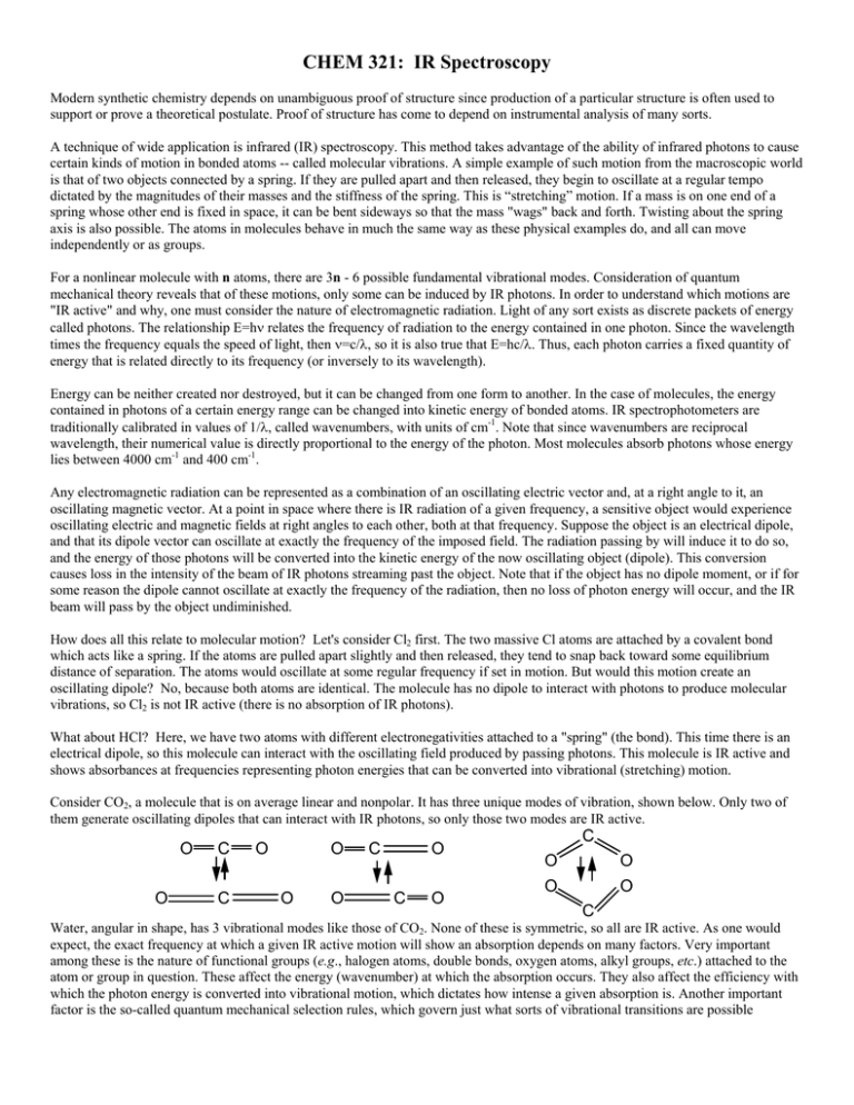
CHEM 321: IR Spectroscopy Modern synthetic chemistry depends on unambiguous proof of structure since production of a particular structure is often used to support or prove a theoretical postulate. Proof of structure has come to depend on instrumental analysis of many sorts. A technique of wide application is infrared (IR) spectroscopy. This method takes advantage of the ability of infrared photons to cause certain kinds of motion in bonded atoms -- called molecular vibrations. A simple example of such motion from the macroscopic world is that of two objects connected by a spring. If they are pulled apart and then released, they begin to oscillate at a regular tempo dictated by the magnitudes of their masses and the stiffness of the spring. This is “stretching” motion. If a mass is on one end of a spring whose other end is fixed in space, it can be bent sideways so that the mass "wags" back and forth. Twisting about the spring axis is also possible. The atoms in molecules behave in much the same way as these physical examples do, and all can move independently or as groups. For a nonlinear molecule with n atoms, there are 3n - 6 possible fundamental vibrational modes. Consideration of quantum mechanical theory reveals that of these motions, only some can be induced by IR photons. In order to understand which motions are "IR active" and why, one must consider the nature of electromagnetic radiation. Light of any sort exists as discrete packets of energy called photons. The relationship E=hν relates the frequency of radiation to the energy contained in one photon. Since the wavelength times the frequency equals the speed of light, then ν=c/λ, so it is also true that E=hc/λ. Thus, each photon carries a fixed quantity of energy that is related directly to its frequency (or inversely to its wavelength). Energy can be neither created nor destroyed, but it can be changed from one form to another. In the case of molecules, the energy contained in photons of a certain energy range can be changed into kinetic energy of bonded atoms. IR spectrophotometers are traditionally calibrated in values of 1/λ, called wavenumbers, with units of cm-1. Note that since wavenumbers are reciprocal wavelength, their numerical value is directly proportional to the energy of the photon. Most molecules absorb photons whose energy lies between 4000 cm-1 and 400 cm-1. Any electromagnetic radiation can be represented as a combination of an oscillating electric vector and, at a right angle to it, an oscillating magnetic vector. At a point in space where there is IR radiation of a given frequency, a sensitive object would experience oscillating electric and magnetic fields at right angles to each other, both at that frequency. Suppose the object is an electrical dipole, and that its dipole vector can oscillate at exactly the frequency of the imposed field. The radiation passing by will induce it to do so, and the energy of those photons will be converted into the kinetic energy of the now oscillating object (dipole). This conversion causes loss in the intensity of the beam of IR photons streaming past the object. Note that if the object has no dipole moment, or if for some reason the dipole cannot oscillate at exactly the frequency of the radiation, then no loss of photon energy will occur, and the IR beam will pass by the object undiminished. How does all this relate to molecular motion? Let's consider Cl2 first. The two massive Cl atoms are attached by a covalent bond which acts like a spring. If the atoms are pulled apart slightly and then released, they tend to snap back toward some equilibrium distance of separation. The atoms would oscillate at some regular frequency if set in motion. But would this motion create an oscillating dipole? No, because both atoms are identical. The molecule has no dipole to interact with photons to produce molecular vibrations, so Cl2 is not IR active (there is no absorption of IR photons). What about HCl? Here, we have two atoms with different electronegativities attached to a "spring" (the bond). This time there is an electrical dipole, so this molecule can interact with the oscillating field produced by passing photons. This molecule is IR active and shows absorbances at frequencies representing photon energies that can be converted into vibrational (stretching) motion. Consider CO2, a molecule that is on average linear and nonpolar. It has three unique modes of vibration, shown below. Only two of them generate oscillating dipoles that can interact with IR photons, so only those two modes are IR active. O O C C O O O O C O C O C O O O O C Water, angular in shape, has 3 vibrational modes like those of CO2. None of these is symmetric, so all are IR active. As one would expect, the exact frequency at which a given IR active motion will show an absorption depends on many factors. Very important among these is the nature of functional groups (e.g., halogen atoms, double bonds, oxygen atoms, alkyl groups, etc.) attached to the atom or group in question. These affect the energy (wavenumber) at which the absorption occurs. They also affect the efficiency with which the photon energy is converted into vibrational motion, which dictates how intense a given absorption is. Another important factor is the so-called quantum mechanical selection rules, which govern just what sorts of vibrational transitions are possible (allowed). Analysis of the equations describing these motions shows that vibrational and rotational states are both quantized, and that only certain transitions between ground and excited states are allowed. What happens when an IR active molecule absorbs a photon? The amplitude of the vibration increases (not the frequency). The IR Tutor software included in the CD ROM that came with your text is very helpful, and I encourage you to use it. To get to it, put the CD in your computer, "Explore" its contents, and go to one of the following: if you use a PC, "OrganicView\edugen\PC_IR_Tutor\Ir_tutor" (double click on Ir_tutor ) if you use a Mac, "OrganicView\edugen\ Mac IR Tutor\IR Tutor" (double click on IR Tutor) The program is very good for learning how assignment of absorptions to functional groups is made. Over the years, a great many compounds have been analyzed by IR spectroscopy, and catalogs of spectra have been published. What emerges from this mass of data is that certain groups always show absorption of energy in certain regions of the spectrum. One can summarize this information in tables like the one found on p. 82 of your text. More extensive tables ("IR Correlation Charts") can be found in some texts and in the CRC Handbook. Experimental Work in groups of 3-4. Choose a solid or a liquid from one of the labeled bottles in a cabinet as your sample. LIQUIDS: Run as a film between plates of NaCl. Samples must be free of water, since the plates dissolve in water. Plates are very brittle - never put them under any bending strain, and do not allow them to hit anything. They are very soft and easily scratched - do not rub them together. Do not touch the polished faces - your fingers are damp and will mar the surface. Work with them right above the desk top, never over the floor. To use, put one plate in the holder, add one drop of organic liquid, and center the other plate on it. Gently/evenly screw down the upper cover just tight enough that the plates don't slip. No air bubbles should be visible. Solids can be run as a KBr pellet. To make one, mix about 5% by weight of your compound with anhydrous KBr (put cap back on ASAP!!) and grind to a very fine powder in a mortar and pestle. Put a little of the powder into a die apparatus and squeeze at high pressure to form the pellet. The perfect KBr pellet is transparent, but in practice most will be more or less cloudy (less cloudy is better). *** Be SUPER careful not to drop any part of the die apparatus, and never force its pieces together!!! *** Other methods that we won't use here: Solids can be ground into a fine powder in mineral oil to produce a paste (a "mull") which is then spread on salt plates and squished into a thin film. A drawback to this method is that the hydrocarbon oil matrix obscures three narrow spectral regions. A liquid or solid may be dissolved in CCl4, which is transparent in most regions of interest, and run as the solution in special cells. If this method is chosen, a concentration of about 10% (weight/volume) is a good starting point. An instructor will show you how to use the IR instrument and print your spectrum. ******************************************************************************************************** Please make it a habit to CLEAN UP every time you finish working with the IR equipment. Salt plates must be carefully cleaned after use. Lay a clean Kimwipe flat on the countertop. Hold a plate only by its edges and be very careful not to bend or drop it. Clean it by rubbing its flat surfaces on the Kimwipe. If the sample is hard to remove, the Kimwipe can be moistened with DRY (meaning water-free) acetone or CCl4 . Wrap clean plates in new Kimwipes and store in the desiccator. KBr pellets: put in the trash. Put KBr reagent bottle in the desiccator. Mortar/pestle and die apparatus: if demand is heavy, they should be disassembled and thoroughly wiped with a clean Kimwipe. At day’s end, disassemble and rinse each part very well with deionized water, then dry and leave disassembled near the instrument on a paper towel. ********************************************************************************************************* REPORT (1) a. Write the name and structure of your compound on the spectrum. b. Identify the exact motion responsible for at least 3 peaks not counting those caused by C-H stretch (examples: O-H stretch, C=O stretch, C-H bend). Use your text and an IR Correlation Chart (the CRC Handbook has good ones). Cite the sources used and turn in your labeled spectrum with all this information on it. Citations should be complete: title, edition if necessary, author or editor, publisher, place of publication, date of publication, page(s). (2) What are two reasons why your sample had to be dry (no water)?
