Fluorescent Cell Imaging Activities for Your Classroom - Bio-Rad
advertisement

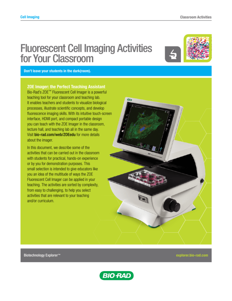
Cell Imaging Classroom Activities Fluorescent Cell Imaging Activities for Your Classroom Don’t leave your students in the dark(room). ZOE Imager: the Perfect Teaching Assistant Bio-Rad’s ZOE™ Fluorescent Cell Imager is a powerful teaching tool for your classroom and teaching lab. It enables teachers and students to visualize biological processes, illustrate scientific concepts, and develop fluorescence imaging skills. With its intuitive touch-screen interface, HDMI port, and compact portable design you can teach with the ZOE Imager in the classroom, lecture hall, and teaching lab all in the same day. Visit bio-rad.com/web/ZOEedu for more details about the imager. In this document, we describe some of the activities that can be carried out in the classroom with students for practical, hands-on experience or by you for demonstration purposes. This small selection is intended to give educators like you an idea of the multitude of ways the ZOE Fluorescent Cell Imager can be applied in your teaching. The activities are sorted by complexity, from easy to challenging, to help you select activities that are relevant to your teaching and/or curriculum. Biotechnology Explorer ™ explorer.bio-rad.com ZOE™ Fluorescent Cell Imager Activities Easy Activities Imaging Cheek Cell Nuclei Have you ever seen your own DNA? You can, and all you need is an empty petri dish, some cheek cells, and UView™ 6x Loading Dye (166-5111EDU). Have students spit into empty petri dishes. Mix in some UView and immediately image on the ZOE Imager using the UV LED (blue channel). Staining Major Cell Organelles Are you teaching cell structure and organelle function? Help your students visualize nuclei, mitochondria, lysosomes, actin filaments, and microtubules using the various channels of the ZOE Imager. Not only will they learn about the different cellular compartments, but they will also be able to generate colorful, vibrant images. You will need fixed eukaryotic cells, PureBlu™ DAPI Nuclear Staining Dye (135-1303EDU) or PureBlu Hoechst 33342 Nuclear Staining Dye (135-1304EDU), a mitochondrial stain, and an F-actin stain. Grow cultured cells on glass coverslips, fix and permeabilize the cells, and incubate them with the various dyes. Capture images of the cells using the different channels (blue channel to view the nuclei, green channel to view the actin filaments, and red channel to view mitochondria) and merge the images into one final image of a stained eukaryotic cell. In addition to these cellular components, you can incubate the cells with a tubulin dye to visualize microtubules and a lysotropic dye to visualize acidic organelles such as lysosomes. The nucleus (blue) of a human cheek cell stained with UView and visualized on the blue channel of the ZOE Imager. Visualizing Cell Division Have your students visualize cells and cell division using the carboxyfluorescein diacetate succinimidyl ester (CFDA-SE) dye (135-1201EDU). This is a cell-permeable dye that fluoresces after its acetate groups are cleaved by intracellular esterase enzymes to form CFSE. CFSE is retained in the cell and is successively halved during cell divisions. You and your students can visualize up to eight cell divisions using this dye. Stain cells with the dye and view the cells using the green channel. Since eukaryotic cells typically divide every 24 hours, keep the cells growing and view them on consecutive days to document cell division. Biotechnology Explorer ™ Use the ZOE Imager to create merged images of cells stained for mitochondria (red), nuclei (blue), and actin (green). explorer.bio-rad.com ZOE™ Fluorescent Cell Imager Activities Moderate Activities Transfection Efficiency Need a way to illustrate experimental optimization? Have your students quantify transfection efficiency by directly viewing GFP expression in treated cells. You will need eukaryotic cells growing in culture, TransFectin™ Lipid Reagent (170-3350EDU), a GFP-expressing plasmid, and VivaFix™ Cell Viability Assay (135-1111EDU). Transfect the eukaryotic cells with the GFP-expressing plasmid using the TransFectin Lipid Reagent. To demonstrate experimental optimization, vary the amount of transfection reagent used. Forty-eight hours after transfection, use the VivaFix Dye to count the number of live cells. Then visualize the cells under the green channel and count the number of green GFP-expressing cells. Calculate transfection efficiency by determining the percentage of total live cells that express GFP. You will notice that, up to a certain point, transfection efficiency increases with increasing amounts of transfection reagent. Your students can evaluate transfection efficiency with HEK 293 cells transfected with enhanced green fluorescent protein (EGFP) and/or red fluorescent protein (RFP) and visualized under the ZOE Imager’s green and red channels. Fluorescent Kinase Assay Protein phosphorylation is an essential process in signaling, enzyme activation/inhibition, and gene regulation. This activity will help your students monitor the difference between the amount of unphosphorylated and phosphorylated protein in a cell. Pick your favorite peptide substrate/kinase pair and carry out a fluorescence-based kinase enzymatic reaction. Phosphorylation of cellular peptides by the kinase will result in a change in fluorescence, which you can visualize by monitoring the cells over time under the appropriate ZOE Imager channel. Drosophila Embryogenesis This activity is a dramatic way to visualize embryogenesis. Treat a fertilized Drosophila egg with PureBlu DAPI Nuclear Staining Dye (135-1303EDU) or PureBlu Hoechst 33342 Nuclear Staining Dye (135-1304EDU) and watch the nucleus of the fertilized egg divide numerous times within the cytoplasm, generating a large number of nuclei. Approximately four hours after fertilization, after 13 mitotic divisions, an estimated 6,000 nuclei will organize themselves at the surface of the oocyte to create the syncytial blastoderm. Worms Feeding on GFP Bacteria This is a great activity for visualizing two very different organisms, with one literally inside the other! Grow a fat lawn of pGLO-transformed Escherichia coli using the pGLO™ Bacterial Transformation Kit (166-0003EDU) or pGLO™ Transformation and Inquiry Kit (166-0335EDU). Transfer Caenorhabditis elegans from the C. elegans Behavior Kit (166-5120EDU) to the pGLO plate. After at least one day, collect the worms and treat them with UView 6x Loading Dye (166-5111EDU). Transfer the worms to an agar plate that has been poured as thinly as possible. You can visualize the worms under the ZOE Imager’s blue channel and the ingested E. coli under the imager’s green channel. Biotechnology Explorer ™ explorer.bio-rad.com ZOE™ Fluorescent Cell Imager Activities Challenging Activities Calcium Flux Organisms spend a great deal of their resources maintaining electrochemical gradients that can then be used for energy generation, muscle contraction, and neuron signaling. You can show your students the fast action flux of these gradients using this activity. You will need to preload cells with a calcium dye (such as eFluor 514 Calcium Sensor Dye) and then treat them with a chemoattractant. This will cause the cells to display a flash of fluorescence upon calcium release and cell signaling. You can monitor this reaction using the ZOE Imager’s green channel. This is a good activity for illustrating what happens in cells and the complicated balance inherent in the internal cellular environment. Zebrafish Injury Help your students see the dynamic and fascinating actions of the immune system. For this activity you will need transgenic zebrafish with GFP-expressing leukocytes. Cut their tails or fins to initiate an immune response. Use the ZOE Imager’s green channel to monitor the GFP-expressing macrophages that collect at the site of injury. This is a good activity for demonstrating what happens to us when we get injured and how the body is able to heal itself. GFP Promoter Activity Differential gene expression is essential to life. Whether you are teaching developmental biology, basic genetics, or introductory biology, this activity will help you demonstrate tissue specificity and promoter activity using the power of GFP. Transfect eukaryotic cells with one of various GFP-expressing plasmids with different promoters to show the differences in expression and/or localization. You can transfect cells with a plasmid in which GFP is linked either to a constitutive promoter, for total cellular GFP expression, or to a specific promoter (for example, endothelial, mitochondrial, nuclear), for GFP expression in that particular region. Use the ZOE Imager’s green channel to visualize the cells. We Want to Hear from You These activities are just a sampling of what can be achieved with the ZOE Fluorescent Cell Imager. With its flexible operation and simplified cell imaging, and because this can all be carried out directly in the classroom, the ZOE Imager is a powerful tool that educators can use to introduce students to the fascinating world of fluorescence imaging and biology in general. In an effort to get the word out and inspire other educators, we encourage you to submit your teaching ideas and your students’ ZOE Imager images for posting at Bio-Rad’s Explorer Community (bio-rad.com/web/educator). With the Explorer Program, we strive to continually improve our teaching resources; your input is extremely important for this. eFluor is a trademark of eBioscience, Inc. Hoechst is a trademark of Hoechst GmbH. Biotechnology Explorer ™ explorer.bio-rad.com Bulletin 6686 Ver B 15-1339 0116


