High-Density Assembly of Nanocrystalline Silicon Quantum Dots 60nm
advertisement

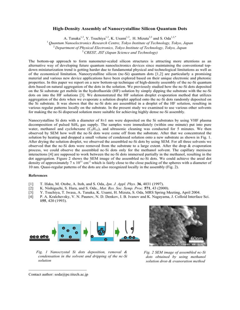
High-Density Assembly of Nanocrystalline Silicon Quantum Dots 1 A. Tanaka1,3, Y. Tsuchiya1,3, K. Usami1,3, H. Mizuta2,3 and S. Oda1,3,* Quantum Nanoelectronics Research Centre, Tokyo Institute of Technology, Tokyo, Japan 2 Department of Physical Electronics, Tokyo Institute of Technology, Tokyo, Japan 3 CREST, JST (Japan Science and Technology) The bottom-up approach to form nanometer-scaled silicon structures is attracting more attentions as an alternative way of developing future quantum nanoelectronics devices since maintaining the conventional topdown miniaturization trend is getting harder due to fundamental physical and technological limitations as well as of the economical limitation. Nanocrystalline silicon (nc-Si) quantum dots [1,2] are particularly a promising material and various new device applications have been explored based on their unique electronic and photonic properties. In this paper we report on a new bottom-up technique of high-density assembly of the nc-Si quantum dots based on natural aggregation of the dots in the solution. We previously studied how the nc-Si dots deposited on the Si substrate get mobile in the hydrofluoride (HF) solution by simply dipping the substrate with the nc-Si dots on into the HF solutions [3]. We demonstrated the HF solution droplet evaporation method that utilizes aggregation of the dots when we evaporate a solution droplet applied onto the nc-Si dots randomly deposited on the Si substrate. It was shown that the nc-Si dots are assembled in a droplet of the HF solution, resulting in various regular patterns locally on the substrate. In the present study we examined to use various other solvents for making the nc-Si dispersed solution more suitable for achieving highly dense nc-Si assembly. Nanocrystalline Si dots with a diameter of 8±1 nm were deposited on the Si substrates by using VHF plasma decomposition of pulsed SiH4 gas supply. The samples were immediately (within one minute) put into pure water, methanol and cyclohexane (C6H12), and ultrasonic cleaning was conducted for 5 minutes. We then observed by SEM how well the nc-Si dots were come off from the substrate. After that we concentrated the solution by heating and dropped a small volume of condensed solution onto a new substrate as shown in Fig. 1. After drying the solution droplet, we observed the assembled nc-Si dots by using SEM. For all three solvents we observed that the nc-Si dots were removed from the substrate to a large extent. After the drop & evaporation process, we could observe the assembled nc-Si dots only for the methanol solvent. The capillary meniscus interactions [4] are supposed to work between the nc-Si dots immersed partially in the methanol, resulting in the dot aggregation. Figure 2 shows the SEM image of the assembled nc-Si dots. We could achieve the areal dot density of approximately 7 x 1011 cm-2 which is fairly close to the close packing of the spheres with a diameter of 10 nm. Quasi-regular patterns of the dots are also recognized locally in the assembly (Fig. 2). References [1] [2] [3] [4] T. Ifuku, M. Otobe, A. Itoh, and S. Oda, Jpn. J. Appl. Phys. 36, 4031 (1997). K. Nishiguchi, S. Hara, and S. Oda., Mat. Res. Soc. Symp. Proc. 571, 43 (2000). Y. Tsuchiya, T. Iwasa, A. Tanaka, K. Usami, H. Mizuta, S. Oda, MRS Spring Meeting, April 2004. P. A. Kralchevsky, V. N. Paunov, N. D. Denkov, I. B. Ivanov and K. Nagayama, J. Colloid Interface Sci. 155, 420 (1993). 60nm Fig. 1 Nanocrystal Si dots deposition, removal & condensation in the solvent and dripping of the nc-Si solution Contact author: soda@pe.titech.ac.jp Fig. 2 SEM image of assembled nc-Si dots obtained by using methanol solution drop & evaporation method


