Images in Cardiovascular Medicine
Incomplete Longitudinal Ventricular Septal Fracture After
Blunt Chest Trauma
François Marcotte, MD, FRCP(C); Jean-Pierre Lavoie, MD, MSC; Annie Dore, MD, FRCP(C);
Paul Khairy, MD, FRCP(C); Lise-Andrée Mercier, MD, FRCP(C); Éléonore Paquet, MD, FRCP(C);
Nancy Poirier, MD; Josephine Pressacco, MD
A
Downloaded from http://circ.ahajournals.org/ by guest on October 1, 2016
currently asymptomatic 31-year-old man was referred
for recent atypical short stabbing chest discomfort
occurring at rest. One month earlier, he had complained of a
prolonged chest pain episode lasting ⬇1 day, temporally
related to the use of inhaled cocaine, for which he did not
seek medical consultation. He had smoked half a pack of
cigarettes a day for 15 years and suffered 2 remote episodes
of blunt chest trauma. The first occurred at 12 years of age
when he was in a low-velocity automobile accident as a
passenger; the second occurred at 18 years of age when he was
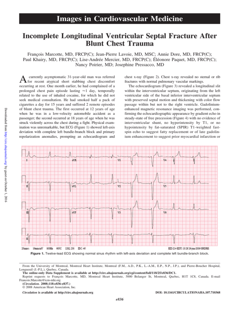
struck violently across the chest during a fight. Physical examination was unremarkable, but ECG (Figure 1) showed left-axis
deviation with complete left bundle-branch block and primary
repolarization anomalies, prompting an echocardiogram and
chest x-ray (Figure 2). Chest x-ray revealed no sternal or rib
fractures with normal pulmonary vascular markings.
The echocardiogram (Figure 3) revealed a longitudinal slit
within the interventricular septum, originating from the left
ventricular side of the basal inferior interventricular septum
with preserved septal motion and thickening with color flow
passage within but not to the right ventricle. Gadoliniumenhanced magnetic resonance imaging was performed, confirming the echocardiographic appearance by gradient echo in
steady-state of free precession (Figure 4) with no evidence of
interventricular shunt, no hyperintensity by T1, or no
hypointensity by fat-saturated (SPIR) T1-weighted fastspin echo to suggest fatty replacement or of late gadolinium enhancement to suggest prior myocardial infarction or
Figure 1. Twelve-lead ECG showing normal sinus rhythm with left-axis deviation and complete left bundle-branch block.
From the University of Montreal, Montreal Heart Institute, Montreal (F.M., A.D., P.K., L.-A.M., E.P., N.P., J.P.), and Pierre-Boucher Hospital,
Longueuil (J.-P.L.), Quebec, Canada.
The online-only Data Supplement is available at http://circ.ahajournals.org/cgi/content/full/118/25/e836/DC1.
Reprint requests to François Marcotte, MD, Montreal Heart Institute, 5000 Belanger St, Montreal, Quebec, H1T 1C8, Canada. E-mail
Francois.Marcotte@icm-mhi.org
(Circulation. 2008;118:e836-e837.)
© 2008 American Heart Association, Inc.
Circulation is available at http://circ.ahajournals.org
DOI: 10.1161/CIRCULATIONAHA.107.710368
e836
Marcotte et al
Posttraumatic Incomplete Septal Fracture
e837
Downloaded from http://circ.ahajournals.org/ by guest on October 1, 2016
Figure 4. Multislice cinematic magnetic resonance imaging in
gradient echo in steady state of free precession in the long-axis
4-chamber view. Abbreviations as in Figure 3, plus Ao indicates
aorta. *Longitudinal septal tear origin or entry site in the left
ventricle.
Figure 2. Posteroanterior chest x-ray showing normal cardiopericardial silhouette and normal pulmonary vascular markings
with absence of visible healed sternal or rib fractures. “G” marks
the left side of the chest.
Figure 5. Multislice magnetic resonance imaging viability study
in gradient echo with inversion recovery preparation in the
short-axis view. Abbreviations as in Figure 3. *Longitudinal septal tear origin or entry site in left ventricle.
fibrosis (Figure 5). The absence of fatty replacement, scar,
or fibrosis made the diagnosis of cocaine abuse–related
infarction with ensuing rupture less likely than a posttraumatic incomplete septal fracture. The patient has thus far
declined surgical repair.
Figure 3. Transthoracic 2-dimensional echocardiogram in the
apical 4-chamber view. IVS indicates interventricular septum;
LV, left ventricle; RV, right ventricle; RA, right atrium; and RV,
right ventricle.
Disclosures
None.
Incomplete Longitudinal Ventricular Septal Fracture After Blunt Chest Trauma
François Marcotte, Jean-Pierre Lavoie, Annie Dore, Paul Khairy, Lise-Andrée Mercier,
Éléonore Paquet, Nancy Poirier and Josephine Pressacco
Downloaded from http://circ.ahajournals.org/ by guest on October 1, 2016
Circulation. 2008;118:e836-e837
doi: 10.1161/CIRCULATIONAHA.107.710368
Circulation is published by the American Heart Association, 7272 Greenville Avenue, Dallas, TX 75231
Copyright © 2008 American Heart Association, Inc. All rights reserved.
Print ISSN: 0009-7322. Online ISSN: 1524-4539
The online version of this article, along with updated information and services, is located on the
World Wide Web at:
http://circ.ahajournals.org/content/118/25/e836
Data Supplement (unedited) at:
http://circ.ahajournals.org/content/suppl/2008/12/29/118.25.e836.DC1.html
Permissions: Requests for permissions to reproduce figures, tables, or portions of articles originally published
in Circulation can be obtained via RightsLink, a service of the Copyright Clearance Center, not the Editorial
Office. Once the online version of the published article for which permission is being requested is located,
click Request Permissions in the middle column of the Web page under Services. Further information about
this process is available in the Permissions and Rights Question and Answer document.
Reprints: Information about reprints can be found online at:
http://www.lww.com/reprints
Subscriptions: Information about subscribing to Circulation is online at:
http://circ.ahajournals.org//subscriptions/


