43 Semi log graph paper
advertisement
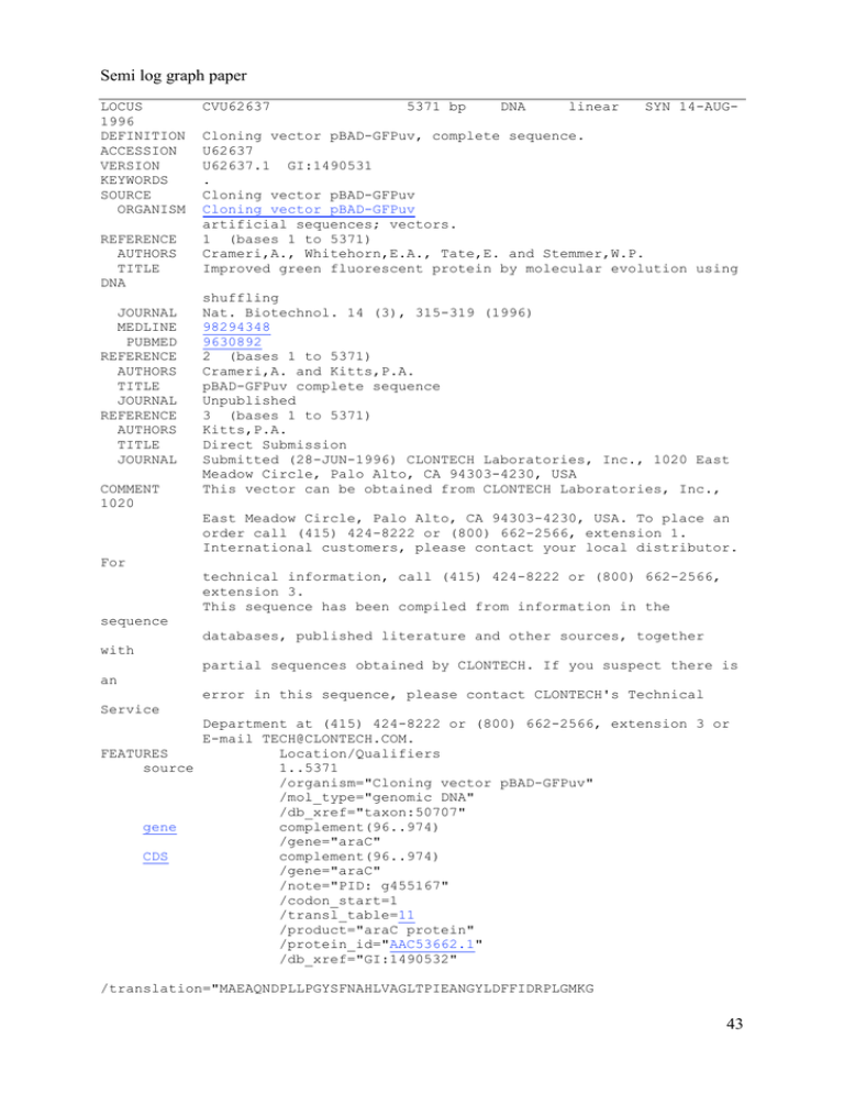
Semi log graph paper LOCUS 1996 DEFINITION ACCESSION VERSION KEYWORDS SOURCE ORGANISM REFERENCE AUTHORS TITLE DNA JOURNAL MEDLINE PUBMED REFERENCE AUTHORS TITLE JOURNAL REFERENCE AUTHORS TITLE JOURNAL COMMENT 1020 CVU62637 5371 bp DNA linear SYN 14-AUG- Cloning vector pBAD-GFPuv, complete sequence. U62637 U62637.1 GI:1490531 . Cloning vector pBAD-GFPuv Cloning vector pBAD-GFPuv artificial sequences; vectors. 1 (bases 1 to 5371) Crameri,A., Whitehorn,E.A., Tate,E. and Stemmer,W.P. Improved green fluorescent protein by molecular evolution using shuffling Nat. Biotechnol. 14 (3), 315-319 (1996) 98294348 9630892 2 (bases 1 to 5371) Crameri,A. and Kitts,P.A. pBAD-GFPuv complete sequence Unpublished 3 (bases 1 to 5371) Kitts,P.A. Direct Submission Submitted (28-JUN-1996) CLONTECH Laboratories, Inc., 1020 East Meadow Circle, Palo Alto, CA 94303-4230, USA This vector can be obtained from CLONTECH Laboratories, Inc., East Meadow Circle, Palo Alto, CA 94303-4230, USA. To place an order call (415) 424-8222 or (800) 662-2566, extension 1. International customers, please contact your local distributor. For technical information, call (415) 424-8222 or (800) 662-2566, extension 3. This sequence has been compiled from information in the sequence databases, published literature and other sources, together with partial sequences obtained by CLONTECH. If you suspect there is an error in this sequence, please contact CLONTECH's Technical Service Department at (415) 424-8222 or (800) 662-2566, extension 3 or E-mail TECH@CLONTECH.COM. FEATURES Location/Qualifiers source 1..5371 /organism="Cloning vector pBAD-GFPuv" /mol_type="genomic DNA" /db_xref="taxon:50707" gene complement(96..974) /gene="araC" CDS complement(96..974) /gene="araC" /note="PID: g455167" /codon_start=1 /transl_table=11 /product="araC protein" /protein_id="AAC53662.1" /db_xref="GI:1490532" /translation="MAEAQNDPLLPGYSFNAHLVAGLTPIEANGYLDFFIDRPLGMKG 43 YILNLTIRGQGVVKNQGREFVCRPGDILLFPPGEIHHYGRHPEAREWYHQWVYFRPRA YWHEWLNWPSIFANTGFFRPDEAHQPHFSDLFGQIINAGQGEGRYSELLAINLLEQLL LRRMEAINESLHPPMDNRVREACQYISDHLADSNFDIASVAQHVCLSPSRLSHLFRQQ LGISVLSWREDQRISQAKLLLSTTRMPIATVGRNVGFDDQLYFSRVFKKCTGASPSEF RAGCEEKVNDVAVKLS" gene 1342..2061 /gene="gfpuv" CDS 1342..2061 /gene="gfpuv" /note="GFPuv is the GFP variant called 'cycle 3'; Allele: AC2; green fluorescent protein variant" /codon_start=1 /transl_table=11 /product="GFPuv" /protein_id="AAC53663.1" /db_xref="GI:1490533" /translation="MASKGEELFTGVVPILVELDGDVNGHKFSVSGEGEGDATYGKLT LKFICTTGKLPVPWPTLVTTFSYGVQCFSRYPDHMKRHDFFKSAMPEGYVQERTISFK DDGNYKTRAEVKFEGDTLVNRIELKGIDFKEDGNILGHKLEYNYNSHNVYITADKQKN GIKANFKIRHNIEDGSVQLADHYQQNTPIGDGPVLLPDNHYLSTQSALSKDPNEKRDH MVLLEFVTAAGITHGMDELYK" gene 2636..3496 /gene="bla" CDS 2636..3496 /gene="bla" /function="confers resistance to ampicillin" /codon_start=1 /transl_table=11 /product="beta-lactamase" /protein_id="AAC53664.1" /db_xref="GI:1490534" /translation="MSIQHFRVALIPFFAAFCLPVFAHPETLVKVKDAEDQLGARVGY IELDLNSGKILESFRPEERFPMMSTFKVLLCGAVLSRVDAGQEQLGRRIHYSQNDLVE YSPVTEKHLTDGMTVRELCSAAITMSDNTAANLLLTTIGGPKELTAFLHNMGDHVTRL DRWEPELNEAIPNDERDTTMPAAMATTLRKLLTGELLTLASRQQLIDWMEADKVAGPL LRSALPAGWFIADKSGAGERGSRGIIAALGPDGKPSRIVVIYTTGSQATMDERNRQIA EIGASLIKHW" ORIGIN 1 atcgatgcat aatgtgcctg tcaaatggac gaagcaggga ttctgcaaac 61 tccgtcaagc cgtcaattgt ctgattcgtt accaattatg acaacttgac 121 ttcacttttt cttcacaacc ggcacggaac tcgctcgggc tggccccggt 181 aatacccgcg agaaatagag ttgatcgtca aaaccaacat tgcgaccgac 241 ggcatccggg tggtgctcaa aagcagcttc gcctggctga tacgttggtc 301 cttaagacgc taatccctaa ctgctggcgg aaaagatgtg acagacgcga 361 caaacatgct gtgcgacgct ggcgatatca aaattgctgt ctgccaggtg 421 tactgacaag cctcgcgtac ccgattatcc atcggtggat ggagcgactc 481 tccatgcgcc gcagtaacaa ttgctcaagc agatttatcg ccagcagctc 541 ccttcccctt gcccggcgtt aatgatttgc ccaaacaggt cgctgaaatg 601 gcttcatccg ggcgaaagaa ccccgtattg gcaaatattg acggccagtt cctatgctac ggctacatca gcatttttta ggtggcgata ctcgcgccag cggcgacaag atcgctgatg gttaatcgct cgaatagcgc cggctggtgc aagccattca 44 661 721 781 841 901 961 1021 1081 1141 1201 1261 1321 1381 1441 1501 1561 1621 1681 1741 1801 1861 1921 1981 2041 2101 2161 2221 2281 2341 2401 2461 2521 2581 2641 2701 2761 2821 2881 2941 3001 3061 3121 3181 3241 3301 3361 3421 3481 3541 3601 3661 3721 3781 3841 3901 3961 4021 4081 4141 4201 4261 4321 4381 tgccagtagg tgacgaccgt acaaattctc taacctttca ggcgttaaac tgcgcttcag tgcatcagac accccgctta aacaaaagtg ctttgctatg tcgcaactct ctttaagaag ccaattcttg ggtgaaggtg ctacctgttc cgttatccgg gtacaggaac aagtttgaag gatggaaaca acggcagaca gatggatccg gtccttttac gaaaagcgtg atggatgagc cctgcaggca gattaaatca ggtggtccca tgtggggtcc gtgcaaagac gacaaatccg aggacgcccg cctttttgcg gctcatgaga tattcaacat tgctcaccca gggttacatc acgttttcca tgacgccggg gtactcacca tgctgccata accgaaggag ttgggaaccg agcaatggca gcaacaatta ccttccggct tatcattgca ggggagtcag gattaagcat cgccctgtag cacttgccag tcgccggctt ctttacggca cgccctgata tcttgttcca ggattttgcc cgaattttaa gataatctca gtagaaaaga caaacaaaaa ctttttccga tagccgtagt ctaatcctgt tcaagacgat cgcgcggacg agtgatgaat gtccctgatt ttcccagcgg ccgccaccag ccatactttt attgccgtca ttaaaagcat tctataatca ccatagcatt ctactgtttc gagatataca ttgaattaga atgctacata catggccaac atcatatgaa gcactatatc gtgataccct ttctcggaca aacaaaagaa ttcaactagc cagacaacca accacatggt tctacaaata tgcaagcttg gaacgcagaa cctgacccca cccatgcgag tgggcctttc ccgggagcgg ccataaactg tttctacaaa caataaccct ttccgtgtcg gaaacgctgg gaactggatc atgatgagca caagagcaac gtcacagaaa accatgagtg ctaaccgctt gagctgaatg acaacgttgc atagactgga ggctggttta gcactggggc gcaactatgg tggtaactgt cggcgcatta cgccctagcg tccccgtcaa cctcgacccc gacggttttt aactggaaca gatttcggcc caaaatatta tgaccaaaat tcaaaggatc aaccaccgct aggtaactgg taggccacca taccagtggc agttaccgga aaagtaaacc ctctcctggc tttcaccacc tcggtcgata atgggcatta catactcccg ctgcgtcttt tctgtaacaa cggcagaaaa tttatccata tccatacccg tatggctagc tggtgatgtt cggaaagctt acttgtcact acggcatgac tttcaaagat tgttaatcgt caaactcgag tggaatcaaa agaccattat ttacctgtcg ccttcttgag atgaattcga gctgttttgg gcggtctgat tgccgaactc agtagggaac gttttatctg atttgaacgt ccaggcatca ctctttgttt gataaatgct cccttattcc tgaaagtaaa tcaacagcgg cttttaaagt tcggtcgccg agcatcttac ataacactgc ttttgcacaa aagccatacc gcaaactatt tggaggcgga ttgctgataa cagatggtaa atgaacgaaa cagaccaagt agcgcggcgg cccgctcctt gctctaaatc aaaaaacttg cgccctttga acactcaacc tattggttaa acgtttacaa cccttaacgt ttcttgagat accagcggtg cttcagcaga cttcaagaac tgctgccagt taaggcgcag cactggtgat gggaacagca ccctgaccgc aaaaaatcga aacgagtatc ccattcagag tactggctct agcgggacca gtccacattg agattagcgg tttttttggg aaaggagaag aatgggcaca acccttaaat actttctctt tttttcaaga gacgggaact atcgagttaa tacaactata gctaacttca caacaaaata acacaatctg tttgtaactg gctcggtacc cggatgagag aaaacagaat agaagtgaaa tgccaggcat ttgtttgtcg tgcgaagcaa aattaagcag atttttctaa tcaataatat cttttttgcg agatgctgaa taagatcctt tctgctatgt catacactat ggatggcatg ggccaactta catgggggat aaacgacgag aactggcgaa taaagttgca atctggagcc gccctcccgt tagacagatc ttactcatat gtgtggtggt tcgctttctt gggggctccc atttgggtga cgttggagtc ctatctcggg aaaatgagct tttaaaagga gagttttcgt cctttttttc gtttgtttgc gcgcagatac tctgtagcac ggcgataagt cggtcgggct accattcgcg aaatatcacc gaatggtgag gataaccgtt ccggcagcag aagaaaccaa tctcgctaac aagccatgac attatttgca atcctacctg ctagaaataa aacttttcac aattttctgt ttatttgcac atggtgttca gtgccatgcc acaagacgcg aaggtattga actcacacaa aaattcgcca ctccaattgg ccctttcgaa ctgctgggat cggggatcct aagattttca ttgcctggcg cgccgtagcg caaataaaac gtgaacgctc cggcccggag aaggccatcc atacattcaa tgaaaaagga gcattttgcc gatcagttgg gagagttttc ggcgcggtat tctcagaatg acagtaagag cttctgacaa catgtaactc cgtgacacca ctacttactc ggaccacttc ggtgagcgtg atcgtagtta gctgagatag atactttaga tacgcgcagc cccttccttt tttagggttc tggttcacgt cacgttcttt ctattctttt gatttaacaa tctaggtgaa tccactgagc tgcgcgtaat cggatcaaga caaatactgt cgcctacata cgtgtcttac gaacgggggg agcctccgga cggtcggcaa attgagaata ggcctcaatc gggatcattt ttgtccatat caaaccggta aaaaacgcgt cggcgtcaca acgcttttta ttttgtttaa tggagttgtc cagtggagag tactggaaaa atgcttttcc cgaaggttat tgctgaagtc ttttaaagaa tgtatacatc caacattgaa cgatggccct agatcccaac tacacatggc ctagagtcga gcctgataca gcagtagcgc ccgatggtag gaaaggctca tcctgagtag ggtggcgggc tgacggatgg atatgtatcc agagtatgag ttcctgtttt gtgcacgagt gccccgaaga tatcccgtgt acttggttga aattatgcag cgatcggagg gccttgatcg cgatgcctgc tagcttcccg tgcgctcggc ggtctcgcgg tctacacgac gtgcctcact ttgatttacg gtgaccgcta ctcgccacgt cgatttagtg agtgggccat aatagtggac gatttataag aaatttaacg gatccttttt gtcagacccc ctgctgcttg gctaccaact ccttctagtg cctcgctctg cgggttggac ttcgtgcaca 45 4441 4501 4561 4621 4681 4741 4801 4861 4921 4981 5041 5101 5161 5221 5281 5341 cagcccagct gaaagcgcca ggaacaggag gtcgggtttc agcctatgga tttgctcaca tttgagtgag gaggaagcgg caccgcatat acactccgct ctgacgcgcc tctccgggag aaggagatgg caagcgctca taggcgccag aggatctaat tggagcgaac cgcttcccga agcgcacgag gccacctctg aaaacgccag tgttctttcc ctgataccgc aagagcgcct ggtgcactct atcgctacgt ctgacgggct ctgcatgtgt cgcccaacag tgagcccgaa caaccgcacc tctcatgttt gacctacacc agggagaaag ggagcttcca acttgagcgt caacgcggcc tgcgttatcc tcgccgcagc gatgcggtat cagtacaatc gactgggtca tgtctgctcc cagaggtttt tcccccggcc gtggcgagcc tgtggcgccg gacagcttat gaactgagat gcggacaggt gggggaaacg cgatttttgt tttttacggt cctgattctg cgaacgaccg tttctcctta tgctctgatg tggctgcgcc cggcatccgc caccgtcatc acggggcctg cgatcttccc gtgatgccgg c acctacagcg atccggtaag cctggtatct gatgctcgtc tcctggcctt tggataaccg agcgcagcga cgcatctgtg ccgcatagtt ccgacacccg ttacagacaa accgaaacgc ccaccatacc catcggtgat ccacgatgcg tgagctatga cggcagggtc ttatagtcct aggggggcgg ttgctggcct tattaccgcc gtcagtgagc cggtatttca aagccagtat ccaacacccg gctgtgaccg gcgaggcagc cacgccgaaa gtcggcgata tccggcgtag 46 BACTERIAL TRANSFORMATION Introduction: Genetic transformation occurs when a cell takes up and expresses a new piece of genetic material. In many bacteria this transformation takes place within the bacterial plasmid DNA eg. Escherichia coli and Agrobacterium tumefaciens. Plasmids are small circular extra-chromosomal bits of DNA contained within the bacteria cell. The insertion of the gene(s) usually provides the organism with a new trait(s) (eg. pest or antibiotic resistance). For example a gene used for the production of insulin in humans has been cloned into a plasmid and transformed into bacteria. Under the right conditions these transformed bacteria will produce authentic human insulin to treat diabetic patients. Green Fluorescent Protein (GFP) is expressed by a gene found in the bioluminescent jellyfish Aequorea victoria. This causes the jellyfish to fluoresce and glow in the dark. Therefore if this gene is cloned into a plasmid and transformed into bacterial cells, the newly transformed bacterial cells should fluoresce a green colour when placed under UV light. Expressions of traits are regulated by specific promoters. If ampicillin is added to the media, the gene for ampicillin resistance on the plasmid is switched on allowing the bacteria to grow. The GFP gene is turned on in presence of the sugar arabinose. In this practical we will supply you with a plasmid containing the genes for expressing GFP and ampicillin resistance and E. coli cells that will be transformed with the new plasmid. If the transformation is successful the E. coli will grow on media containing ampicillin and fluoresce with UV light. Laboratory technique Sterile conditions are an important factor with all facets of molecular biology. An attempt should be made to minimise contamination of the experiment. Contamination can be found on hands, bench tops and equipment etc. It is important that gloves are worn during the practical and equipment used is sterile and not touched by any contaminated surface. The host organism is E. coli K-12 strain, which is a non-pathogenic bacteria. All used disposable pieces of equipment (i.e. loops, pipette tips, plates and gloves) should be placed in the bins to be autoclaved. Ultra violet light can cause damage to eyes and skin, so always make sure that a perspex barrier is between you and the UV light. 47 Experimental Procedure: 1. Label one of the micro tubes with +DNA and the other -DNA. Both tubes should have your group name. 2. Transfer, using a pipette and sterile tip, 200µL of the transformation solution to each micro tube. (The transformation solution is 50mM CaCl2, pH7.4 which neutralises the repulsive negative charges of the phosphate backbone of the DNA and the phospholipids of the cell membrane allowing the DNA to pass through the cell membrane.) 3. Place the micro tubes on ice. 4. Using a sterile loop pick a single colony of E. coli from the culture plate and immerse the loop into the +DNA micro tube. Spin the loop once immersed in the transformation solution to disperse the bacteria thoroughly. Replace back on ice. Repeat for the -DNA micro tube. 5. Immerse a sterile loop into the plasmid DNA making sure that there is a film of plasmid solution across the loop. Transfer to the +DNA micro tube, spinning once more to mix the plasmid and bacteria. Return to ice. DO NOT add plasmid DNA to the -DNA micro tube. 6. Incubate the micro tubes on ice for 10 minutes. 7. While you are waiting label the bottom of the four agar plates with your group name as follows; LB plate LB/amp LB/amp LB/amp/ara -DNA -DNA +DNA +DNA 8. After 10 minutes on ice, transfer the foam rack containing both micro tubes into the water bath at 42°C for exactly 50 seconds. Remove micro tubes and replace on ice for 2 minutes. (The heat shock increases the permeability of the cell membrane to increase the efficiency that the plasmid enters the. The time of heat shock is crucial for optimum transformation.) 9. After 2 minutes on ice remove micro tubes and place on the bench top. Add 200µL of the LB broth solution to both micro tubes. Incubate for 10 minutes at room temperature. (This 10 minute incubation while allow the transformed cells to grow and express the ampicillin resistance protein, beta-lactamase.) 48 10. Tap the base of the closed micro tubes with your finger to mix the bacterial suspension. Add 100µL of the suspension from both micro tubes to the appropriate plates as follows; 100µL 100µL 100µL 100µL -DNA -DNA +DNA +DNA LB plate LB/amp plate LB/amp plate LB/amp/ara plate 11. Using a sterile loop for each plate, spread the suspension evenly over the surface of the media. 12. Wrap each plate with parafilm, taping them in a stack upside down. Incubate in the 25°C room for one week. Check colony formation at the beginning of next weeks practical, for growth and fluorescence. (Colony formation can be achieved over night by using a 37°C incubation temperature.) Question 1. We have given you a plasmid with the gene that encodes the GFP already in it. The gene for GFP was not originally present in the plasmid. Briefly explain what processes you think we used to get the GFP gene into the plasmid. Question 2. Describe how you could use two LB/agar plates, some E. coli and some ampicillin to determine how E. coli cells are affected by ampicillin. Question 3. Explain the reasoning behind plating the -DNA transformation onto LB and LB/amp plates. Question 4. Why do you think the bugs are incubated for 10 minutes after the Lbroth has been added at step nine ? Question 5. Assuming everything has gone according to plan in this prac, what do you expect to find on each of your four plates next week ? When answering this, make sure you give the reasons why you these results. 49 Lab Requirements Group of 20 students 10 X LB plate with E. coli 20 X LB plate 10g LB per 500mlDI 8g Agar 40 X LB/Amp plate 10g LB 0.0375g Amp per 500mL DI 8g Agar 20 X LB/Amp/Ara plate 10g LB 0.0375g Amp 2.5g L+Arabanose per 500mL 8g Agar 60 X Petri dishes 100 X Micro tubes 20 X Micro tube racks 20 X Marker pen fine 20 X pipette P200 & tips 20 X disposable sterile loops packs of 25 20 X 1mL transformation solution (50mM CaCl2 MM 147.02) 20 X 1mL LB broth 20 X timer 20 X paintbrushes 20 X floaties Each Bench 4 X Esky of ice 4 X Parafilm 4 X scissors Class Plasmid DNA 1 X Waterbath 42°C UV transilluminator 25°C or 37°C incubator Microwave 6 X autoclave bags waste beakers 50 Bacterial Practical Bacteria belong to the Prokaryotes group, and are the oldest in evolutionary terms. They are the most abundant and smallest cellular organisms in the world. Some bacteria have such a rapid cell division they can double their population in short periods of time eg. E. coli at 20 minutes under optimum conditions. The main differences between bacteria and other cellular organisms are; the lack of a nuclear membrane and the composition of their cell wall. The classification of bacteria has mainly been based on their size, shape and cell wall. They come in rod, filamentous, spherical, etc. The chemical assay of their cell wall sorts them into Gram -ve or Gram +ve. As a group we will discuss the growth habits and safety concerns in dealing with bacterial cultures. Also the possibilities of painting with bacteria using the agar plates as the 'canvas'. Things to consider are: • Bacteria can be found almost everywhere in life. • Many bacteria can cause health and safety issues. • The source of the bacteria being it from body fluids or the environment. • The speed in which bacteria multiply. • Use of antibiotics for the selection of bacterial strains. • The disposal of the bacterial cultures after use. Lab requirements. 100 X Petri dishes LB broth Nutrient broth Agar Microwave Nutrient broth 500ml schott X 2 Cotton buds autoclaved Microscopes compound X 3 Microscopes dissect X 3 Streptomycin Slides X 10 coverslips X 10 loops parafilm X 4 scissors X 4 razor blades 51 Fungal Practical Fungi, once thought of as plants, are now classified as their own kingdom. Most fungi are multicellular, however the yeasts are unicellular. Yeasts have a growth habit similar to that of bacteria, forming colonies on an agar plate. Filamentous fungi produce masses of strands known as mycelium. When this mycelium is tightly packed it can produce complex spore-producing structures such as mushrooms (Basidiomycetes) and puffballs (Ascomycetes). Fungi can reproduce sexually with + and - mating strains. They also reproduce asexually with the production of spores and budding _____________________________________________________________________ Demonstration: Under the compound and dissecting microscopes are various plates and slides showing some of the features of fungus. Observe the growth habits, colours and structures that are produced by fungi. Making a slide. Using a dissection needle, remove a thin slice of the fungal culture and place on to a new microscope slide. Chop up into smaller pieces and add a drop of the biological dyes. Gently push on a cover slip and blot off any excess dye with a tissue or filter paper. Observe the slide under a compound microscope. An alternative method of producing a slide is by using sticky tape. Open the culture and press gently a piece of sticky tape onto the fungal mycelium. Remove and stick onto a new glass slide. Observe under a compound microscope. Dyeing using extracted fungal pigments. * Provided is a solution of dye extracted from puffball fungi. Using this dye and fabrics supplied, ranging from silk to cotton create a new design. * Donna Franklin, SymbioticA Resident during her Masters of Creative Art (Textiles Major) is the source for much of the material and knowledge associated with this section. 52 Lab requirements. 100 X Petri dishes Puff balls Range of fabrics Aluminum sulphate Pots Silk Gas cooker Detergent Vinegar White wool raw demo posters Donna fashion Dissecting needles Aniline blue Gongo red Fusarium Aureobasidium Yellow culture Orange culture parafilm scissors razor blades demo slides sticky tape PHOTOS OF DONNAS FUNGI AND MICRO DETAILS 53 Plant Tissue Culture Plant tissue culture has many valuable uses in agricultural research. It is used for the micro propagation of selected cultivars in fruit crops, floriculture and the nursery industry. Plant tissue culture material can also be used for genetic engineering i.e. cloning. Tissue culture involves growing cells, tissues or plantlets on a nutrient medium (which has usually been solidified with agar), in a sterile environment such as glass jar (in vitro means “in glass”) under favourable conditions of temperature and light. The nutrient medium consists of minerals, vitamins and sugar supplemented with growth regulators that control the development of the plants. Many Western Australian native plants produce seeds that are difficult to germinate artificially. Therefore the use of tissue culture in the form of an embryo rescue may increase the chances of germinating the seed successfully. This is done by excising the seed embryo to overcome any chemical and/or physical dormancy. Once removed the embryo can be grown on a tissue culture media. _____________________________________________________________________ You should wear a lab coat and gloves. (There is no eating, drinking, etc. in a lab!) PRACTICAL Part 1 Embryo Rescue • Spray the bench and under the culture hood with 70% ethanol and wipe dry with the tissue paper. • Light the Bunsen burner or ethanol burner outside the hood. • Place a sterile Petri dish into the hood. • Tear open the sterile instruments in the laminar flow and attach the blade to the scalpel handle. (If you fill uncomfortable please ask for help). Lay the instruments on an open Petri dish. • Dip the instruments in ethanol and flame, holding the scalpel horizontally. • Allow the tools to cool. • With sterile forceps and working under the hood, excise the embryo from the seed, using a Petri dish as a cutting board. • In the hood, open a new vial containing nutrient media. Place the embryo on the filter paper. 54 • Replace the lid tightly, label and date the new tissue culture tube. Part 2 Lateral bud micro propagation. • Setting up the culture hoods as described in part 1. • Remove, under the hood, a section of cultured plant material and place it on a newly opened petri dish. • Cut a single node piece of stem, removing the leaf. • Making sure the node is the right way up, push the base into a new tube of media. • Replace the lid tightly, label and date the new tissue culture tube. Lab requirements 6 X Culture cabinets (Botany) 3 X spray bottles of 70% ethanol 2 X boxes of tissues 6 X Bunsen burner on gas or ethanol burners 100% ethanol 200 X Petri dishes Petunia cultures African Violet cultures Pea seeds 10 X pre autoclaved tools (scalpel and forceps) Box scalpel blades Glass jars for 100% ethanol 5 X M&S nutrient mix pacs sprinkle of BAP 4 X parafilm 4 X scissors 100 X pre autoclaved 250ml vials 100 X pre autoclaved 30ml vials with filter paper 20ml 1M NaOH Litmus paper 2 X 2L beaker Portable balance Weigh boats 10 X Marker pens 10 X plastic spoons 100g Sucrose 50g Agar Microwave Gloves S M L Hand soap FIRE EXTINGUISHER 55 Mammalian Tissue Culture Tissue Culture is the technique for growing cells from multicellular organisms in a liquid medium for research. Tissue culture is often a generic term that refers to both organ culture and cell culture and the terms are often used interchangeably. Cell cultures are derived from either primary tissue explants or cell suspensions. Primary cell cultures typically will have a finite life span in culture whereas continuous cell lines are, by definition, abnormal and are often transformed cell lines. A. Growth pattern. Cells will initially go through a quiescent or lag phase that depends on the cell type, the seeding density, the media components, and previous handling. The cells will then go into exponential growth where they have the highest metabolic activity. The cells will then enter into stationary phase where the number of cells is constant, this is characteristic of a confluent population (where all growth surfaces are covered). B. Harvesting. Cells are harvested when the cells have reached a population density which suppresses growth. Ideally, cells are harvested when they are in a semi-confluent state and are still in log phase. Cells that are not passaged and are allowed to grow to a confluent state can sometime lag for a long period of time and some may never recover. It is also essential to keep your cells as happy as possible to maximize the efficiency of transformation. Most cells are passaged Proteolytic enzymes - Trypsin, collagenase, or pronase, usually in combination with EDTA, causes cells to detach from the growth surface. This method is fast and reliable but can damage the cell surface by digesting exposed cell surface proteins. The proteolysis reaction can be quickly terminated by the addition of complete medium containing serum C. Media and growth requirements 1. Physiological parameters A. temperature 37C for cells from homeotherm B. pH - 7.2-7.5 and osmolality of medium must be maintained C. humidity is required D. gas phase - bicarbonate conc. and CO2 tension in equilibrium E. visible light - can have an adverse effect on cells; light induced production of toxic compounds can occur in some media; cells should be cultured in the dark and exposed to room light as little as possible; 2. Medium requirements: A. Nutrient media (DMEM) B. serum - contains a large number of growth promoting activities such as buffering toxic nutrients by binding them, neutralizes trypsin and other proteases, has undefined effects on the interaction between cells and substrate, and contains peptide hormones or hormone-like growth factors that promote healthy growth. C. antibiotics - although not required for cell growth, antibiotics are often used to control the growth of bacterial and fungal contaminants. 3. Feeding - 2-3 times/week. 56 SAFETY CONSIDERATIONS Assume all cultures are hazardous since they may harbor latent viruses or other organisms that are uncharacterized. The following safety precautions should also be observed: pipetting: use pipette aids to prevent ingestion and keep aerosols down to a minimum no eating, drinking, or smoking wash hands after handling cultures and before leaving the lab decontaminate work surfaces with disinfectant (before and after) autoclave all waste use biological safety cabinet (laminar flow hood) when working with hazardous organisms. The cabinet protects worker by preventing airborne cells and viruses released during experimental activity from escaping the cabinet; there is an air barrier at the front opening and exhaust air is filtered with a HEPA filter make sure cabinet is not overloaded and leave exhaust grills in the front and the back clear (helps to maintain a uniform airflow) use aseptic technique dispose of all liquid waste after each experiment and treat with bleach Tissue Culture of Mammalian Cells PASSAGING CELLS -Cells should be kept at 37°C in 75cm2 flasks (up to 1x10⁷ cells) in 5% CO2 atmosphere (incubator). -Remove medium from flask of confluent cells (60-70% for myoblasts). -Wash 2x with 10ml of PBS and remove. (Medium contains trypsin inhibitor - FCS) -Add 1-2ml of trypsin/EDTA and rock gently to cover all cells. -Incubate cells at 37°C for 2-3 minutes. -Check for cells lifting off the flask. Tilt and knock the side of the flask until the cells purge and turn the film of trypsin/EDTA cloudy. Check for maximum cell “lift off” under the microscope. -Add about 5ml of medium plus serum and gently resuspend cells to break up cell clumps. -Ideally a cell count should be made of this suspension so that you know how much to add to a new flask that will seed a new flask and reach confluency in about 3-4 days. You may alter this volume so that the number of cells varies to give you a confluent flask in a day or a week, although it is recommended to seed at least 1x10⁵ cells for healthy growth. -Add to a final volume of 15 ml of medium and update passage number and date on the side of the flask. -Incubate at 37°C. N.B. Sterilize hood and all possible apparatus before and after use with alcohol. Wear gloves. 57 References: Sambrook, J., Fritsch,E. F. & Maniatis, T. (1989) Molecular Cloning; A Laboratory Manual 2nd Ed. Cold Spring Harbor Laboratory Press. Vandemark, P. J. & Batzing, B.L. (1987) The Microbes: An Introduction to Their Nature and Importance. Benjamin/Cummings Publishing Co. Inc. Raven, M., Evert, R. F. & Curtis, H. (1981) Biology of Plants, 3rd Ed. Worth Publishing. Brundrett, M., Bougher, N., Dell, B., Grove, T. & Malajczvk, N. (1996) Working with Mycorrhizas in Forestry and Agriculture. ACIAR. Curtis, H. (1983) Biology, 4th Ed. Worth Publishing. Bold, H. C., Alexopoulos, C. J. & Delevoryas, T. (1980) Morphology of Plants and Fungi, 4th Ed. Harper and Row Publishing Inc. Kimball, J. W. (1984) Cell Biology, 3rd Ed. Addison-Wesley Publishing Co. Inc. George, E.F. (1993) Plant Propagation By Tissue Culture (Part I: The Technology). Exegetics Ltd. U.K. George, E.F. (1996) Plant Propagation By Tissue Culture (Part II: In Practice). Exegetics Ltd. U.K. 58 BioTech Art Reader: • The Ethics and Aesthetics of Biological Art, Art Press #276 2002 • Jens Hauser, Genes, Geneies, Genes, in L'art Biotech Catalogue, Trĕzĕlan France: Filigranes Ēditions, 2003 9 (translated by the Author). • George Gessert, Notes on the Art of Plant Breeding, in L'art Biotech Catalogue, Trĕzĕlan France: Filigranes Ēditions, 2003 • Joe Davis, Lóriginie du Monde, in L'art Biotech Catalogue, Trĕzĕlan France: Filigranes Ēditions, 2003 • Oron Catts & Ionat Zurr, Growing Semi-Living Sculptures: The Tissue Culture & Art Project, LEONARDO, Vol 35 No. 4 pp.365-370, 2002. • Marta de Menezes, The Artificial Natural: Manipulating Butterfly Wing Pattern for Artistic Purposes, LEONARDO, Vol 36 No. 1 pp.29-32, 2003 • Eduardo Kac, GFP Bunny, LEONARDO, Vol 36 No. 2 pp.97-102, 2003 • Donna Haraway, A Manifesto for Cyborgs: Science, Technology, and Socialist Feminism in the 1980s, The Haraway Reader, Routledge NY and London, pp.7-41 2004 • Sharon Begley, The Science Wars, in Science and Gender,, Routlrdge, London and NY pp. 114-118 2001 • Jackie Stevens, Why are Defense Contractors Suddenly Sponsering Art About Games (Shift-Ctrl)? Why are Biotech Companies Suddenly Sponsoring Art About Genes (Paradise Now)? http://www.rtmark.com/rockwell.html • Orlan: This is my Body…This is my Software, Black Dog Publishing Limited, pp.4-5 1996 • Bogdan Robert, Conclusion: Freak Encounter, Notes on the Sociology of Deviance and Disability, in Freak Show: Presenting Human Oddities for Amusement and Profit, the University of Chicago Press Chicago and London pp.267281 1988 • Lori Andrews & Dorothy Nelkin, Biocollectibles and Body Displays, in Body Bazaar: the Market for Human Tissue in the Biotechnology Age, Crown Publishers NY pp. 126-142 2001 • Roy Porter, The Body Grotesque and Monstrous, Bodies Politic: Disease, Death and Doctors in Britain, 1650-1900, Reaktion Books pp. 35-62 2001 • Jeremy Rifkin, The Biotech Century: Harnessing the Gene and Remaking the World, Jeremy P. Tercher/Puntnam NY pp.1-36 1998 • Genetic Savings and Clone – the Leading Provider of Pet Gene Banking and Pet Cloning http://www.savingsandclone.com/ • Twix Apes and angeles, NewScientist 20/27 December 2003 • They Came, they Glowed, , NewScientist 20/27 December 2003 • Growing Human Organs on the Farm, , NewScientist 20/27 December 2003 • Art and Genetics Bibliography, compiled by George Gessert, Novewmber 1996, Updated May 2003. • James Elkins, Monsters by Analogy in Pictures of the Body, Pain and Metamorphosis, Stanford University Press pp.212-230 1999 • Peter Singer, All Animals Are Equal in Animal Liberation, Lowe & Brydone Ltd, GB, pp1-26 1976 • Steve Baker, The Incalculabe in The Postmodern Animal, Reaktion Books pp.76-78 2000 • Julian Huxley, The Tissue Culture King in Great Science Fictions by Scientists, Groff Conklin Ed., Collier Books NY pp.147-170 1946 • Susan M. Squier, Life and Death at Strangeway: The Tissue Culture Point of View in Biotechnology and Culture, Paul E. Brodwin Ed., Indiana University Press pp27-45 2000 • Eric Frank Russel, SymbioticA in Famous Science Fiction Stories: Adventures in Time and Space, The Modern Library NY pp. 205-248 1946 • Tess Williams (SymbioticA), How Green was their Love 59
