EDITORIAL
Europace (2012) 14, 773–775
doi:10.1093/europace/eus057
The frontal plane QRS-T angle
Peter W. Macfarlane*
Institute of Cardiovascular and Medical Sciences, University of Glasgow, Scotland
Received 9 February 2012; accepted after revision 15 February 2012; online publish-ahead-of-print 20 April 2012
This editorial refers to ‘QRS-T angle as a predictor of
sudden cardiac death in a middle-aged general population’
by A.L. Aro et al., on page 872
Downloaded from by guest on September 30, 2016
Availability of the limb leads of the 12-lead electrocardiogram
(ECG) allows calculation of a mean QRS axis and a mean T axis,
as is well known. These axes lie in the frontal plane of the body
and can be used to calculate a very simple difference in angle
between the two axes, namely the planar QRS-T axis. In contrast,
it is possible to calculate the mean direction of ventricular activation in three-dimensional space. This is sometimes termed the
orientation of the mean spatial QRS vector. Similarly, there is a
mean spatial T vector and the science of vectorcardiography
revolves around the study of spatial P, QRS, and T vectors as
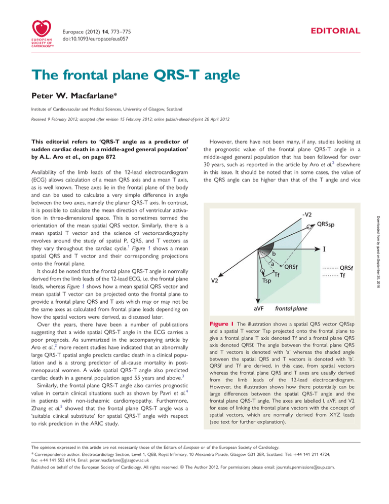
they vary throughout the cardiac cycle.1 Figure 1 shows a mean
spatial QRS and T vector and their corresponding projections
onto the frontal plane.
It should be noted that the frontal plane QRS-T angle is normally
derived from the limb leads of the 12-lead ECG, i.e. the frontal plane
leads, whereas Figure 1 shows how a mean spatial QRS vector and
mean spatial T vector can be projected onto the frontal plane to
provide a frontal plane QRS and T axis which may or may not be
the same axes as calculated from frontal plane leads depending on
how the spatial vectors were derived, as discussed later.
Over the years, there have been a number of publications
suggesting that a wide spatial QRS-T angle in the ECG carries a
poor prognosis. As summarized in the accompanying article by
Aro et al.,2 more recent studies have indicated that an abnormally
large QRS-T spatial angle predicts cardiac death in a clinical population and is a strong predictor of all-cause mortality in postmenopausal women. A wide spatial QRS-T angle also predicted
cardiac death in a general population aged 55 years and above.3
Similarly, the frontal plane QRS-T angle also carries prognostic
value in certain clinical situations such as shown by Pavri et al. 4
in patients with non-ischaemic cardiomyopathy. Furthermore,
Zhang et al.5 showed that the frontal plane QRS-T angle was a
‘suitable clinical substitute’ for spatial QRS-T angle with respect
to risk prediction in the ARIC study.
However, there have not been many, if any, studies looking at
the prognostic value of the frontal plane QRS-T angle in a
middle-aged general population that has been followed for over
30 years, such as reported in the article by Aro et al.2 elsewhere
in this issue. It should be noted that in some cases, the value of
the QRS angle can be higher than that of the T angle and vice
Figure 1 The illustration shows a spatial QRS vector QRSsp
and a spatial T vector Tsp projected onto the frontal plane to
give a frontal plane T axis denoted Tf and a frontal plane QRS
axis denoted QRSf. The angle between the frontal plane QRS
and T vectors is denoted with ‘a’ whereas the shaded angle
between the spatial QRS and T vectors is denoted with ‘b’.
QRSf and Tf are derived, in this case, from spatial vectors
whereas the frontal plane QRS and T axes are usually derived
from the limb leads of the 12-lead electrocardiogram.
However, the illustration shows how there potentially can be
large differences between the spatial QRS-T angle and the
frontal plane QRS-T angle. The axes are labelled I, aVF, and V2
for ease of linking the frontal plane vectors with the concept of
spatial vectors, which are normally derived from XYZ leads
(see text for further explanation).
The opinions expressed in this article are not necessarily those of the Editors of Europace or of the European Society of Cardiology.
* Correspondence author. Electrocardiology Section, Level 1, QEB, Royal Infirmary, 10 Alexandra Parade, Glasgow G31 2ER, Scotland. Tel: +44 141 211 4724;
fax: +44 141 552 6114, Email: peter.macfarlane@glasgow.ac.uk
Published on behalf of the European Society of Cardiology. All rights reserved. & The Author 2012. For permissions please email: journals.permissions@oup.com.
774
versa. Aro et al.2 chose to take the absolute value of the difference
between the two angles.
The normal limits of the frontal plane QRS-T angle are well
known. For example, this writer published age- and sex-related
normal limits some years ago.6 In young male individuals under
29 years of age, the normal range was from 239 to 718 (i.e. a
span of 1108). In young women of the same age, the range was
246 to 598 (i.e. a span of 1058). In older male individuals over
50 years of age, the range was 282 to 408 (i.e. 1208). In older
female individuals in the same age bracket, the range was 289
to 268 (i.e. a range of 1178). In their study, Aro et al.2 arbitrarily
chose 1008 as a threshold for an abnormal planar QRS-T angle
but it should be noted that they measured angles to the nearest
108 so that if a planar QRS-T angle exceeded 1008 in their
study, it had a minimum value of 1108.
These frontal plane angles are easily calculated whereas a spatial
QRS-T angle requires an additional electrocardiographic dimension
to allow the mean spatial QRS vector to be derived, as does the
mean spatial T vector. The angle between these two vectors is
the spatial QRS-T vector (see Figure 1).
Editorial
In order to do this, authors generally transform the 12-lead ECG
into the three orthogonal XYZ lead ECG, from which the vectorcardiogram is derived, using equations7 that are best applied via
computer techniques. With the use of these three XYZ leads,
the spatial mean QRS and T vectors can be derived and hence
the spatial QRS-T angle can be calculated. For those readers not
familiar with the three orthogonal lead ECG, leads X, Y, and Z
can very broadly be likened to leads I, aVF, and 2V2, which is
why axes have been labelled in this way in Figure 1. Indeed,
spatial mean QRS and T vectors could be calculated less accurately
using I, aVF, and V2, in which case their projections onto the frontal
plane would produce planar QRS and T vectors very close to
those derived directly from the limb leads.
It is clearly simpler for the practising physician to assess QRS-T
in the frontal plane but on the other hand, with the widespread
availability of automated ECG interpretation machines nowadays,
a spatial QRS-T angle could easily be calculated and incorporated
into the output measurements matrix.
Figure 1 has been designed deliberately to show that although
there may be a large spatial QRS-T angle, it is theoretically feasible
Downloaded from by guest on September 30, 2016
Figure 2 An example of limb leads which give rise to a frontal plane QRS-T angle of 108 due to a QRS axis of 238 and a T axis of 2138. The
electrocardiogram was recorded from an apparently healthy 44-year-old man.
775
Editorial
and those with T axis ≥1008 treated separately still had an
increased risk of an adverse outcome.
The message from Aro et al. is that a wide frontal plane QRS-T
angle carries a considerably increased risk of sudden arrhythmic
death and all-cause mortality but not of non-arrhythmic cardiac
mortality. However, the authors stress that, in the main, this
result is a reflection of an abnormal T axis, which reflects some
of the argument above. Indeed, patients with an abnormal T axis
≤ 210 or ≥1008 had an increased risk of arrhythmic death with
a relative risk of 2.13, which is very similar to the relative risk of
2.26 associated with a wide QRS-T angle.
It could be argued that the limitations of the study such as
(i) measuring angles to the nearest 108 and (ii) perhaps some uncertainly over an arrhythmic vs. non-arrhythmic death, even allowing for clearly specified definitions, could have influenced results in
some way but the overall conclusion is in keeping with other
studies, which suggests that these limitations did not have a significant effect on the overall result of the study.
The conclusion is that a simple check on QRS-T angle in the
frontal plane provides a further indicator of cardiac risk, although
with an abnormal angle having a prevalence of 2%, at least in a
general population, it may be of limited value to the clinician.
Conflict of interest: none declared.
References
1. Chou TC, Helm RA. Clinical Vectorcardiography. New York: Grune and Stratton;
1967.
2. Aro AL, Huikuri HV, Tikkanen JT, Juntilla MJ, Rissanen HA, Reunanen A et al.
QRS-T angle as a predictor of sudden cardiac death in a middle-aged general population. Europace 2012;14:872 –876.
3. Kardys I, Kors J, van der Meer I, Hofman A, van der Kuip DAM, Witteman JCM.
Spatial QRS-T angle predicts cardiac death in a general population. Eur Heart J
2003;24:1357 –64.
4. Pavri BB, Hillis MB, Subacius H, Brumberg GE, Schaechter A, Levine J et al. Prognostic value and temporal behaviour of the planar QRS-T angle in patients with
nonischemic cardiomyopathy. Circulation 2008;117:3181 –866.
5. Zhang ZM, Prineas RJ, Case D, Soliman EZ, Rautaharju PM. For the ARIC Research
Group. Comparison of the prognostic significance of the electrocardiographic
QRS/T angles in predicting incident coronary heart disease and total mortality
(from the atherosclerosis risk in communities study). Am J Cardiol 2007;100:844 –9.
6. Macfarlane PW, Lawrie TDV (eds) Comprehensive Electrocardiology. Oxford: Pergamon Press; 1989;Vol 3:xp1459.
7. Edenbrandt L, Pahlm O. Vectorcardiogram synthesized from a 12 lead ECG: superiority of the inverse Dower matrix. J Electrocardiol 1988;21:361 –7.
8. Prineas RJ, Crow RS, Zhang ZM. The Minnesota Code Manual of Electrocardiographic
Findings. London: Springer; 2010.
Downloaded from by guest on September 30, 2016
for the projections of the mean spatial QRS and T vectors onto the
frontal plane to produce a narrow QRS-T frontal planar angle,
which may be minimally different from the frontal QRS-T angle
derived from limb leads. The spatial QRS-T angle therefore
might be thought to be inherently different compared with the
frontal planar QRS-T angle, though they were equivalent with
respect to risk prediction in one study.5
What does an abnormally wide planar QRS-T imply from an
electrocardiographic standpoint? Aro et al. selected a value of
≥1008 as being abnormal. This implies that, at one extreme,
there could be an upright QRS in aVF (QRS axis ¼ 908) and an
inverted T wave in the same lead (T axis ≤ 2108 say). At the
other extreme, if the QRS axis were to be at 08, then there
would be an upright R wave in lead I and an inverted T wave in
the same lead, e.g. T axis ≥1008. Thus, the implication of the
wide QRS-T angle in the frontal plane is that there must be a relatively flat or inverted T wave in the inferior lead aVF or the lateral
lead I. It might therefore be argued at this point that wide QRS-T
angle is not adding to the presence of a more basic T-wave abnormality in the inferior or lateral leads.
Aro et al. indicated that 212 individuals in their study, i.e. 2%, had
a QRS-T angle ≥1008. However, 0.7% (46) had a T-wave axis
≥1008 implying T-wave inversion in lead I while 4.4% (509) had
a T axis ≤ 2108 suggesting T-wave inversion in aVF. Thus, many
more individuals had abnormal T axes than abnormal QRS-T
angles. This can be explained relatively easily with reference to
Figure 2 where the QRS axis ¼ 238 and the T axis ¼ 2138. In
this example from a 44-year-old apparently healthy man, the
T-wave changes could be regarded as non-specific because of
the narrow QRS-T angle. The Minnesota Code recognized 50
years ago that T-wave inversion in the inferior lead aVF was not
a codeable finding if the QRS in aVF was not mainly upright as in
Figure 2 and the latest version of the code8 maintains this stance.
The code completely ignores T-wave inversion in lead III. Aro
et al. indicate that, in their study, the T axis was the main contributor to the risk associated with an abnormal QRS-T angle. It would
have been interesting to compare those individuals with a
T axis ≤ 2108 and a normal QRS-T angle versus those with T
axis ≤ 2108 and an abnormal QRS-T angle. Furthermore, would
there have been any difference in risk with QRS axis.T axis compared with QRS axis ,T axis? This question might partially have
been answered by noting that individuals with T axis ≤ 2108


