Fixing Stained Cells for Subsequent Flow Cytometric
advertisement

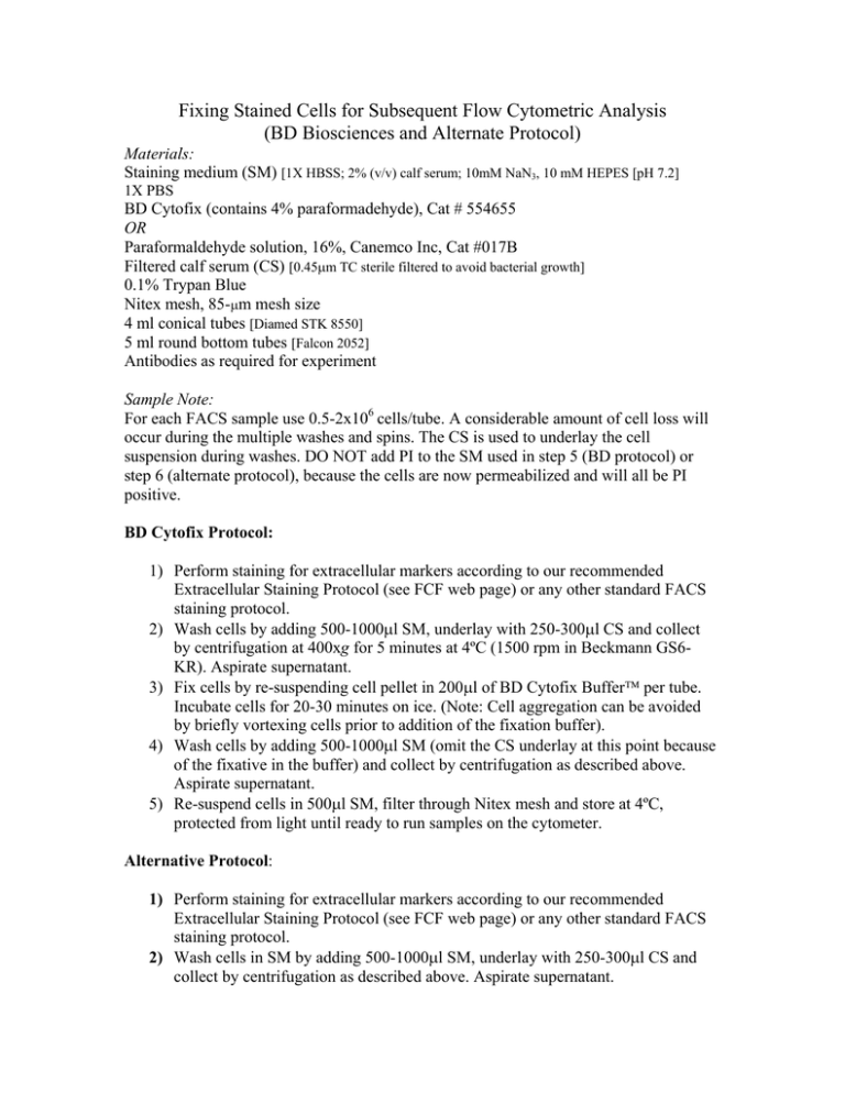
Fixing Stained Cells for Subsequent Flow Cytometric Analysis (BD Biosciences and Alternate Protocol) Materials: Staining medium (SM) [1X HBSS; 2% (v/v) calf serum; 10mM NaN3, 10 mM HEPES [pH 7.2] 1X PBS BD Cytofix (contains 4% paraformadehyde), Cat # 554655 OR Paraformaldehyde solution, 16%, Canemco Inc, Cat #017B Filtered calf serum (CS) [0.45µm TC sterile filtered to avoid bacterial growth] 0.1% Trypan Blue Nitex mesh, 85-µm mesh size 4 ml conical tubes [Diamed STK 8550] 5 ml round bottom tubes [Falcon 2052] Antibodies as required for experiment Sample Note: For each FACS sample use 0.5-2x10⁶ cells/tube. A considerable amount of cell loss will occur during the multiple washes and spins. The CS is used to underlay the cell suspension during washes. DO NOT add PI to the SM used in step 5 (BD protocol) or step 6 (alternate protocol), because the cells are now permeabilized and will all be PI positive. BD Cytofix Protocol: 1) Perform staining for extracellular markers according to our recommended Extracellular Staining Protocol (see FCF web page) or any other standard FACS staining protocol. 2) Wash cells by adding 500-1000µl SM, underlay with 250-300µl CS and collect by centrifugation at 400xg for 5 minutes at 4ºC (1500 rpm in Beckmann GS6KR). Aspirate supernatant. 3) Fix cells by re-suspending cell pellet in 200µl of BD Cytofix Buffer per tube. Incubate cells for 20-30 minutes on ice. (Note: Cell aggregation can be avoided by briefly vortexing cells prior to addition of the fixation buffer). 4) Wash cells by adding 500-1000µl SM (omit the CS underlay at this point because of the fixative in the buffer) and collect by centrifugation as described above. Aspirate supernatant. 5) Re-suspend cells in 500µl SM, filter through Nitex mesh and store at 4ºC, protected from light until ready to run samples on the cytometer. Alternative Protocol: 1) Perform staining for extracellular markers according to our recommended Extracellular Staining Protocol (see FCF web page) or any other standard FACS staining protocol. 2) Wash cells in SM by adding 500-1000µl SM, underlay with 250-300µl CS and collect by centrifugation as described above. Aspirate supernatant. 3) In the meantime, prepare 2% paraformaldehyde solution in 1xPBS (from16% paraformaldehyde stock). This working solution is stable for a week when stored at 4ºC, in an airtight container, protected from light, provided that the solution remains at pH 7. 4) Fix cells by re-suspending the pellet in 500-1000µl of 2% paraformaldehyde. Alternatively, clumping may be best avoided first resuspending cells in 900µl 1xPBS and then adding 100µl 16% parafromaldehyde for a final concentration of 1.6%. Incubate at room temperature for 10 minutes or for 20-30 minutes on ice. 5) Wash cells in 2ml of 1xHBSS or 1xPBS SM (omit the CS underlay at this point because of the fixative in the buffer) and collect by centrifugation. Aspirate supernatant. 6) Re-suspend cells in 500µl SM, filter thru Nitex mesh and store at 4ºC, protected from light until ready to run samples on the cytometer. Note: works well with all fluorochromes, including PE and APC based tandem conjugates. It is recommended that fixed samples be run on the cytometer within 1 week, preferably the next day. Autofluorescence tends to increase and sample quality generally declines with long-term storage of fixed samples.


